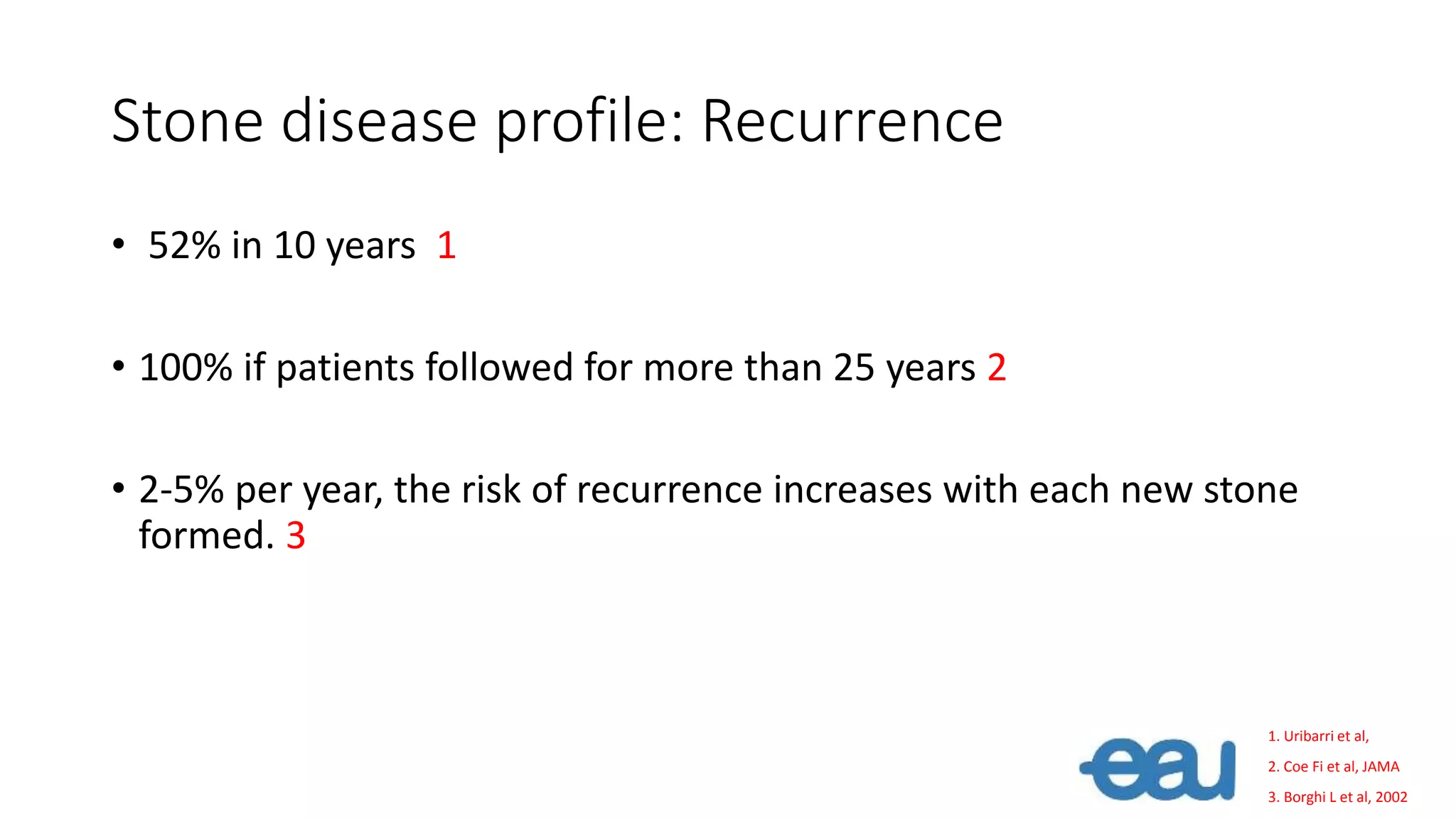
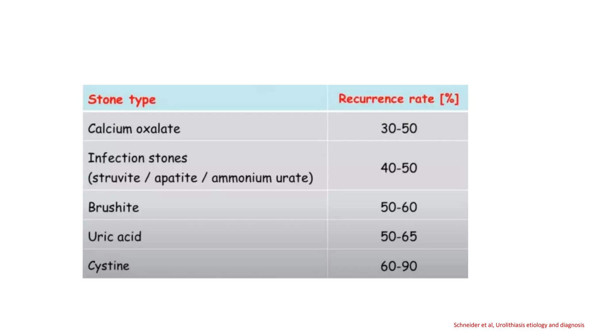
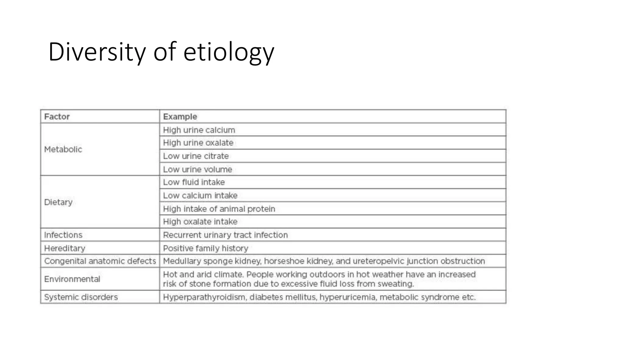
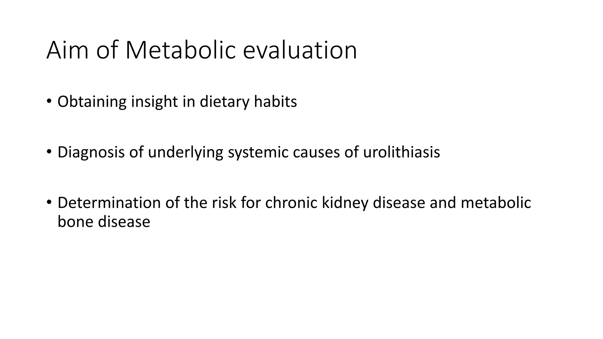
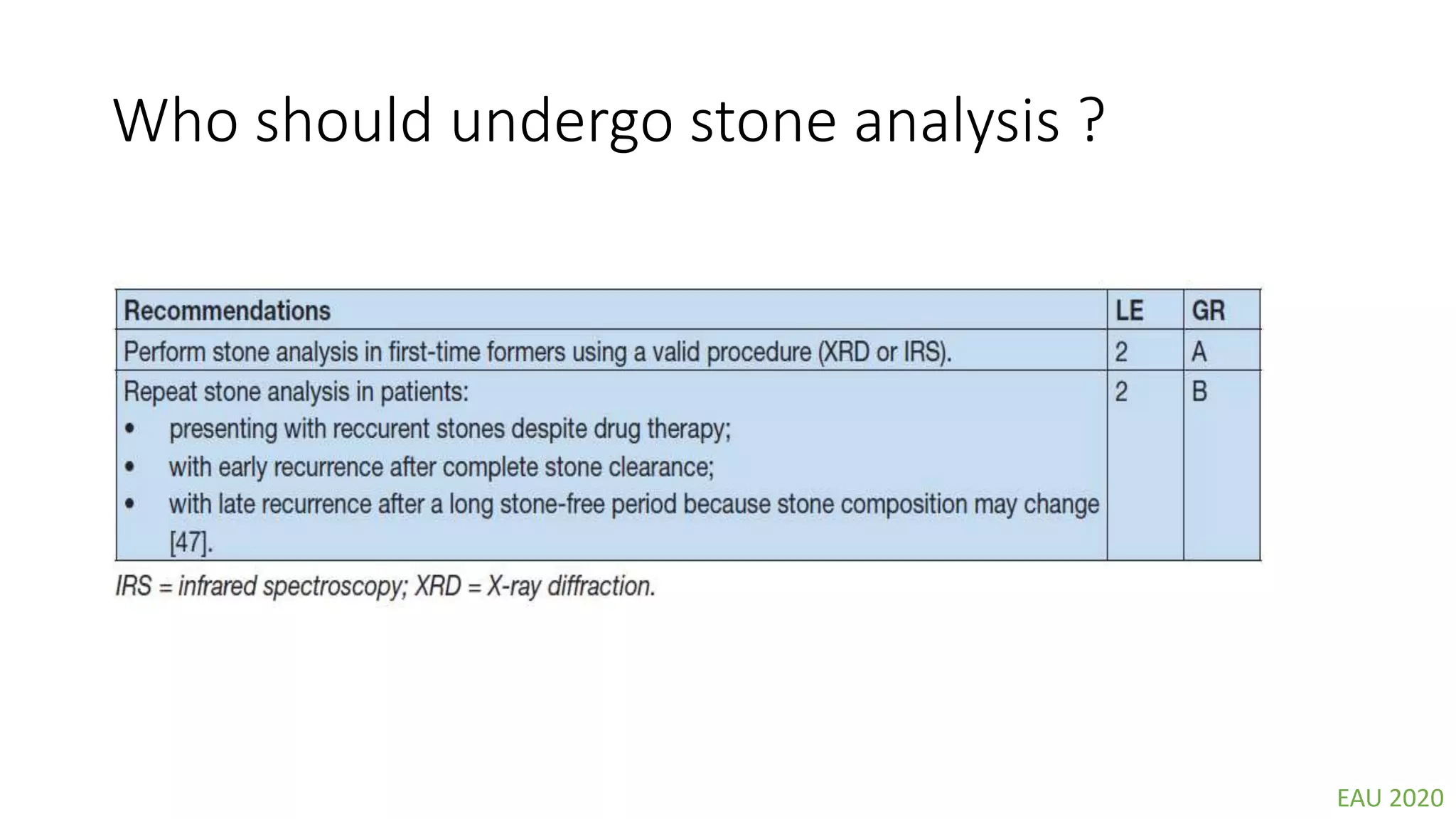
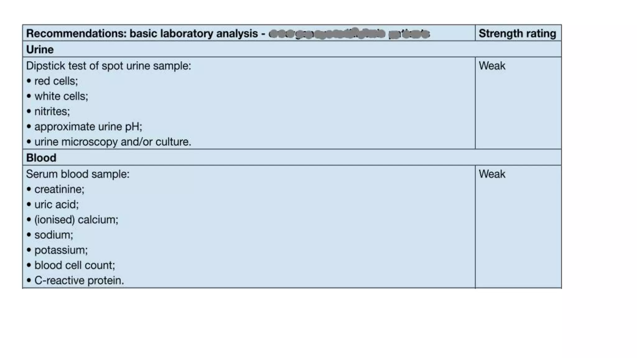
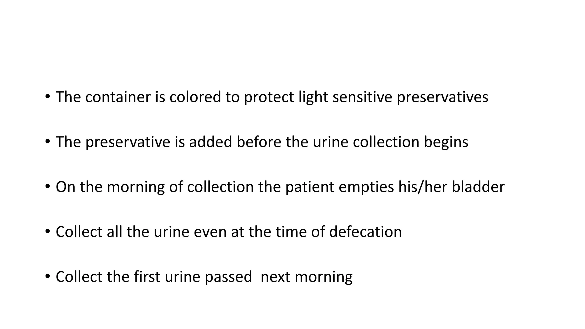
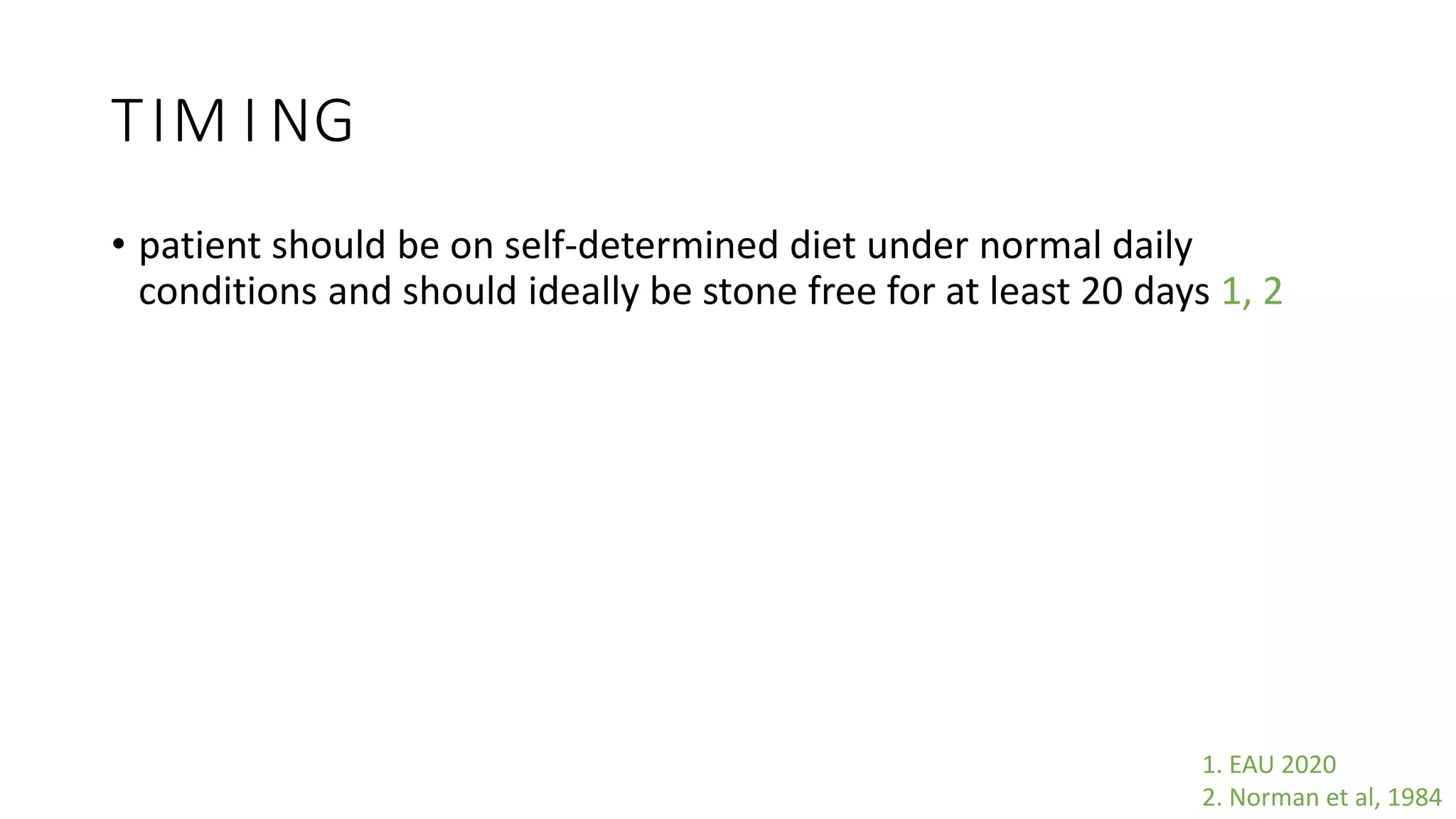
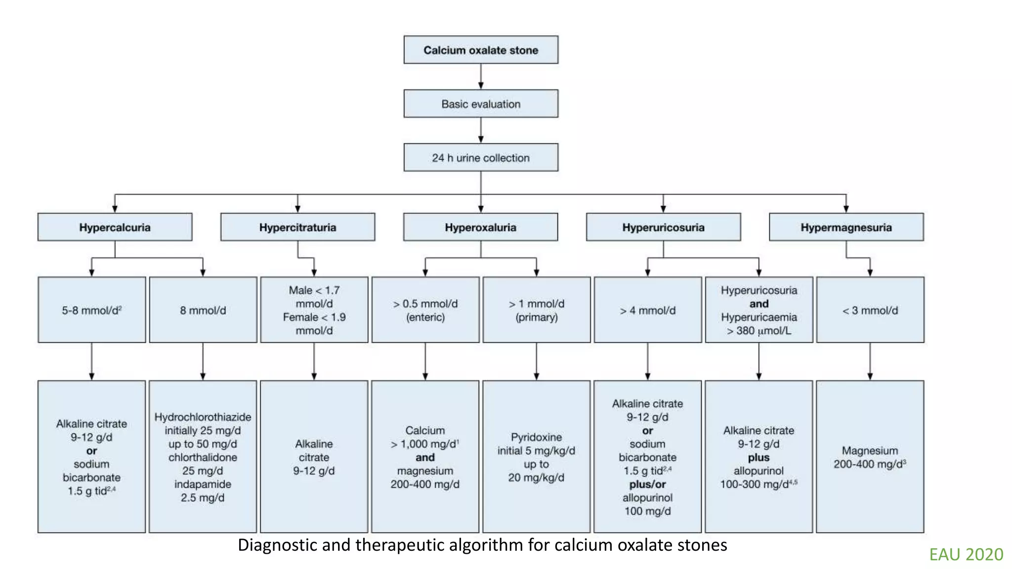
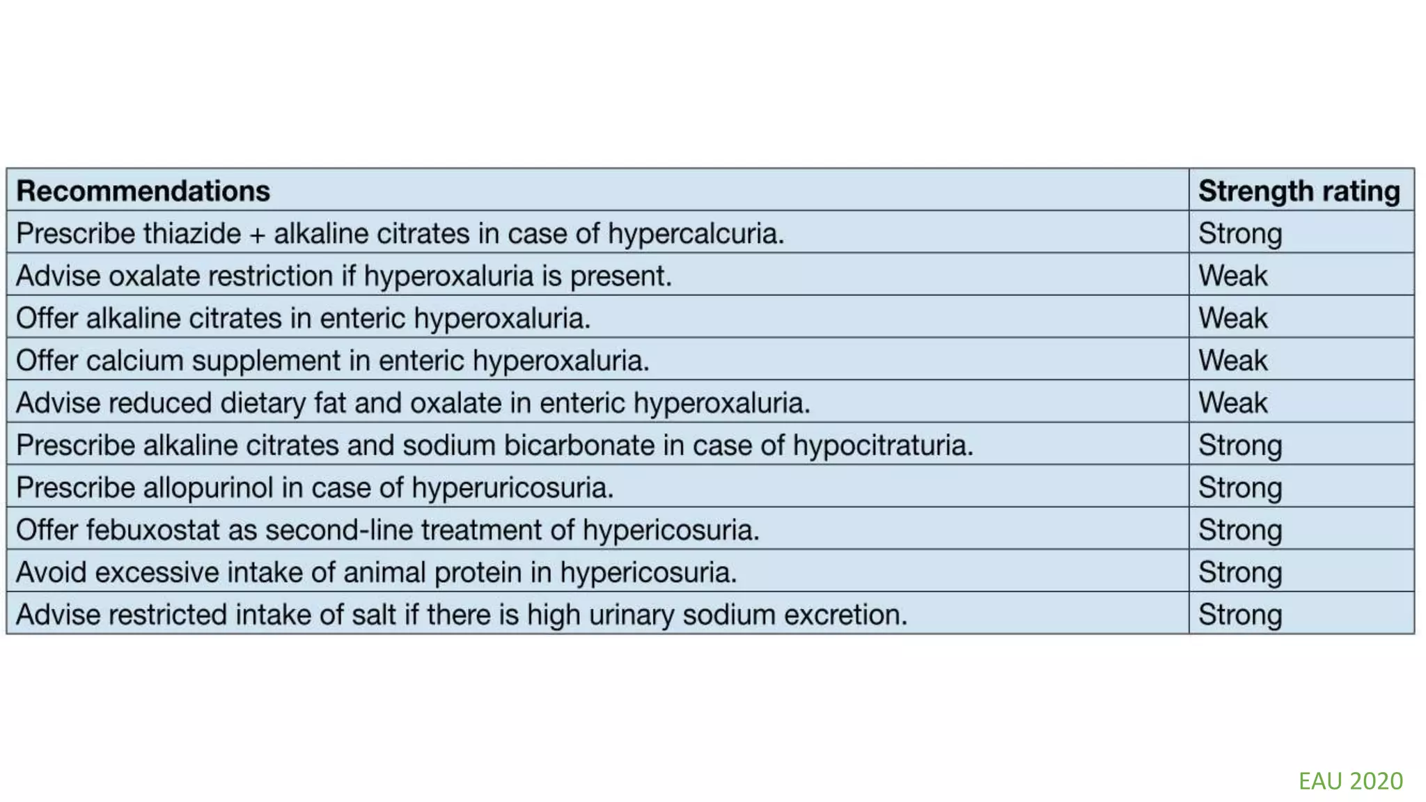
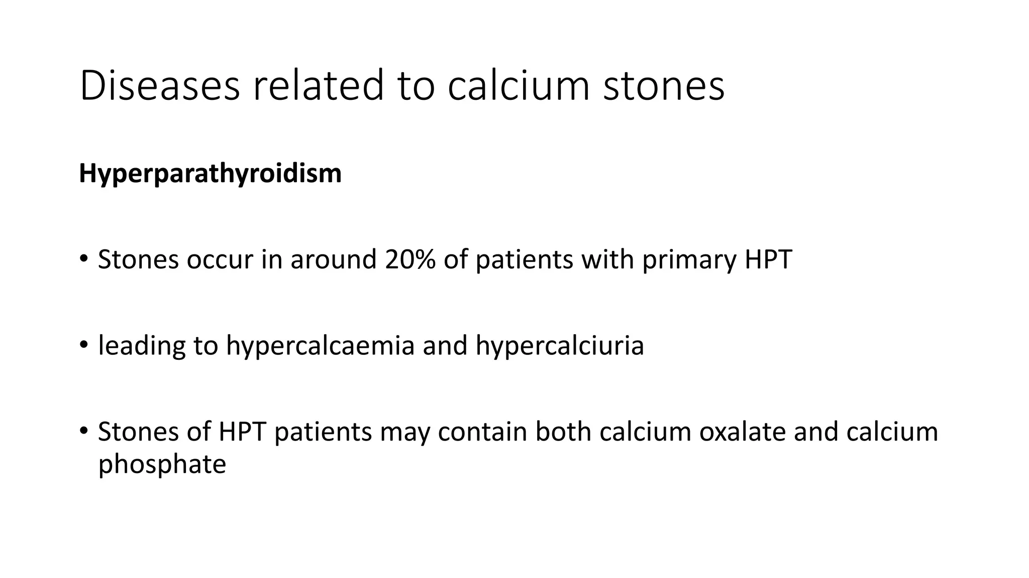
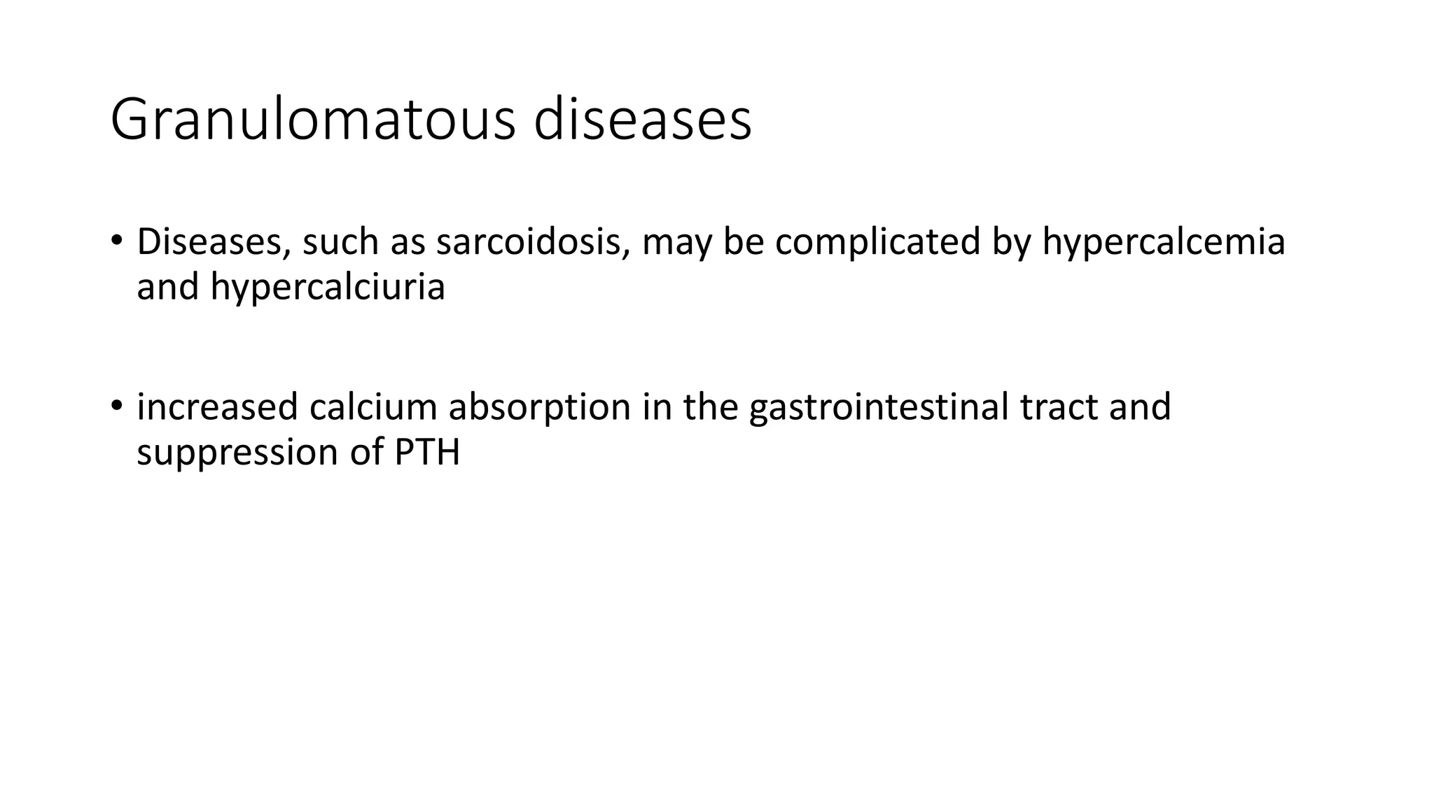
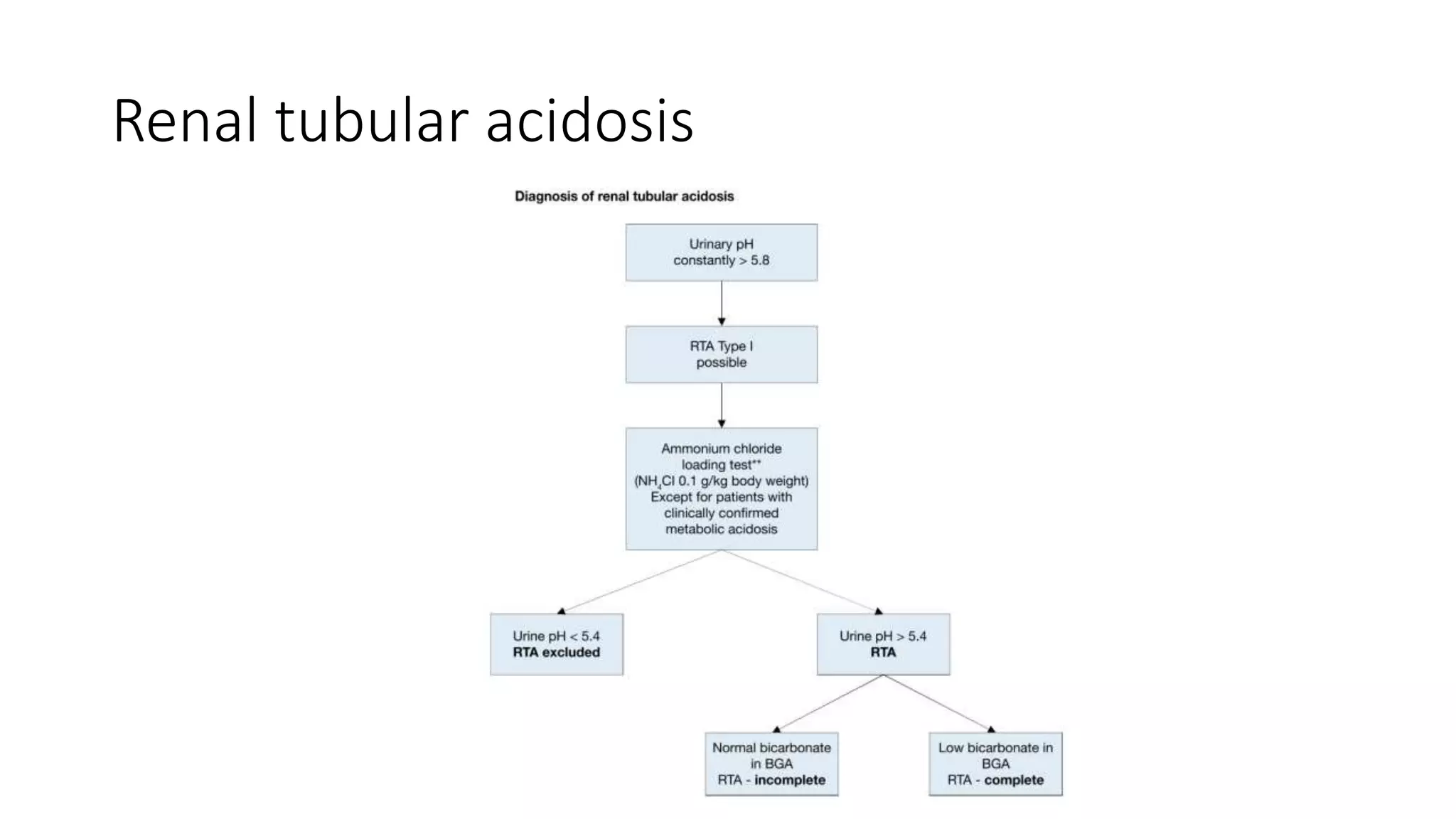
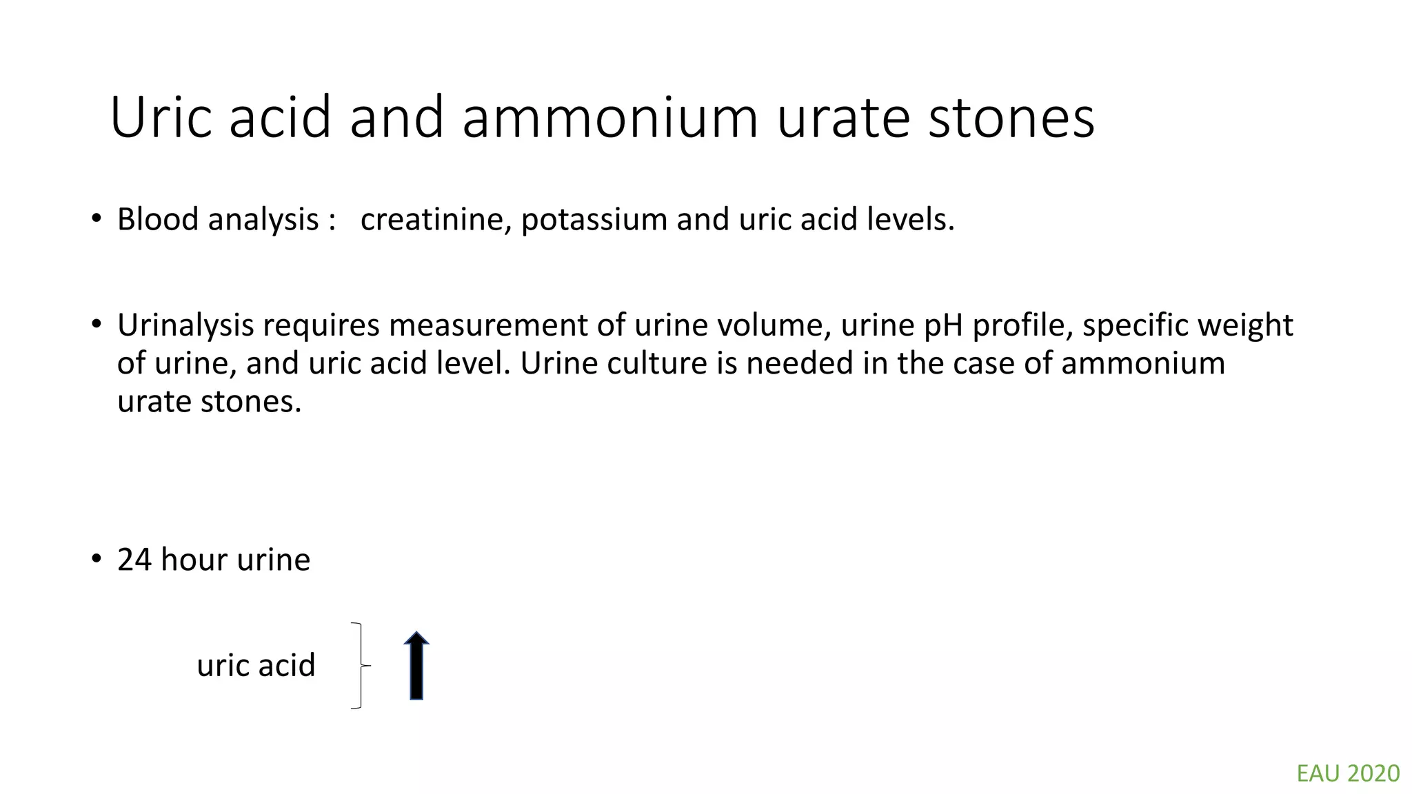
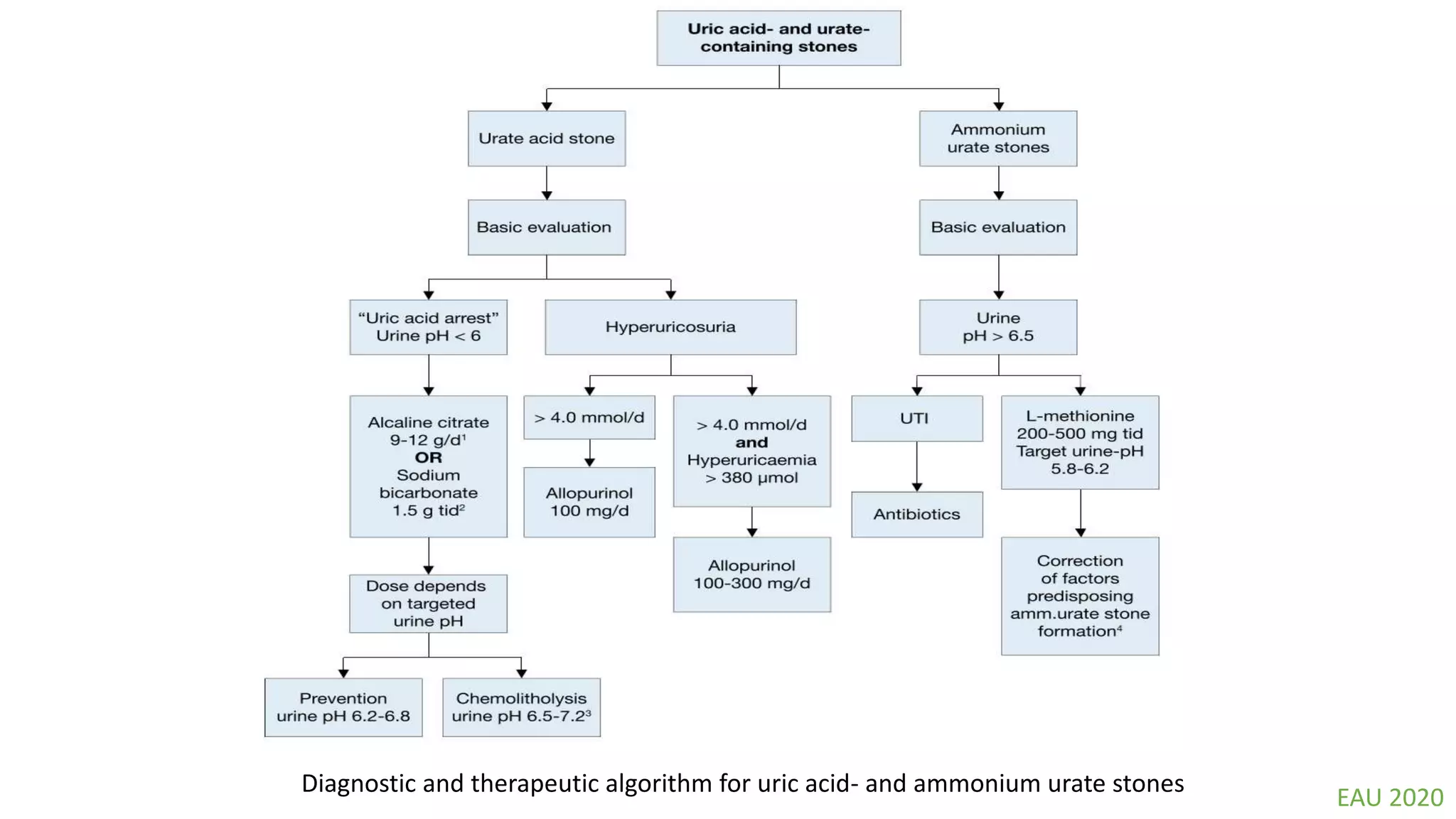
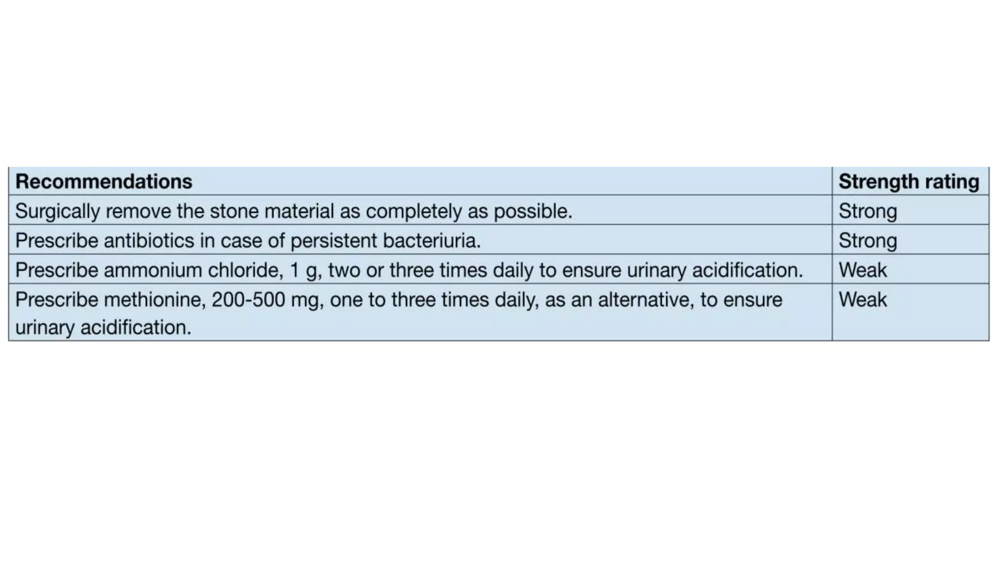
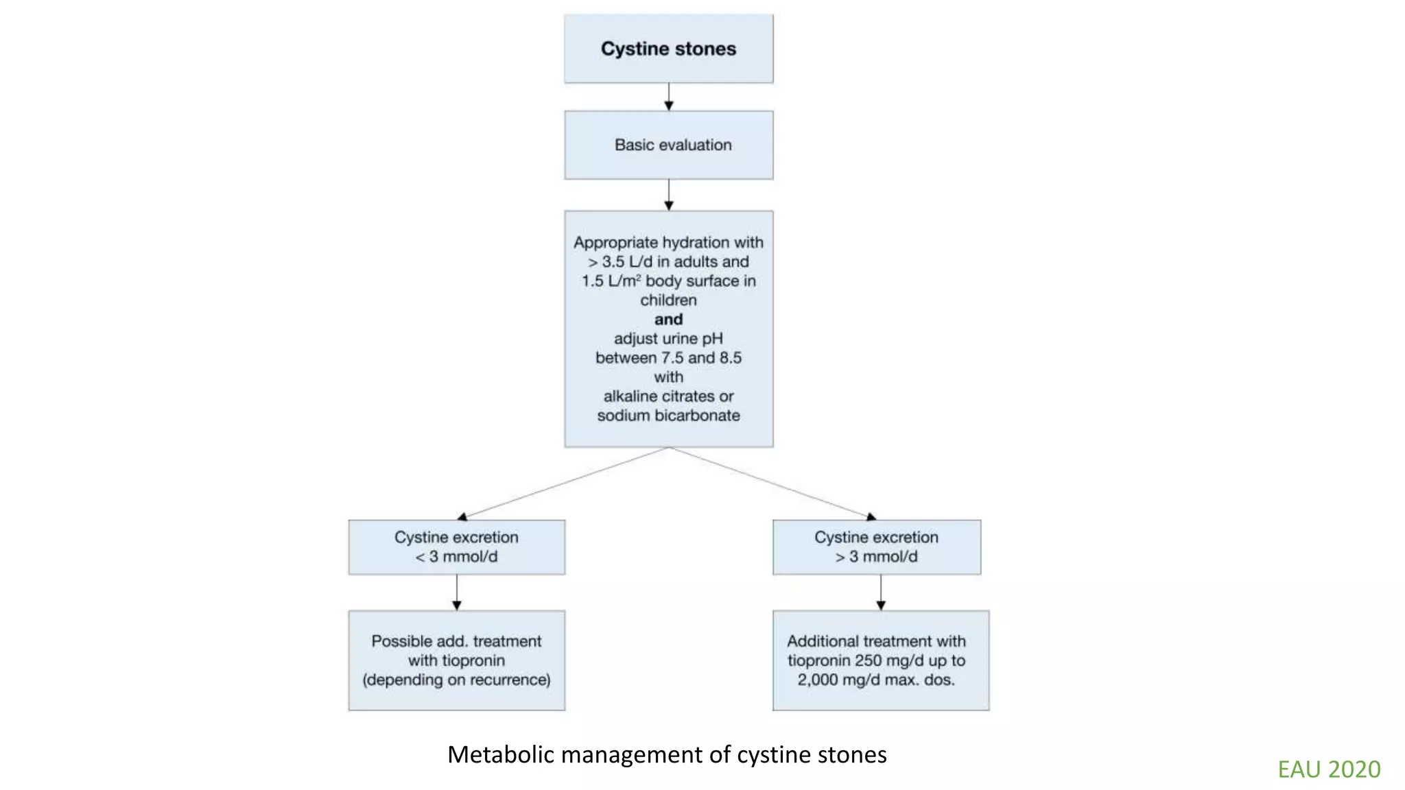
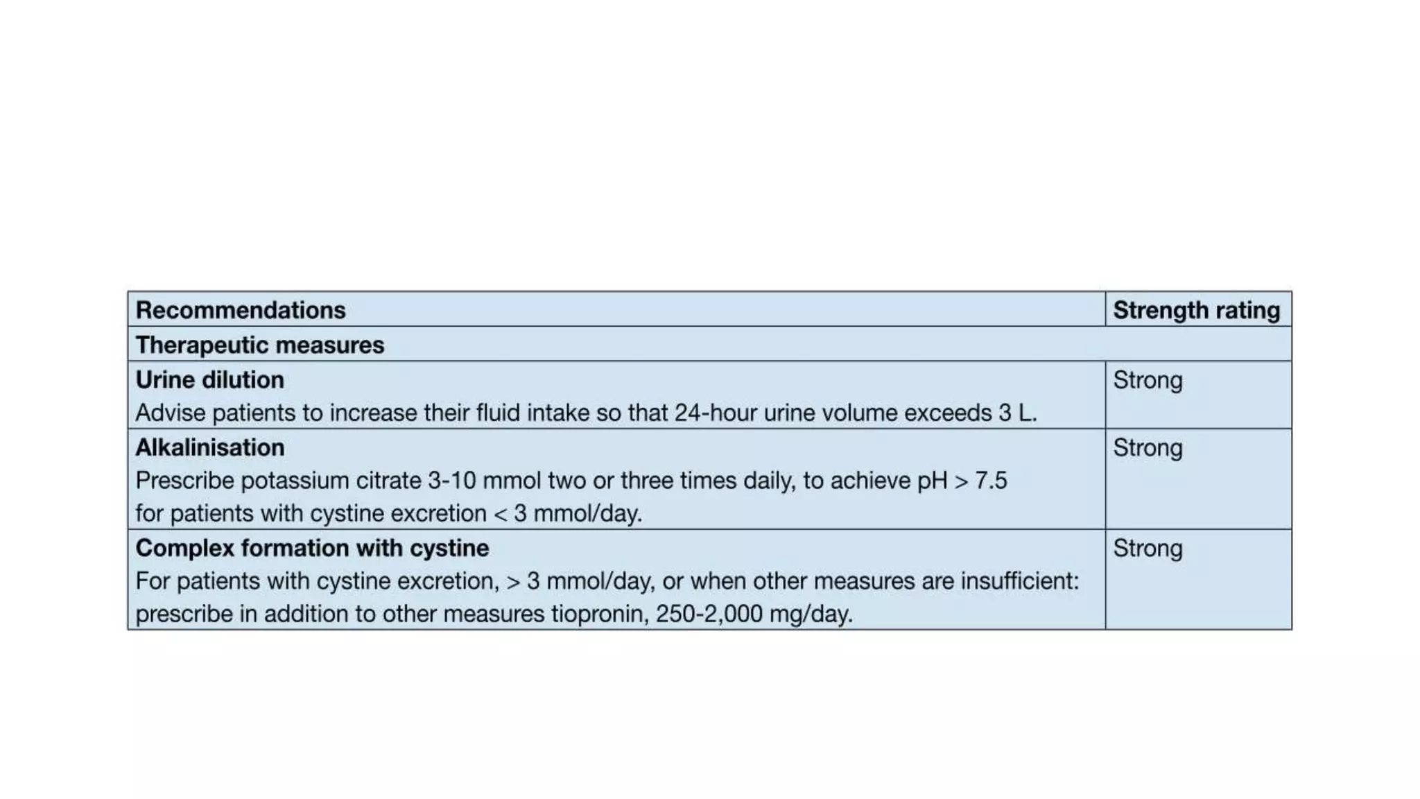
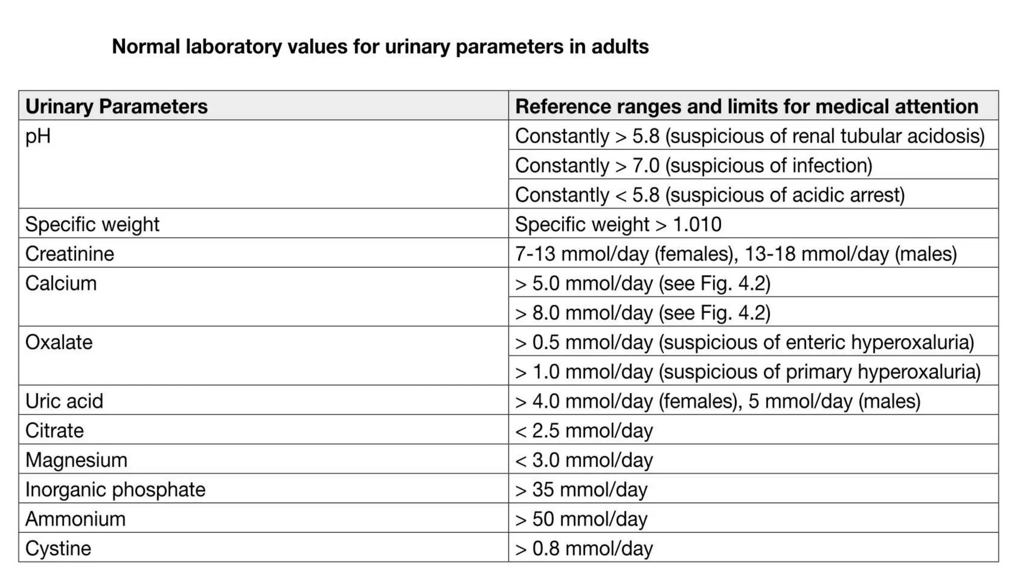
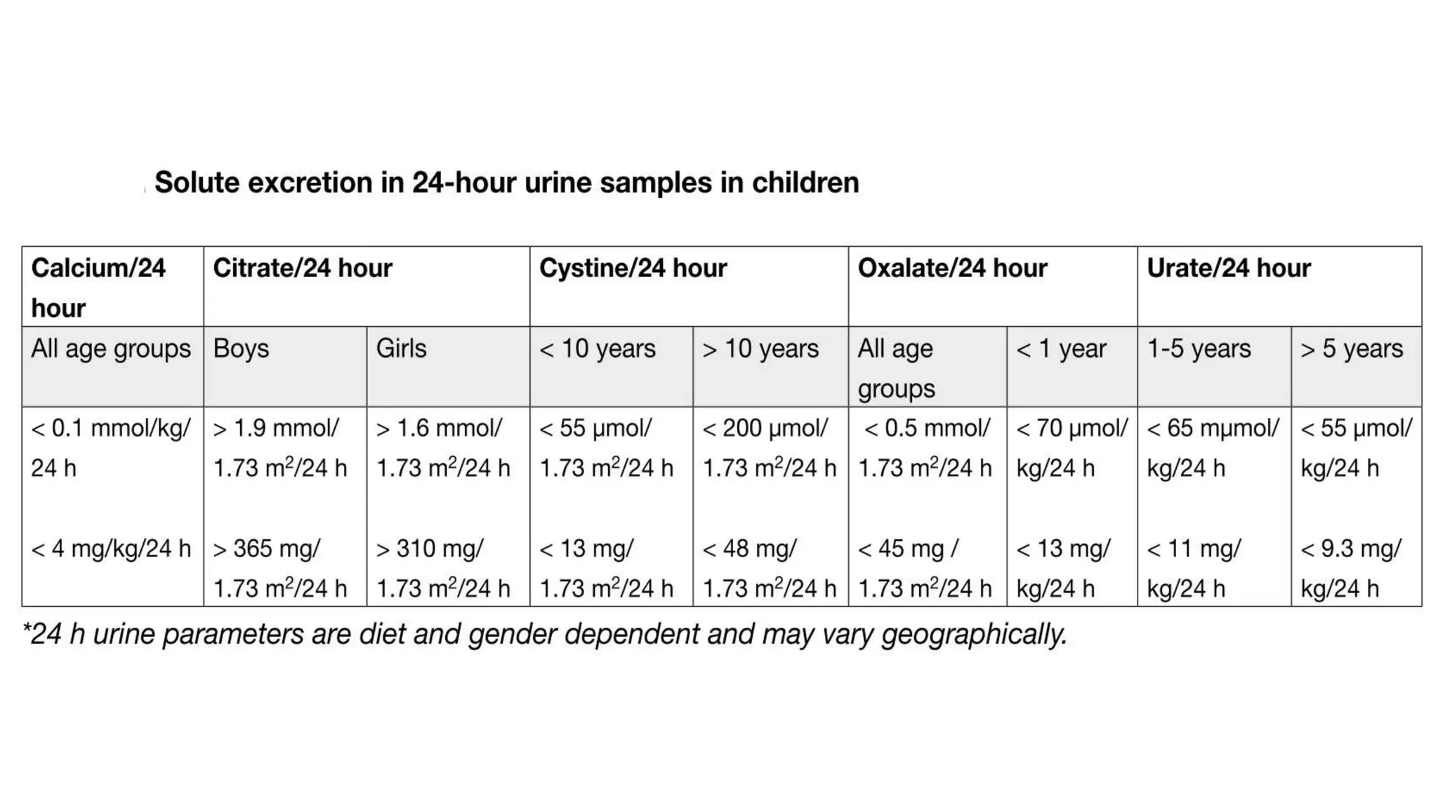
Metabolic Evaluation
&
Prevention Strategies
Presented by:
Rojan Adhikari
FCPS II Urology Resident
SDNTCSpecific Metabolic
evaluation
Basic
evaluation
Stone
Analysis
Stone disease profile: Recurrence
• 52% in 10 years 1
• 100% if patients followed for more than 25 years 2
• 2-5% per year, the risk of recurrence increases with each new stone
formed. 3
1. Uribarri et al,
2. Coe Fi et al, JAMA
3. Borghi L et al, 2002
Schneider et al, Urolithiasis etiology and diagnosis
GOALS OF Metabolic Evaluation
• to prevent recurrent stone formation in high-risk stone producers,
• to prevent extrarenal complications in associated systemic disorders
Why metabolic evaluation??
Renal stone and renal function loss
R Todd Alexander et al. BMJ 2012;345:bmj.e5287
Diversity of etiology
Stone Recurrence
Specific metabolic evaluation and
metaphylaxis can lower the recurrence rate
by 46%
Nolde et al, Bonn team
Walter et al, 2012
• Included 28 RCT of which 20 pharmacological and 8 diatery
management . In the review 23 were of calcium stone , 2 struvite
stone and 3 of other types
• Treatment duration 1-5 years
Aim of Metabolic evaluation
• Obtaining insight in dietary habits
• Diagnosis of underlying systemic causes of urolithiasis
• Determination of the risk for chronic kidney disease and metabolic
bone disease
EAU 2020
Who should undergo stone analysis ?
EAU 2020
FT-IR
• Fourier Transformation – Infrared Spectroscopy
Risk classification
• After stone passage, every patient should be assigned to a group
• For correct classification, reliable stone analysis and basic evaluation
of every patient are required.
Low
Risk High
RIsk
General Preventive Measures
High risk stone formers
• Stone-specific metabolic evaluation
Urine sampling
• two consecutive 24-hour urine samples. 1, 2
• Collecting bottles
5% thymol in isopropanol or
boric acid (10 g powder per urine container)
+
stored at < 8°C during collection to prevent the risk of spontaneous
crystallisation in the urine. 1
• Urine pH should be assessed during collection of freshly voided urine four times
daily using sensitive pH-dipsticks or a pH-meter 1, 3
1. EAU 2020,
2. Elsheemy et al 2014
3. Ferraz et al 2006
• The container is colored to protect light sensitive preservatives
• The preservative is added before the urine collection begins
• On the morning of collection the patient empties his/her bladder
• Collect all the urine even at the time of defecation
• Collect the first urine passed next morning
TIM I NG
• patient should be on self-determined diet under normal daily
conditions and should ideally be stone free for at least 20 days 1, 2
1. EAU 2020
2. Norman et al, 1984
Calcium oxalate
• Blood analysis: creatinine, sodium, potassium, ionised calcium (or total
calcium + albumin), uric acid, and parathyroid hormone (PTH) (and vitamin
D) in the case of increased calcium levels.
• Urinalysis requires measurement of urine volume, urine pH profile, specific
weight, calcium, oxalate, uric acid, citrate, sodium and magnesium.
• 24 hour urine
calcium citrate
oxalate magnesium
uric acid
EAU 2020
Diagnostic and therapeutic algorithm for calcium oxalate stones
EAU 2020
EAU 2020
Calcium phosphate
• Blood analysis : creatinine, sodium, potassium, chloride, ionised
calcium (or total calcium + albumin), and PTH (in the case of increased
calcium levels).
• Urinalysis includes measurement of: volume, urine pH profile, specific
weight, calcium, phosphate and citrate.
• 24 hour urine
Calcium phosphate
Citrate
EAU 2020
Diagnostic and therapeutic algorithm for calcium phosphate stones
EAU 2020
Diseases related to calcium stones
Hyperparathyroidism
• Stones occur in around 20% of patients with primary HPT
• leading to hypercalcaemia and hypercalciuria
• Stones of HPT patients may contain both calcium oxalate and calcium
phosphate
Granulomatous diseases
• Diseases, such as sarcoidosis, may be complicated by hypercalcemia
and hypercalciuria
• increased calcium absorption in the gastrointestinal tract and
suppression of PTH
Renal tubular acidosis
Uric acid and ammonium urate stones
• Blood analysis : creatinine, potassium and uric acid levels.
• Urinalysis requires measurement of urine volume, urine pH profile, specific weight
of urine, and uric acid level. Urine culture is needed in the case of ammonium
urate stones.
• 24 hour urine
uric acid
EAU 2020
Diagnostic and therapeutic algorithm for uric acid- and ammonium urate stones
EAU 2020
Struvite stone
• 24 hour urine not required
URINE CULTURE
EAU 2020
Diagnostic and therapeutic algorithm for infection stones EAU 2020
Factors predisposing to struvite stone
formation
Cystine stones
Blood analysis includes measurement of creatinine, and urinalysis
includes measurement of urine volume, pH profile, specific weight, and
cystine.
• 24 hour urine
cystine
EAU 2020
Metabolic management of cystine stones
EAU 2020
Xanthine stones
• high risk of recurrence
• Rare
• Fluid intake and diet is recommended for general preventive
measures
Summary of specific metabolic
evaluation
24 hour urine
Oxalate stone Phosphate
stone
Uric acid
stone
Struvite stone Cystine stone
Calcium
Oxalate
Uric acid
Citrate
Magnesium
Calcium
Phosphate
Uric acid - Cystine
Total urine volume
Urine pH
Specific weight
Follow - up
• first follow-up 24-hour urine evaluation is done after 8-12 weeks
after initiation of therapy
• 24 hour urine evaluation every 12 months is enough
1. EAU 2020
2. Norman et al, 1984
Summary
• Kidney stones are preventable if we take an holistic approach
• To maximize the efficacy of preventable regimens, appropriate data should be
gathered through proper stone analysis and basic metabolic evaluation
• Stone analysis should be done in every stone patients
• All patients should be classified into a high risk or low risk group at the time of
discharge
• High risk patients should undergo evaluation and should be given opportunity for
preventing recurrence
Thank You
1. EAU
2. Assimos et al 2007
3. Hesse et al, 2002
Diagnostic classification of stones
?
?
?
?
?
?
?
?
?
?
?
?
Indications for a Metabolic Stone Evaluation
• Recurrent stone formers
• Strong family history of stones
• Intestinal disease (particularly chronic diarrhea)
• Pathologic skeletal fractures
• Osteoporosis
• History of urinary tract infection with calculi
• Personal history of gout
• Infirm health (unable to tolerate repeat stone episodes)
• Solitary kidney
• Anatomic abnormalities
• Renal insufficiency
• Stones composed of cystine, uric acid, struvite
• Children

Metabolic evaluation of stone

  • 1.
    Metabolic Evaluation & Prevention Strategies Presentedby: Rojan Adhikari FCPS II Urology Resident SDNTCSpecific Metabolic evaluation Basic evaluation Stone Analysis
  • 3.
    Stone disease profile:Recurrence • 52% in 10 years 1 • 100% if patients followed for more than 25 years 2 • 2-5% per year, the risk of recurrence increases with each new stone formed. 3 1. Uribarri et al, 2. Coe Fi et al, JAMA 3. Borghi L et al, 2002
  • 4.
    Schneider et al,Urolithiasis etiology and diagnosis
  • 5.
    GOALS OF MetabolicEvaluation • to prevent recurrent stone formation in high-risk stone producers, • to prevent extrarenal complications in associated systemic disorders
  • 6.
    Why metabolic evaluation?? Renalstone and renal function loss R Todd Alexander et al. BMJ 2012;345:bmj.e5287
  • 7.
  • 8.
    Stone Recurrence Specific metabolicevaluation and metaphylaxis can lower the recurrence rate by 46% Nolde et al, Bonn team Walter et al, 2012
  • 9.
    • Included 28RCT of which 20 pharmacological and 8 diatery management . In the review 23 were of calcium stone , 2 struvite stone and 3 of other types • Treatment duration 1-5 years
  • 11.
    Aim of Metabolicevaluation • Obtaining insight in dietary habits • Diagnosis of underlying systemic causes of urolithiasis • Determination of the risk for chronic kidney disease and metabolic bone disease
  • 12.
  • 13.
    Who should undergostone analysis ? EAU 2020
  • 14.
    FT-IR • Fourier Transformation– Infrared Spectroscopy
  • 15.
    Risk classification • Afterstone passage, every patient should be assigned to a group • For correct classification, reliable stone analysis and basic evaluation of every patient are required. Low Risk High RIsk
  • 17.
  • 18.
  • 20.
  • 21.
    Urine sampling • twoconsecutive 24-hour urine samples. 1, 2 • Collecting bottles 5% thymol in isopropanol or boric acid (10 g powder per urine container) + stored at < 8°C during collection to prevent the risk of spontaneous crystallisation in the urine. 1 • Urine pH should be assessed during collection of freshly voided urine four times daily using sensitive pH-dipsticks or a pH-meter 1, 3 1. EAU 2020, 2. Elsheemy et al 2014 3. Ferraz et al 2006
  • 22.
    • The containeris colored to protect light sensitive preservatives • The preservative is added before the urine collection begins • On the morning of collection the patient empties his/her bladder • Collect all the urine even at the time of defecation • Collect the first urine passed next morning
  • 23.
    TIM I NG •patient should be on self-determined diet under normal daily conditions and should ideally be stone free for at least 20 days 1, 2 1. EAU 2020 2. Norman et al, 1984
  • 24.
    Calcium oxalate • Bloodanalysis: creatinine, sodium, potassium, ionised calcium (or total calcium + albumin), uric acid, and parathyroid hormone (PTH) (and vitamin D) in the case of increased calcium levels. • Urinalysis requires measurement of urine volume, urine pH profile, specific weight, calcium, oxalate, uric acid, citrate, sodium and magnesium. • 24 hour urine calcium citrate oxalate magnesium uric acid EAU 2020
  • 25.
    Diagnostic and therapeuticalgorithm for calcium oxalate stones EAU 2020
  • 26.
  • 27.
    Calcium phosphate • Bloodanalysis : creatinine, sodium, potassium, chloride, ionised calcium (or total calcium + albumin), and PTH (in the case of increased calcium levels). • Urinalysis includes measurement of: volume, urine pH profile, specific weight, calcium, phosphate and citrate. • 24 hour urine Calcium phosphate Citrate EAU 2020
  • 28.
    Diagnostic and therapeuticalgorithm for calcium phosphate stones EAU 2020
  • 30.
    Diseases related tocalcium stones Hyperparathyroidism • Stones occur in around 20% of patients with primary HPT • leading to hypercalcaemia and hypercalciuria • Stones of HPT patients may contain both calcium oxalate and calcium phosphate
  • 31.
    Granulomatous diseases • Diseases,such as sarcoidosis, may be complicated by hypercalcemia and hypercalciuria • increased calcium absorption in the gastrointestinal tract and suppression of PTH
  • 32.
  • 34.
    Uric acid andammonium urate stones • Blood analysis : creatinine, potassium and uric acid levels. • Urinalysis requires measurement of urine volume, urine pH profile, specific weight of urine, and uric acid level. Urine culture is needed in the case of ammonium urate stones. • 24 hour urine uric acid EAU 2020
  • 35.
    Diagnostic and therapeuticalgorithm for uric acid- and ammonium urate stones EAU 2020
  • 36.
    Struvite stone • 24hour urine not required URINE CULTURE EAU 2020
  • 37.
    Diagnostic and therapeuticalgorithm for infection stones EAU 2020
  • 39.
    Factors predisposing tostruvite stone formation
  • 40.
    Cystine stones Blood analysisincludes measurement of creatinine, and urinalysis includes measurement of urine volume, pH profile, specific weight, and cystine. • 24 hour urine cystine EAU 2020
  • 41.
    Metabolic management ofcystine stones EAU 2020
  • 43.
    Xanthine stones • highrisk of recurrence • Rare • Fluid intake and diet is recommended for general preventive measures
  • 44.
    Summary of specificmetabolic evaluation 24 hour urine Oxalate stone Phosphate stone Uric acid stone Struvite stone Cystine stone Calcium Oxalate Uric acid Citrate Magnesium Calcium Phosphate Uric acid - Cystine Total urine volume Urine pH Specific weight
  • 45.
    Follow - up •first follow-up 24-hour urine evaluation is done after 8-12 weeks after initiation of therapy • 24 hour urine evaluation every 12 months is enough 1. EAU 2020 2. Norman et al, 1984
  • 46.
    Summary • Kidney stonesare preventable if we take an holistic approach • To maximize the efficacy of preventable regimens, appropriate data should be gathered through proper stone analysis and basic metabolic evaluation • Stone analysis should be done in every stone patients • All patients should be classified into a high risk or low risk group at the time of discharge • High risk patients should undergo evaluation and should be given opportunity for preventing recurrence
  • 47.
  • 49.
    1. EAU 2. Assimoset al 2007 3. Hesse et al, 2002
  • 54.
    Diagnostic classification ofstones ? ? ? ? ? ? ? ? ? ? ? ?
  • 55.
    Indications for aMetabolic Stone Evaluation • Recurrent stone formers • Strong family history of stones • Intestinal disease (particularly chronic diarrhea) • Pathologic skeletal fractures • Osteoporosis • History of urinary tract infection with calculi • Personal history of gout • Infirm health (unable to tolerate repeat stone episodes) • Solitary kidney • Anatomic abnormalities • Renal insufficiency • Stones composed of cystine, uric acid, struvite • Children

Editor's Notes

  • #2 Metabolic evaluation revolves around stone analysis, basic evaluation and specific metabolic evaluation
  • #6 It should be simple to perform, economically viable, provide information that can be applied toward a selective, rational therapy of stone disease.
  • #7  Forest plot of multivariable adjusted hazard ratios for kidney stones and ESRD. Absolute rates are ESRD rates per 1 000 000 person days The hazard ratio is a comparison between the probability of events in a treatment group, compared to the probability of events in a control group. It's used to see if patients receiving a treatment progress faster (or slower) than those not receiving treatment
  • #8 If a patient have calcium oxalate stone, obiously the etiology differs for stone formation , so to find the etiology and treat the condition we need to do metabolic evaluation
  • #13 Patients identified as high-risk stone formers include those with a family history of stone disease, those with obesity and/or metabolic syndrome, and patients with medical conditions that predispose to stone formation such as gastrointestinal (GI) disease or prior surgical resection resulting in malabsorption, primary hyperparathyroidism, renal tubular acidosis, sarcoidosis, gout, type 2 diabetes mellitus, and urinary tract infection (UTI). Stone
  • #14 1. IR Spectroscopy FT-IR is the best method 2. X-ray diffraction method Also another option if available 3. Polarisation microscopy Only in centers with expertise 4. Chemical analysis (wet chemistry) Obsolete (not valid)
  • #15 Costs around $10,000 The FT is an algorithm that transforms wave sine change function into frequency, giving us a typical graph for particular molecule
  • #19 Only high-risk stone formers require specific metabolic evaluation. Stone type is the deciding factor for further diagnostic tests.
  • #25 Acid arrest (pH < 6) may promote cocrystallisation of uric acid & ca oxalate Blood analysis requires measurement of creatinine, sodium, potassium, chloride, ionised calcium (or total calcium + albumin), uric acid, and parathyroid hormone (PTH) (and vitamin D) in the case of increased calcium levels. Urinalysis requires measurement of urine volume, urine pH profile, specific weight, calcium, oxalate, uric acid, citrate, sodium and magnesium
  • #26 The most common metabolic abnormalities associated with calcium stone formation are hypercalciuria, affects 30-60% of adult stone formers, and hyperoxaluria (26-67%), followed by hyperuricosuria (15-46%), hypomagnesiuria (7-23%), and hypocitraturia (5-29%). Elevated levels of ionised calcium in serum (or total calcium and albumin) require assessment of intact PTH to confirm or exclude suspected hyperparathyroidism (HPT). • • “Acidic arrest” (urine pH constantly < 5.8) may promote co-crystallisation of uric acid and calcium oxalate. Similarly, increased uric acid excretion (> 4 mmol/day in adults or > 12 mg/kg/day in children) can act as a promoter. • Urine pH levels constantly > 5.8 in the day profile indicate RTA, provided UTI has been excluded. An ammonium chloride loading test confirms RTA and identifies RTA subtype (Section 4.6.5). Hypercalciuria may be associated with normocalcemia (idiopathic hypercalciuria, or granulomatous diseases) or hypercalcaemia (hyperparathyroidism, granulomatous diseases, vitamin D excess, or malignancy). Hypocitraturia (male < 1.7 mmol/d, female < 1.9 mmol/d) may be idiopathic or secondary to metabolic acidosis or hypokalaemia. Oxalate excretion > 0.5 mmol/day in adults (> 0.37 mmol/1.73 m2/day in children) confirms hyperoxaluria. oo primary hyperoxaluria (oxalate excretion mostly > 1 mmol/day), appears in three genetically determined forms; oo oo secondary hyperoxaluria (oxalate excretion > 0.5 mmol/day, usually < 1 mmol/day), occurs due to intestinal hyperabsorption of oxalate or extreme dietary oxalate intake; mild hyperoxaluria (oxalate excretion 0.45-0.85 mmol/day), commonly found in idiopathic calcium oxalate stone formers. • Hypomagnesiuria (< 3.0 mmol/day) may be related to poor dietary intake or to reduced intestinal absorption (chronic diarrhoea).
  • #28 Diagnosis requires blood analysis for: creatinine, sodium, potassium, chloride, ionised calcium (or total calcium + albumin), and PTH (in the case of increased calcium levels). Urinalysis includes measurement of: volume, urine pH profile, specific weight, calcium, phosphate and citrate.
  • #29 HPT = hyperparathyroidism; RTA = renal tubular acidosis; UTI = urinary tract infection
  • #32 diseases, such as sarcoidosis, may be complicated by hypercalcaemia and hypercalciuria secondary to increased calcitriol production
  • #33 Renal tubular acidosis is caused by severe impairment of proton or bicarbonate handling along the nephron. Kidney stone formation most probably occurs in patients with distal RTA type I. Renal tubular acidosis can be acquired or inherited. Reasons for acquired RTA can be obstructive uropathy, recurrent pyelonephritis, acute tubular necrosis, renal transplantation, analgesic nephropathy, sarcoidosis, idiopathic hypercalciuria, and primary parathyroidism; it may also be drug-induced (e.g. zonisamide).
  • #34 The main therapeutic aim of RTA treatment is restoring a normal acid-base equilibrium. Despite the alkaline pH of urine in RTA, alkalinisation using alkaline citrates or sodium bicarbonate is important for normalising the metabolic changes (intracellular acidosis) responsible for stone formation (Table 4.8). The alkali load reduces tubular reabsorption of citrate, which in turn normalises citrate excretion and simultaneously reduces calcium turnover. Therapeutic success can be monitored by venous blood gas analysis (base excess: ± 2.0 mmol/L) in complete RTA. If excessive calcium excretion (> 8 mmol/day) persists after re-establishing acid-base equilibrium, thiazides may lower urinary calcium excretion.
  • #35 considered to be at high risk of recurrence Uric acid nephrolithiasis is responsible for approximately 10% of renal stones [562] and associated with hyperuricosuria or low urinary pH. Hyperuricosuria may be a result of dietary excess, endogenous overproduction (enzyme defects), myeloproliferative disorders, tumour lysis syndrome, drugs, gout or catabolism . Uric acid and ammonium urate stones form under completely different biochemical conditions. Acidic arrest (urine pH constantly < 5.8) promotes uric acid crystallisation
  • #37 All infection-stone formers are deemed at high risk of recurrence. Struvite stones represent 2-15% of the stones sent for analysis. Stones that contain struvite may originate de novo or grow on pre-existing stones, which are infected with urea-splitting bacteria [576]. There are several factors predisposing patients to struvite stone formation Diagnosis Blood analysis requires measurement of creatinine, and urinalysis requires repeat urine pH measurements and urine culture.
  • #38 Infection stones contain the following minerals: struvite and/or carbonate apatite and/or ammonium urate. Urine culture typically provides evidence for urease-producing bacteria, which increase ammonia ions and develop alkaline urine. Carbonate apatite starts to crystallise at a urine pH level of 6.8. Struvite only precipitates at pH > 7.2 . Proteus mirabilis accounts for more than half of all urease-positive UTIs
  • #41 Cystine stones account for 1-2% of all urinary stones in adults and 6-8% of the stones reported in paediatric studies [32, 586]. All cystine stone formers are deemed at high risk of recurrence
  • #42 Cystine is poorly soluble in urine and crystallises spontaneously within the physiological urinary pH range. Cystine solubility depends strongly on urine pH: at pH 6.0, the limit of solubility is 1.33 mmol/L. Routine analysis of cystine is not suitable for therapeutic monitoring. Regardless of phenotype or genotype of the cystinuric patient, the clinical manifestations are the same . There is no role for genotyping patients in the routine management of cystinuria . Reductive therapy targets the disulphide binding in the cysteine molecule. For therapy monitoring, it is essential to differentiate between cystine, cysteine and drug-cysteine complexes. Only high-performance liquid chromatography (HPLC)-based analysis differentiates between the different complexes formed by therapy. Diagnosis is established by stone analysis. The typical hexagonal crystals are detectable in only 20-25% of urine specimens from patients with cystinuria [590]. The cyanide nitroprusside colorimetric qualitative test detects the presence of cystine at a threshold concentration of 75 mg/L, with a sensitivity of 72% and specificity of 95%. False-positive results in patients with Fanconi’s syndrome, homocystinuria, or those taking various drugs, including infection stones [591]. • Quantitative 24-hour urinary cystine excretion confirms the diagnosis in the absence of stone analysis. • Levels above 30 mg/day are considered abnormal
  • #46 first follow-up 24-hour urine measurement is suggested eight to twelve weeks after starting pharmacological prevention of stone recurrence i.e. to enable drug dosage to be adjusted 1 Once urinary parameters have been normalised, it is sufficient to perform 24-hour urine evaluation every twelve months 1
  • #55 If we consider even upto 50 percent attributable to low urine volume or no no abnormality, rest 50 percent will have some form of metabolic defect