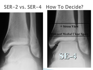
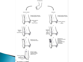
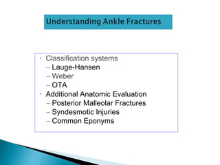
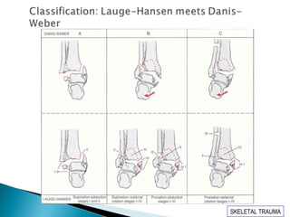
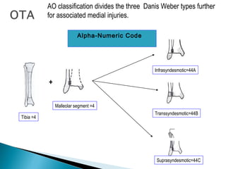
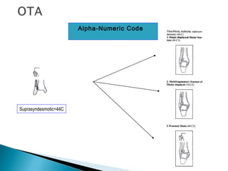
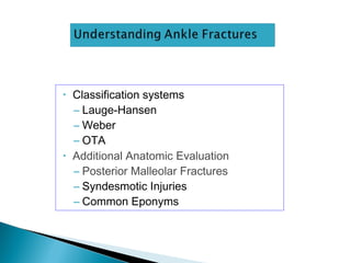
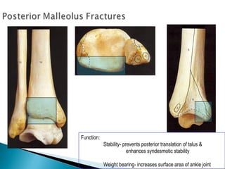
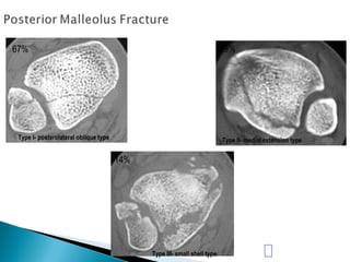
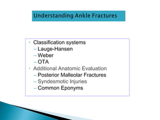
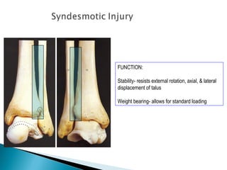
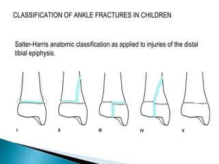
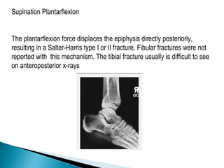
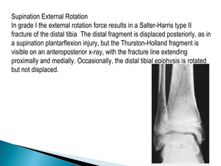
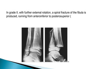
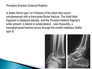
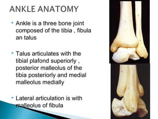
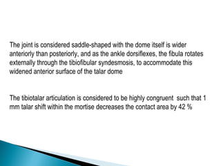
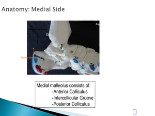
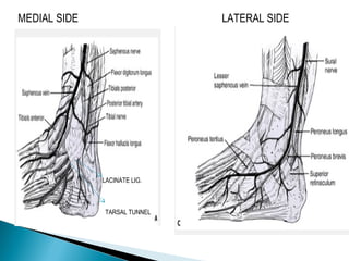
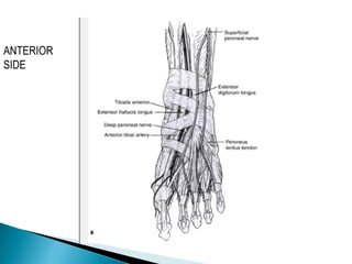
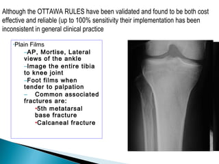
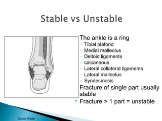
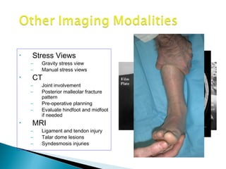
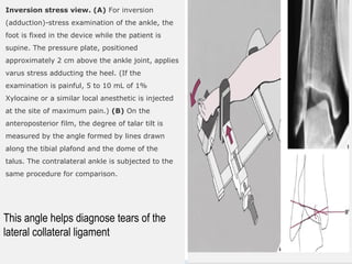
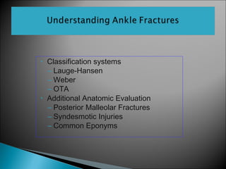
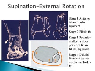
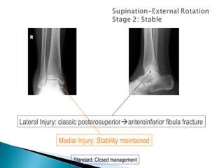
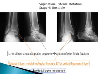
Mpfl tech - MPFL Reconstruction for Patellar InstabilityDelhiArthroscopy
MPFL Rec onstruction for Patellar Instability - By Dr Shekhar Srivastav .
Surgical Technique
- Diagnostic Arthroscopy
- Look for any Osteochondral fragment
(Loose body)
- Look for any Chondral damage
- Patellar tracking though Supero-lateral portal
Post-op Protocol
Ambulation with stick and Knee Brace- 3 wks
ROM exer – Next day upto 300 and progress
Review every 2 wks,6 wks,3 mnths,6 mnths and
yearly thereafter
Post-op assessment (Crosby-Insall criteria)
Excellent- No pain,normal activity
Good- Occasional pain,discomfort
Fair/Poor- Pain,loss of flexion,recurrent
dislocation/subluxation
Worse- Pain increased,displacement more
frequent
Caution
Must avoid overtightening-
Medial instability
Medial patellar arthritis
Patellar fractures
Preexisting Chondromalacia
Details @ http://www.delhiarthroscopy.com/
i present this lovely topic at Notional Guard Hospital in Al-Ahsa in the Orthopedic department.
hope you enjoy
Fahad Al Hulaibi
Orthopedic Resident
NGH-A
Can read freely here
https://sethiortho.blogspot.com/
Challenges and Solutions in
Management of Distal Humerus Fractures
Epidemiology
Anatomy
Classification
Controversies and Recent studies
Approach
Implants selection
Plate configuration
Ulnar nerve transposition
Role of total elbow arthroplasty in DHF
Role of hemiarthroplasty in DHF
Metaphyseal comminution –
Anatomic complexity of the distal humerus
Positioning of the plates
TBW –
Skin closure
Osteoporotic nature of the bone –
Less BMD/Thin metaphysis
Screw Pullout strength is low
DHF account for 2% of all adult fractures
The common pattern of fracture
Intraarticular and involves both columns
Bimodal distribution
Peak incidence in young male and in older female patients
Young male – High-velocity injury
Older female - Osteoporosis
The distal humerus is flattened and expanded bony structure
It is composed of lateral and medial columns with the trochlea situated between these columns.
The location of the trochlea is central rather than medial
Formed by Medial SCR + M/Epicondyle
The distal end has 450 angulation with humeral shaft
M/ Epicondyle gives attachment for MCL & Common Flexor Origin
The MCL originates from the undersurface of the medial epicondyle where it is vulnerable to excessive dissection
Ulnar nerve
Formed by Lateral SCR and L/Epicondyle and Capitulum
Distal end has 200 with humeral shaft
L/ epicondyle gives attachment for LCL & common extensor origin
Its posterior surface is non articular and can be used as a site for a plate fixation
The lateral column curves anteriorly
Placement of a straight plate on the posterolateral surface of the humerus risks straightening of distal humerus.
The medial column including the medial epicondyle is in line with the humeral shaft.
It forms the center of the triangle
It has 30 - 80 – external rotation & 250 anterior divergent with the shaft
It forms a 40 - 80 degree valgus direction
X-ray -
Anterior-posterior view
lateral View
Traction View – This can help to define articular fragments and aid in pre-operative classification of the fracture.
NCCT – Elbow
Articular surfaces
Position of the fracture fragments
useful for identifying impacted fracture fragments that make reduction challenging
Olecranon Osteotomy Approach – 52-57%
Triceps sparing VS Olecranon osteotomy approach
The lateral column was often the first to fail as a result of excessive varus forces acting on the elbow during normal activities of daily living. Small anterior-posterior diameter
Smaller diameter of the humerus, permitting only one or two short screws for fixation.
Interruption of blood supply to the lateral column
blood supply to the lateral column is also derived from posterior segmental vessels. Sagittal plane plating has less risk of injuring these structures, which may improve the chances of union
1) Subtrochanteric Fracture
Subtrochanteric typically defined as area from lesser trochanter to 5cm distal fractures with an associated intertrochanteric component may be called peritrochanteric fracture.
*Unique Aspect
Blood loss is greater than with femoral neck or trochanteric fractures – covered with anastomosing branches of the medial and lateral circumflex femoral arteries branch of profunda femoris trunk.
2) Femoral Shaft Fracture
Femoral shaft fracture is defined as a fracture of the diaphysis occurring between 5 cm distal to the lesser trochanter and 5 cm proximal to the adductor tubercle
The femoral shaft is padded with large muscles.
- reduction can be difficult as muscle contraction displaces the fracture
- healing potential is improved by having this well-vascularized
*Age
-usually a fracture of young adults and results from a high energy injury
-elderly patients should be considered ‘pathological’ until proved otherwise
-children under 4 years the suspected possibility of physical abuse
*FRACTURES ASSOCIATED WITH VASCULAR INJURY
Warning signs of an associated vascular injury are
(1) excessive bleeding or haematoma formation; and
(2) paraesthesia, pallor or pulselessness in the leg and foot.
~Warm ischemia in 2-3H
~If > 6H – salvage not possible
*‘FLOATING KNEE’
Ipsilateral fractures of the femur and tibia may leave the knee joint ‘floating’
3) Distal Femoral Fracture
Defined as fractures from articular surface to 5cm above metaphyseal flare
*clinical feature
The knee is swollen because of a haemarthrosis – this can be severe enough to cause blistering later
Movement is too painful to be attempted
The tibial pulses should always be checked to ensure the popliteal artery was not injured in the fracture.
Reference: Apley's System of Orthopaedic and Fracture (9th edition)
This is the Presentation on the topic "Pathomechanics of Knee Joint".
The presentation includes images and a clip for proper understanding. The sentences are framed in the way that you can learn it in a easy way.
Similar to Anklefracturesfinal 120314092959-phpapp01 (20)
ARTIFICIAL INTELLIGENCE IN HEALTHCARE.pdfAnujkumaranit
Artificial intelligence (AI) refers to the simulation of human intelligence processes by machines, especially computer systems. It encompasses tasks such as learning, reasoning, problem-solving, perception, and language understanding. AI technologies are revolutionizing various fields, from healthcare to finance, by enabling machines to perform tasks that typically require human intelligence.
These simplified slides by Dr. Sidra Arshad present an overview of the non-respiratory functions of the respiratory tract.
Learning objectives:
1. Enlist the non-respiratory functions of the respiratory tract
2. Briefly explain how these functions are carried out
3. Discuss the significance of dead space
4. Differentiate between minute ventilation and alveolar ventilation
5. Describe the cough and sneeze reflexes
Study Resources:
1. Chapter 39, Guyton and Hall Textbook of Medical Physiology, 14th edition
2. Chapter 34, Ganong’s Review of Medical Physiology, 26th edition
3. Chapter 17, Human Physiology by Lauralee Sherwood, 9th edition
4. Non-respiratory functions of the lungs https://academic.oup.com/bjaed/article/13/3/98/278874
TEST BANK for Operations Management, 14th Edition by William J. Stevenson, Ve...kevinkariuki227
TEST BANK for Operations Management, 14th Edition by William J. Stevenson, Verified Chapters 1 - 19, Complete Newest Version.pdf
Knee anatomy and clinical tests 2024.pdfvimalpl1234
This includes all relevant anatomy and clinical tests compiled from standard textbooks, Campbell,netter etc..It is comprehensive and best suited for orthopaedicians and orthopaedic residents.
Tom Selleck Health: A Comprehensive Look at the Iconic Actor’s Wellness Journeygreendigital
Tom Selleck, an enduring figure in Hollywood. has captivated audiences for decades with his rugged charm, iconic moustache. and memorable roles in television and film. From his breakout role as Thomas Magnum in Magnum P.I. to his current portrayal of Frank Reagan in Blue Bloods. Selleck's career has spanned over 50 years. But beyond his professional achievements. fans have often been curious about Tom Selleck Health. especially as he has aged in the public eye.
Follow us on: Pinterest
Introduction
Many have been interested in Tom Selleck health. not only because of his enduring presence on screen but also because of the challenges. and lifestyle choices he has faced and made over the years. This article delves into the various aspects of Tom Selleck health. exploring his fitness regimen, diet, mental health. and the challenges he has encountered as he ages. We'll look at how he maintains his well-being. the health issues he has faced, and his approach to ageing .
Early Life and Career
Childhood and Athletic Beginnings
Tom Selleck was born on January 29, 1945, in Detroit, Michigan, and grew up in Sherman Oaks, California. From an early age, he was involved in sports, particularly basketball. which played a significant role in his physical development. His athletic pursuits continued into college. where he attended the University of Southern California (USC) on a basketball scholarship. This early involvement in sports laid a strong foundation for his physical health and disciplined lifestyle.
Transition to Acting
Selleck's transition from an athlete to an actor came with its physical demands. His first significant role in "Magnum P.I." required him to perform various stunts and maintain a fit appearance. This role, which he played from 1980 to 1988. necessitated a rigorous fitness routine to meet the show's demands. setting the stage for his long-term commitment to health and wellness.
Fitness Regimen
Workout Routine
Tom Selleck health and fitness regimen has evolved. adapting to his changing roles and age. During his "Magnum, P.I." days. Selleck's workouts were intense and focused on building and maintaining muscle mass. His routine included weightlifting, cardiovascular exercises. and specific training for the stunts he performed on the show.
Selleck adjusted his fitness routine as he aged to suit his body's needs. Today, his workouts focus on maintaining flexibility, strength, and cardiovascular health. He incorporates low-impact exercises such as swimming, walking, and light weightlifting. This balanced approach helps him stay fit without putting undue strain on his joints and muscles.
Importance of Flexibility and Mobility
In recent years, Selleck has emphasized the importance of flexibility and mobility in his fitness regimen. Understanding the natural decline in muscle mass and joint flexibility with age. he includes stretching and yoga in his routine. These practices help prevent injuries, improve posture, and maintain mobilit
Acute scrotum is a general term referring to an emergency condition affecting the contents or the wall of the scrotum.
There are a number of conditions that present acutely, predominantly with pain and/or swelling
A careful and detailed history and examination, and in some cases, investigations allow differentiation between these diagnoses. A prompt diagnosis is essential as the patient may require urgent surgical intervention
Testicular torsion refers to twisting of the spermatic cord, causing ischaemia of the testicle.
Testicular torsion results from inadequate fixation of the testis to the tunica vaginalis producing ischemia from reduced arterial inflow and venous outflow obstruction.
The prevalence of testicular torsion in adult patients hospitalized with acute scrotal pain is approximately 25 to 50 percent
These lecture slides, by Dr Sidra Arshad, offer a quick overview of physiological basis of a normal electrocardiogram.
Learning objectives:
1. Define an electrocardiogram (ECG) and electrocardiography
2. Describe how dipoles generated by the heart produce the waveforms of the ECG
3. Describe the components of a normal electrocardiogram of a typical bipolar leads (limb II)
4. Differentiate between intervals and segments
5. Enlist some common indications for obtaining an ECG
Study Resources:
1. Chapter 11, Guyton and Hall Textbook of Medical Physiology, 14th edition
2. Chapter 9, Human Physiology - From Cells to Systems, Lauralee Sherwood, 9th edition
3. Chapter 29, Ganong’s Review of Medical Physiology, 26th edition
4. Electrocardiogram, StatPearls - https://www.ncbi.nlm.nih.gov/books/NBK549803/
5. ECG in Medical Practice by ABM Abdullah, 4th edition
6. ECG Basics, http://www.nataliescasebook.com/tag/e-c-g-basics
micro teaching on communication m.sc nursing.pdfAnurag Sharma
Microteaching is a unique model of practice teaching. It is a viable instrument for the. desired change in the teaching behavior or the behavior potential which, in specified types of real. classroom situations, tends to facilitate the achievement of specified types of objectives.
HOT NEW PRODUCT! BIG SALES FAST SHIPPING NOW FROM CHINA!! EU KU DB BK substit...GL Anaacs
Contact us if you are interested:
Email / Skype : kefaya1771@gmail.com
Threema: PXHY5PDH
New BATCH Ku !!! MUCH IN DEMAND FAST SALE EVERY BATCH HAPPY GOOD EFFECT BIG BATCH !
Contact me on Threema or skype to start big business!!
Hot-sale products:
NEW HOT EUTYLONE WHITE CRYSTAL!!
5cl-adba precursor (semi finished )
5cl-adba raw materials
ADBB precursor (semi finished )
ADBB raw materials
APVP powder
5fadb/4f-adb
Jwh018 / Jwh210
Eutylone crystal
Protonitazene (hydrochloride) CAS: 119276-01-6
Flubrotizolam CAS: 57801-95-3
Metonitazene CAS: 14680-51-4
Payment terms: Western Union,MoneyGram,Bitcoin or USDT.
Deliver Time: Usually 7-15days
Shipping method: FedEx, TNT, DHL,UPS etc.Our deliveries are 100% safe, fast, reliable and discreet.
Samples will be sent for your evaluation!If you are interested in, please contact me, let's talk details.
We specializes in exporting high quality Research chemical, medical intermediate, Pharmaceutical chemicals and so on. Products are exported to USA, Canada, France, Korea, Japan,Russia, Southeast Asia and other countries.
Lung Cancer: Artificial Intelligence, Synergetics, Complex System Analysis, S...Oleg Kshivets
RESULTS: Overall life span (LS) was 2252.1±1742.5 days and cumulative 5-year survival (5YS) reached 73.2%, 10 years – 64.8%, 20 years – 42.5%. 513 LCP lived more than 5 years (LS=3124.6±1525.6 days), 148 LCP – more than 10 years (LS=5054.4±1504.1 days).199 LCP died because of LC (LS=562.7±374.5 days). 5YS of LCP after bi/lobectomies was significantly superior in comparison with LCP after pneumonectomies (78.1% vs.63.7%, P=0.00001 by log-rank test). AT significantly improved 5YS (66.3% vs. 34.8%) (P=0.00000 by log-rank test) only for LCP with N1-2. Cox modeling displayed that 5YS of LCP significantly depended on: phase transition (PT) early-invasive LC in terms of synergetics, PT N0—N12, cell ratio factors (ratio between cancer cells- CC and blood cells subpopulations), G1-3, histology, glucose, AT, blood cell circuit, prothrombin index, heparin tolerance, recalcification time (P=0.000-0.038). Neural networks, genetic algorithm selection and bootstrap simulation revealed relationships between 5YS and PT early-invasive LC (rank=1), PT N0—N12 (rank=2), thrombocytes/CC (3), erythrocytes/CC (4), eosinophils/CC (5), healthy cells/CC (6), lymphocytes/CC (7), segmented neutrophils/CC (8), stick neutrophils/CC (9), monocytes/CC (10); leucocytes/CC (11). Correct prediction of 5YS was 100% by neural networks computing (area under ROC curve=1.0; error=0.0).
CONCLUSIONS: 5YS of LCP after radical procedures significantly depended on: 1) PT early-invasive cancer; 2) PT N0--N12; 3) cell ratio factors; 4) blood cell circuit; 5) biochemical factors; 6) hemostasis system; 7) AT; 8) LC characteristics; 9) LC cell dynamics; 10) surgery type: lobectomy/pneumonectomy; 11) anthropometric data. Optimal diagnosis and treatment strategies for LC are: 1) screening and early detection of LC; 2) availability of experienced thoracic surgeons because of complexity of radical procedures; 3) aggressive en block surgery and adequate lymph node dissection for completeness; 4) precise prediction; 5) adjuvant chemoimmunoradiotherapy for LCP with unfavorable prognosis.
Ozempic: Preoperative Management of Patients on GLP-1 Receptor Agonists Saeid Safari
Preoperative Management of Patients on GLP-1 Receptor Agonists like Ozempic and Semiglutide
ASA GUIDELINE
NYSORA Guideline
2 Case Reports of Gastric Ultrasound
NVBDCP.pptx Nation vector borne disease control programSapna Thakur
NVBDCP was launched in 2003-2004 . Vector-Borne Disease: Disease that results from an infection transmitted to humans and other animals by blood-feeding arthropods, such as mosquitoes, ticks, and fleas. Examples of vector-borne diseases include Dengue fever, West Nile Virus, Lyme disease, and malaria.
2. Ankle is a three bone joint
composed of the tibia , fibula
an talus
Talus articulates with the
tibial plafond superiorly ,
posterior malleolus of the
tibia posteriorly and medial
malleolus medially
Lateral articulation is with
malleolus of fibula
3. The joint is considered saddle-shaped with the dome itself is wider
anteriorly than posteriorly, and as the ankle dorsiflexes, the fibula rotates
externally through the tibiofibular syndesmosis, to accommodate this
widened anterior surface of the talar dome
The tibiotalar articulation is considered to be highly congruent such that 1
mm talar shift within the mortise decreases the contact area by 42 %
11. INTRODUCTION
Ankle fractures are among the most common injuries and
management of these fractures depends upon careful
identification of the extent of bony injury as well as soft tissue
and ligamentous damage.
Once defined, the key to successful outcome following
rotational ankle fractures is anatomic restoration and healing of
ankle mortise.
12. IMAGING AND DIAGNOSTIC MODALITIES
OTTAWA ANKLE RULES
To manage the large volume of ankle injuries of patients who
presented to emergency certain criteria has been established for
requiring ankle radiographs.
Pain exists near one or both of the malleoli PLUS one or more of the
following:
•Age > 55 yrs old
•Inability to bear weight
•Bone tenderness over the posterior edge or tip of either malleolus .
13. •Plain Films
–AP, Mortise, Lateral
views of the ankle
–Image the entire tibia
to knee joint
–Foot films when
tender to palpation
– Common associated
fractures are:
•5th metatarsal
base fracture
•Calcaneal fracture
Although the OTTAWA RULES have been validated and found to be both cost
effective and reliable (up to 100% sensitivity their implementation has been
inconsistent in general clinical practice
14. An initial evaluation of the radiograph should 1st
focus on
•Tibiotalar articulation and access for fibular shortening
•Widening of joint space
•Malrotation of fibula
•Talar tilt
15. Identifies fractures of
◦ malleoli
◦ distal tibia/fibula
◦ plafond
◦ talar dome
◦ body and lateral process
of talus
◦ calcaneous
16.
17.
18. On the anteroposterior view,
the distal tibia and fibula, including the
medial and lateral malleoli, are well
demonstrated .
important note is that the fibular
(lateral) malleolus is longer than the tibial
(medial) malleolus.
This anatomic feature, important for maintaining ankle stability, is crucial
for reconstruction of the fractured ankle joint. Even minimal displacement
or shortening of the lateral malleolus allows lateral talar shift to occur and
may cause incongruity in the ankle joint, possibly leading to posttraumatic
arthritis.
19. Quantitative analysis
◦Tibiofibular overlap
◦<10mm is abnormal - implies
syndesmotic injury
◦Tibiofibular clear space
◦>5mm is abnormal - implies
syndesmotic injury
◦Talar tiltTalar tilt
◦>2mm is considered abnormal
Consider a comparison with
radiographs of the normal side if there
are unresolved concerns of injury
20. Lateral malleolar fracture
Tib/fib clear space <5mm
Tib/fib overlap >10 mm
No evidence of
syndesmotic injury
21. Taken with ankle in
15-25 degrees of
internal rotation
Useful in evaluation
of articular surface
between talar dome
and mortise
24. Medial clear space
◦ Between lateral border of
medial malleous and
medial talus
◦ <4mm is normal
◦ >4mm suggests lateral shift
of talus
25. •Abnormal findings:
–Medial joint space
widening
–Talocrural angle: <8 or
>15 degrees
–Tibia/fibula
overlap:<1mm
Consider a comparison with
radiographs of the normal side if there
are unresolved concerns of injury
27. •Posterior mallelolar fractures
•AP talar subluxation
•Distal fibular translation &/or
angulation
•Syndesmotic relationship
•Associated or occult injuries
–Lateral process talus
–Posterior process talus
–Anterior process calcaneus
28. The ankle is a ring
◦ Tibial plafond
◦ Medial malleolus
◦ Deltoid ligaments
◦ calcaneous
◦ Lateral collateral ligaments
◦ Lateral malleolus
◦ Syndesmosis
Fracture of single part usually
stable
Fracture > 1 part = unstable
Source: Rosen
30. Some ligament injuries may be diagnosed on the basis of disruption of the ankle
mortise and displacement of the talus; others can be deduced from the
appearance of fractured bones.
For example,
fibular fracture above the level of the ankle joint indicates that the distal anterior
tibiofibular ligament is torn.
Fracture of the fibula above its anterior tubercle strongly suggests that the
tibiofibular syndesmosis is completely disrupted.
Fracture of the fibula above the level of the ankle joint without accompanying
fracture of the medial malleolus indicates rupture of the deltoid ligament.
31. Transverse fracture of the medial malleolus indicates that the deltoid
ligament is intact.
High fracture of the fibula associated with a fracture of the medial
malleolus or tear of the tibiofibular ligament, the so-called Maisonneuve
fracture (see later), indicates rupture of the interosseous membrane up to
the level of the fibular fracture
32. When radiographs of the ankle are normal,
however, stress views are extremely important in
evaluating ligament injuries .
Inversion (adduction) and anterior-draw stress
films are most frequently obtained; only rarely is
an eversion (abduction)-stress examination
required.
33. Inversion stress view. (A) For inversion
(adduction)-stress examination of the ankle, the
foot is fixed in the device while the patient is
supine. The pressure plate, positioned
approximately 2 cm above the ankle joint, applies
varus stress adducting the heel. (If the
examination is painful, 5 to 10 mL of 1%
Xylocaine or a similar local anesthetic is injected
at the site of maximum pain.) (B) On the
anteroposterior film, the degree of talar tilt is
measured by the angle formed by lines drawn
along the tibial plafond and the dome of the
talus. The contralateral ankle is subjected to the
same procedure for comparison.
This angle helps diagnose tears of the
lateral collateral ligament
34. The anterior-draw stress film, obtained in the lateral projection, provides a
useful measurement for determining injury to the anterior talofibular ligament
Values of up to 5 mm of
separation between the
talus and the distal tibia
are considered normal;
values between 5 and 10
mm may be normal or
abnormal, and the opposite
ankle should be stressed
for comparison. Values
above 10 mm always
indicate abnormality.
35. Radiography after reduction should be studied with
following requirements in mind:
•Normal relationship of ankle mortise must be restored.
•Weight bearing alignment of ankle must be at right angle to the
longitudinal axis of leg
•Counters of the articular surface must be as smooth as possible
36. • Classification systems
– Lauge-Hansen
– Weber
– OTA
• Additional Anatomic Evaluation
– Posterior Malleolar Fractures
– Syndesmotic Injuries
– Common Eponyms
37. Based on cadaveric study
• First word: position of foot at time of injury
• Second word: force applied to foot relative to tibia at
time of injury
Types:
Supination External Rotation
Supination Adduction
Pronation External Rotation
Pronation Abduction
38. • In each type there are several stages of injury
• Imperfect system:
– Not every fracture fits exactly into one category
– Even mechanismspecific pattern has been questioned
– Inter and intraobserver variation not ideal
– Still useful and widely used
Remember the injury starts on the tight side of the ankle!
The lateral side is tight in supination, while the medial
side is tight in pronation.
39. Primary advantage :
Characteristic fibular # pattern
useful for reconstructing the mechanism of injury
a guide for the closed reduction
Sequential pattern – inference of ligament injuries
Disadvantages:
complicated, variable inter observer reliability
doesn’t signify prognosis
internal rotation injuries (Weber A3) missed
doesn’t indicate stability
40.
41. 1
23
4
Stage 1 Anterior
tibio- fibular
ligament
Stage 2 Fibula fx
Stage 3 Posterior
malleolus fx or
posterior tibio-
fibular ligament
Stage 4 Deltoid
ligament tear or
medial malleolus
fx
52. • Must x-ray knee to ankle to assess injury
• Syndesmosis is disrupted in most cases
– Eponym: Maissoneuve Fracture
• Restore:
– Fibular length and rotation
– Ankle mortise
– Syndesmotic stability
53. Stage 1 Transverse
medial malleolus fx distal
to mortise
Stage 2 Posterior
malleolus fx or posterior
tibio-fibular ligament
Stage 3 Fibula fracture,
typically proximal to
mortise, often with a
butterfly fragment
1
2 3
54. Medial injury: tranverse to short oblique medial malleolar fracture
Lateral Injury: comminuted impaction type distal lateral malleolar fracture
55. • Classification systems
– Lauge-Hansen
– Weber
– OTA
• Additional Anatomic Evaluation
– Posterior Malleolar Fractures
– Syndesmotic Injuries
– Common Eponyms
56. Based on location of fibula
fracture relative to mortise and
appearance
Weber A fibula distal to
mortise
Weber B fibula at level of
mortise
Weber C fibula proximal to
mortise
Concept - the higher the fibula
the more severe the injury
69. • Classification systems
– Lauge-Hansen
– Weber
– OTA
• Additional Anatomic Evaluation
– Posterior Malleolar Fractures
– Syndesmotic Injuries
– Common Eponyms
70. • Maisonneuve Fracture
– Fracture of proximal fibula with
syndesmotic disruption
• Volkmann Fracture
– Fracture of tibial attachment of
PITFL
– Posterior malleolar fracture type
• Tillaux-Chaput Fracture
– Fracture of tibial attachment of
AITFL
71. Pott fracture.
In the Pott fracture, the fibula is
fractured above the intact distal
tibiofibular syndesmosis, the deltoid
ligament is ruptured, and the talus is
subluxed laterally
72. Dupuytren fracture.
(A) This fracture usually
occurs 2 to 7 cm above
the distal tibiofibular
syndesmosis, with
disruption of the medial
collateral ligament and,
typically, tear of the
syndesmosis leading to
ankle instability. (B) In
the low variant, the
fracture occurs more
distally and the
tibiofibular ligament
remains intact.
73. Wagstaffe-LeFort fracture.
In the Wagstaffe-LeFort
fracture, seen here
schematically on the
anteroposterior view, the
medial portion of the fibula is
avulsed at the insertion of the
anterior tibiofibular ligament.
The ligament, however,
remains intact.
74. •Collicular Fractures
–Avulsion fracture of distal
portion of medial malleolus
–Injury may continue and
rupture the deep deltoid
ligament
•Bosworth fracture dislocation
–Fibular fracture with posterior
dislocation of proximal fibular
segment behind tibia
POSTERIOR COLLICULUS ANTERIOR COLLICULUS
INTERCOLLICULAR GROOVE
75. Tibial Pilon Fractures
The terms tibial plafond fracture, pilon fracture, and distal tibial
explosion fracture all have been used to describe intraarticular fractures
of the distal tibia.
These terms encompass a spectrum of skeletal injury ranging from
fractures caused by low-energy rotational forces to fractures caused by
high-energy axial compression forces arising from motor vehicle
accidents or falls from a height.
Rotational variants typically have a more favorable prognosis, whereas
high-energy fractures frequently are associated with open wounds or
severe, closed, soft-tissue trauma.
77. Rotational fracture of the ankle can be viewed as a continuum,
progressing from single malleolar fractures to bimalleolar fractures to
fractures involving the distal tibial articular surface.
Lauge-Hansen described a pronation-dorsiflexion injury that produces
an oblique medial malleolar fracture, a large anterior lip fracture, a
supraarticular fibular fracture, and a posterior tibial fracture.
Giachino and Hammond described a fracture caused by a combination
of external rotation, dorsiflexion, and abduction that consisted of an
oblique fracture of the medial malleolus and an anterolateral tibial
plafond fracture..
78. These fractures generally have little comminution, no significant
metaphyseal involvement, and minimal soft-tissue injury. They can be
treated similarly to other ankle fractures with internal fixation of the
fibula and lag screw fixation of the distal tibial articular surface through
limited surgical approaches
79. CLASSIFICATION OF ANKLE FRACTURES IN CHILDREN
Salter-Harris anatomic classification as applied to injuries of the distal
tibial epiphysis.
81. Supination Inversion
grade I adduction or inversion force avulses the distal fibular epiphysis
(Salter-Harris type I or II fracture). Occasionally, the fracture is
transepiphyseal; rarely, the lateral ligaments fail.
grade II further inversion produces a tibial fracture, usually a Salter-Harris
type III or IV and, rarely, a Salter-Harris type I or II injury, or the fracture
passes through the medial malleolus below the physis
82. A.Salter-Harris I fracture of the distal tibia
and fibula.
B. B. Salter-Harris I fracture of the fibula,
Salter-Harris II tibial fracture.
C.C. Salter-Harris I fibular fracture, Salter-
Harris III tibial fracture.
D.D. Salter-Harris I fibular fracture, Salter-
Harris IV tibial fracture.
Variants of grade II supination inversion injuries (Dias-Tachdjian
classification).
83. Supination Plantarflexion
The plantarflexion force displaces the epiphysis directly posteriorly,
resulting in a Salter-Harris type I or II fracture. Fibular fractures were not
reported with this mechanism. The tibial fracture usually is difficult to see
on anteroposterior x-rays
84. Supination External Rotation
In grade I the external rotation force results in a Salter-Harris type II
fracture of the distal tibia The distal fragment is displaced posteriorly, as in
a supination plantarflexion injury, but the Thurston-Holland fragment is
visible on an anteroposterior x-ray, with the fracture line extending
proximally and medially. Occasionally, the distal tibial epiphysis is rotated
but not displaced.
85. In grade II, with further external rotation, a spiral fracture of the fibula is
produced, running from anteroinferior to posterosuperior (
86. Pronation Eversion External Rotation
A Salter-Harris type I or II fracture of the distal tibia occurs
simultaneously with a transverse fibular fracture. The distal tibial
fragment is displaced laterally, and the Thurston-Holland fragment,
when present, is lateral or posterolateral . Less frequently, a
transepiphyseal fracture occurs through the medial malleolus (Salter
type II).
Editor's Notes
How do you tell AP from mortise?
AP defined as long axis of foot in true vertical position.
Tib fib overlap defined by Pettrone in classic article [JBJS 1983]
Tibiofibular clear space defined in the same article. It has subsequently been reevaluated multiple times [Harper Foot Ankle 1993; Park et al JOT 2006…]
Talar tilt originated ??? One early reference is Joy et al JBJS 1974. In this it was defined by measuring the distance between the articular surfaces of the tibia and talus in the medial and lateral parts of the joint as seen on the AP.
“In the adult, the coronal plane of the ankle is oriented in about 15 – 20 degrees of ER with reference to the coronal plane of the knee, and therefore the lateral malleolus is slightly posterior to the medial malleolus. To obtain a true AP of the tibiotalar articulation [i.e. a mortise view], the ankle must be positioned with the medial and lateral malleoli parallel to the tabletop; that is, in about 15-20 degrees of internal rotation.” This was best achieved by internally rotating the foot so that the lateral border of the fifth metatarsal was 10 degrees internally rotated with respect to a vertical line.
The medial clear space has been defined as the distance between the lateral border of the medial malleolus and the medial border of the talus at the level of the talar dome [Joy et al JBJS 1974]. The idea dates back at least to the 1940s [Burns 1943]. It is considered to be representative of the status of the deep deltoid ligament. It varies depending on the position of the radiograph, the stress on the ankle, and the injury to the ankle. Historically a space wider than 4mm was considered to be abnormal. More recently, a medial clear space of greater than or equal to 5mm on radiographs taken in dorsiflexion with an external rotation stress was found to be most predictive of deep deltoid ligament transection after distal fibular fracture [Park et al. JOT 2006].
The talocrural angle is the superomedial angle formed by the intersection of a line joining the tips of both malleoli and of a line perpendicular to the distal tibial articular surface. This originated in 1976 [Sarkisian , Cody, J Trauma].
Note tib fib overlap is measured on both the AP and the mortise view. [Pettrone et al. JBJS 1983]. The number revealing likely instability is different by a factor of ten.
Fibular length can be defined by:
Shenton’s line of the ankle
The dime test
Other measurements [eg bimalleolar angular measurements [Rolfe et al Foot and Ankle 1989]
Comparison radiographs always useful
Widened anterior joint space on true lateral radiograph should increase suspicion for external rotation/posterior translation of talus which can occur with syndesmotic widening
Recently even this has been questioned [Koval Presentation OTA 2006]. It is plausible that the degree of instability makes a difference in functional outcome. That is, incomplete deep deltoid injuries could lead to a widened medial joint space with stress…but still heal with nonoperative treatment in a stable position, with no apparent functional problems in the short term [average 18 months].
A medial injury is thought to be required for a syndesmotic injury to alter loading [Boden JBJS 1989]