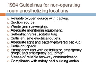
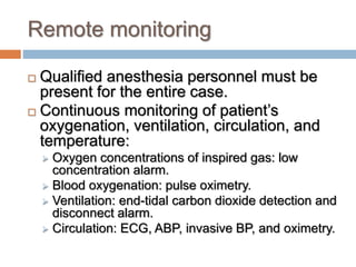
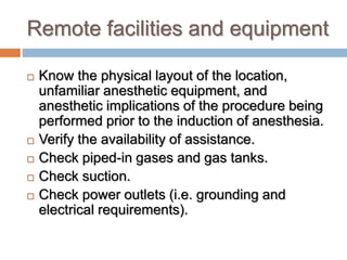
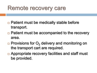
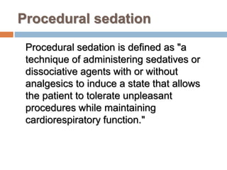
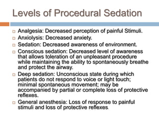
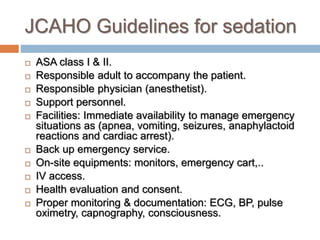
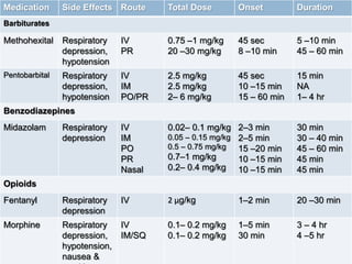
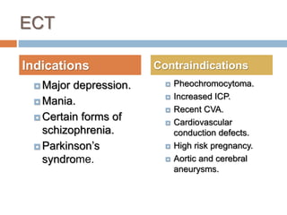
ANESTHESIA OUTSIDE THE
OPERATING ROOM
By
Hala S. El-Ozairy,MD.
Lecturer of anesthesia and
ICU
Objectives
 Understanding that the standards of anesthesia care
and patient monitoring are the same regardless of
location (There are cases of minor surgery, but there
are no cases of minor anesthesia).
 Remember that the key to efficient and safe remote
anesthetic relies on open communication between
the anesthesiologist and non-operating room
personnel.
 Realize that remote locations have different safety
concerns, such as radiation and powerful magnetic
fields.
Remote anesthesia
 Anesthesiologists are increasingly being asked to
provide anesthetic care in locations outside of the
OR.
 These locations include: radiology suites, cardiac
labs, psychiatric units, GI lab, MRI, dental,
ophthalmic, ENT and urology clinics.
 It is the responsibility of the anesthesiologist to
ensure that the location meets the ASA guidelines
for safety.
 The anesthesia needed can range from local
anesthetics, MAC, or general anesthesia.
Problems related to ‘isolated’
environment
 Equipment might be old, not regularly serviced and
not in standard use as in the rest of the hospital.
 Monitoring standards may not be adequate.
 Piped medical gases may not be supplied.
 Other personnel may be unaware of the problems
facing the anesthetist.
 Space may be limited by bulky equipment making
access to the patient difficult.
 Poor environmental conditions (e.g. Lighting,
temperature).
 Recovery facilities may not be available.
 Inadequate ventilation/scavenging causing pollution.
 Problem related to transferring patients.
Problems related to patient
Patients who require general anesthesia are:
 Infants or uncooperative children.
 Older children or adults with psychological,
behavioral or movement disorders.
 Intubated patients such as acute trauma
victims and patients receiving intensive care.
 Interventional procedures under radio-
guidance or painful procedures like ECT,
cardioversion which require amnesia.
Problems related to the procedure
 MRI related problems.
 Bleeding.
 Conversion from sedation to
anesthesia.
 Contrast related problems.
 Radiation.
1994 Guidelines for non-operating
room anesthetizing locations.
 Reliable oxygen source with backup.
 Suction source.
 Waste gas scavenging.
 Adequate monitoring equipment.
 Self-inflating resuscitator bag.
 Sufficient safe electrical outlets.
 Adequate light and battery-powered backup.
 Sufficient space.
 Emergency cart with defibrillator, emergency
drugs, and emergency equipment.
 Means of reliable two-way communication.
 Compliance with safety and building codes.
Remote monitoring
 Qualified anesthesia personnel must be
present for the entire case.
 Continuous monitoring of patient’s
oxygenation, ventilation, circulation, and
temperature:
 Oxygen concentrations of inspired gas: low
concentration alarm.
 Blood oxygenation: pulse oximetry.
 Ventilation: end-tidal carbon dioxide detection and
disconnect alarm.
 Circulation: ECG, ABP, invasive BP, and oximetry.
Remote facilities and equipment
 Know the physical layout of the location,
unfamiliar anesthetic equipment, and
anesthetic implications of the procedure being
performed prior to the induction of anesthesia.
 Verify the availability of assistance.
 Check piped-in gases and gas tanks.
 Check suction.
 Check power outlets (i.e. grounding and
electrical requirements).
Remote personnel
 Nurses and radiology techs are often less
familiar with the management of anesthesia,
therefore they are often unable to provide
skilled assistance in an emergency.
Remote recovery care
 Patient must be medically stable before
transport.
 Patient must be accompanied to the recovery
area.
 Provisions for O2 delivery and monitoring on
the transport cart are required.
 Appropriate recovery facilities and staff must
be provided.
Procedural sedation
Procedural sedation is defined as "a
technique of administering sedatives or
dissociative agents with or without
analgesics to induce a state that allows
the patient to tolerate unpleasant
procedures while maintaining
cardiorespiratory function."
Levels of Procedural Sedation
 Analgesia: Decreased perception of painful Stimuli.
 Anxiolysis: Decreased anxiety.
 Sedation: Decreased awareness of environment.
 Conscious sedation: Decreased level of awareness
that allows toleration of an unpleasant procedure
while maintaining the ability to spontaneously breathe
and protect the airway.
 Deep sedation: Unconscious state during which
patients do not respond to voice or light touch;
minimal spontaneous movement; may be
accompanied by partial or complete loss of protective
reflexes.
 General anesthesia: Loss of response to painful
stimuli and loss of protective reflexes.
JCAHO Guidelines for sedation
 ASA class I & II.
 Responsible adult to accompany the patient.
 Responsible physician (anesthetist).
 Support personnel.
 Facilities: Immediate availability to manage emergency
situations as (apnea, vomiting, seizures, anaphylactoid
reactions and cardiac arrest).
 Back up emergency service.
 On-site equipments: monitors, emergency cart,..
 IV access.
 Health evaluation and consent.
 Proper monitoring & documentation: ECG, BP, pulse
oximetry, capnography, consciousness.
Medication Side Effects Route Total Dose Onset Duration
Barbiturates
Methohexital Respiratory
depression,
hypotension
IV
PR
0.75 –1 mg/kg
20 –30 mg/kg
45 sec
8 –10 min
5 –10 min
45 – 60 min
Pentobarbital Respiratory
depression,
hypotension
IV
IM
PO/PR
2.5 mg/kg
2.5 mg/kg
2– 6 mg/kg
45 sec
10 –15 min
15 – 60 min
15 min
NA
1– 4 hr
Benzodiazepines
Midazolam Respiratory
depression
IV
IM
PO
PR
Nasal
0.02– 0.1 mg/kg
0.05 – 0.15 mg/kg
0.5 – 0.75 mg/kg
0.7–1 mg/kg
0.2– 0.4 mg/kg
2–3 min
2–5 min
15 –20 min
10 –15 min
10 –15 min
30 min
30 – 40 min
45 – 60 min
45 min
45 min
Opioids
Fentanyl Respiratory
depression
IV 2 μg/kg 1–2 min 20 –30 min
Morphine Respiratory
depression,
hypotension,
nausea &
IV
IM/SQ
0.1– 0.2 mg/kg
0.1– 0.2 mg/kg
1–5 min
30 min
3 – 4 hr
4 –5 hr
Other sedative agents
Chloral
hydrate
Prolonged
sedation
PO/PR 50 –75 mg/kg 30 – 60 min 1–8 hr
Ketamine Post-
emergence
delirium
IV
IM
PO
PR
Nasal
0.5 –1 mg/kg
4 mg/kg
5 –10 mg/kg
5 –10 mg/kg
3 – 6 mg/kg
1 min
5 min
30 – 40 min
5 –10 min
5 –10 min
15 min
15 –30 min
2– 4 hr
15 –30 min
15 –30 min
Propofol Respiratory
depression,
hypotension
IVI
IV bolus
0.05 – 0.1 mg/kg/min
1 mg/kg
30 sec
30 sec
8 –10 min
Reversal agents
Flumazenil Withdrawal
symptoms
(agitation)
IV 0.2 mg
0.01 mg/kg
(pediatrics)
1–3 min 30 – 45 min
Naloxone Withdrawal
symptoms
(agitation)
IV/IM/SQ 0.1–2 mg 1–2 min (IV) 15 – 45 min
Radiology suite
 Includes: US, CT, RFA, and neuro-coiling.
 The rooms are often crowded with bulky equipment.
 Patients are often required to hold still for long
periods of time.
 Unique hazard: radiation exposure.
 Leukemia and fetal abnormalities.
 Dosimeters are required (maximum exposure 50 mSv
annually).
 Lead aprons, thyroid shields, leaded glass screens, and
video monitoring.
Radiology suite, contd.
 Iodinated contrast media.
 Older ionized contrast media were hyperosmolar
and toxic.
 Newer non-ionized contrast media have lower
osmolality and improved side-effects.
 Predisposing factors to adverse reactions from
contrast media include a history of:
bronchospasm, allergy, cardiac disease,
hypovolemia, hematologic disease, renal
dysfunction, extremes of age, anxiety, and
medications (beta-blockers, aspirin, and NSAIDs).
Radiology suite, contd.
 Reactions to iodinated contrast media.
 Mild: nausea, perception of warmth, headache,
itchy rash, and mild urticaria.
 Severe: vomiting, rigors, feeling faint, chest pain,
severe urticaria, bronchospasm, dyspnea,
arrythmias, and renal failure.
 Life-threatening: glottic edema/bronchospasm,
pulmonary edema, arrythmias, cardiac arrest, and
seizures/unconsciousness.
 Treatment: O2, bronchodilators, epinephrine,
corticosteroids, and antihistamines.
CT
 Two-dimensional, cross-sectional image.
 Each cross-section requires a few seconds of
radiation exposure.
 Pt immobility is required.
 It is often noisy, warm, and claustrophobic.
 CT can be used for diagnostic and therapeutic
purposes.
 Number one problem: inaccessibility to the
patient.
Anesthesia for CT
 Anesthetist can remain in the room wearing X-
ray protection or view the patient and monitors
from the control room.
 The CT scanner does not interfere with
monitoring equipment.
 The scans are short and can be interrupted.
 The patient couch moves during examination.
 Temporarily interruption of ventilation to
improve image quality – immediately re-
ventilate.
 Patient positioning.
Radiology RFA
 Often done in CT but occasionally MRI.
 Kidney, lung, and liver.
 Currently requesting general anesthesia
with ETT secondary to prone positioning
and the need to lay still for extended
periods of time.
 It is our job to check pressure points and
padding. Radiology techs are not trained
to be concerned.
Interventional Radiology
 Embolization of cerebral and dural AVM’s,
coiling of cerebral aneurysms, angioplasty of
sclerotic lesions, and thrombolysis of acute
thromboembolic stroke.
 These procedures often require deliberate
hypotension and deliberate hypocapnia.
 Radiologist may request rapid transition
between deep sedation and an awake
responsive state.
Cerebral Coiling
 The anesthetist should prepare:
 Arterial line set up.
 Fluid warmer.
 Infusion pump.
 Medications: NTG, nipride, esmolol, labetalol, heparin,
and protamine.
 ACT machine.
 Radiologist may request anything from deep IV sedation to
GA with ETT.
 Always have 2 large-gauge IV’s in place. One for drug
infusion and one for rapid fluid administration.
 Stay in constant communication with OR in case of an
emergency.
 Pt often transported to the ICU post-op.
Remote Cardiac Lab
 Elective cardioversion:
 Cart with emergency drugs.
 Induction drug (Etomidate).
 Standard monitoring.
 Preoxygenate.
 Give small incremental doses of etomidate until the
eyelash reflex is abolished.
 Remove the mask immediately before the shock and
confirm no one is touching the pt.
 Ventilate with 100% O2 post-shock until consciousness is
regained.
 Consider RSI with ETT if high risk for aspiration.
Remote Cardiac Lab contd.
 Cardiac RFA
IV sedation to GA with ETT depending
on the pt’s co-morbidities.
Possible need for an arterial line setup.
Propofol is oftenly used.
Midazolam and fentanyl are used to
titrate in during the more painful parts of
the procedure (esp. the ablation).
Remote Cardiac Lab contd.
 Pacemaker/ ICD placement:
 We are often called just for the ICD check, in which
case proceed like an elective cardioversion.
 If the pt. is very sick, they may require GA. Therefore,
proceed like RFA.
 These pt’s will often need an arterial line for BP monitoring.
 These ICD checks are not without risk. Check pulses
and watch the ECG, pulse oximetry and arterial wave-
forms closely. People have been known to code and
require CPR.
GI Lab
 Endoscopy, Colonoscopy and ERCP.
 Pt’s are often uncooperative or very sick.
 Current rooms in the GI lab are very small.
Anesthesia for GI Procedures
 Pre anesthetic assessment: Age, cooperative,
anxiety, allergies, fluid status, electrolytes, cardiac
history, GERD.
 Type of anesthesia:
 Moderate sedation- midazolam and Fentanyl.
 Deep sedation- Addition of propofol.
 Some cases may require general anesthesia.
 Anesthetic considerations:
 Strong vagal nerve stimulation as result of stimulation
to colon.
 Most patients tolerate these procedures well.
ECT
 Major depression.
 Mania.
 Certain forms of
schizophrenia.
 Parkinson’s
syndrome.
 Pheochromocytoma.
 Increased ICP.
 Recent CVA.
 Cardiovascular
conduction defects.
 High risk pregnancy.
 Aortic and cerebral
aneurysms.
Indications Contraindications
Physiologic effects of ECT
 Electrical stimulus: brief period of muscular
contraction followed by the tonic and then clonic
phases of the seizures.
 Cardiovascular effects of ECT: immediate
parasympathetic response followed within
seconds by a sympathetic response.
 The muscular activity of the seizure and the
increased sympathetic activity causes a rise in
myocardial oxygen consumption, increases
CMRO2, cerebral blood flow, intracranial, intra-
ocular intra-gastric pressure briefly.
Anesthesia for ECT
 General anesthesia is used to provide a
brief period of amnesia and modify the
motor effects of the seizure to protect the
patient.
 Don’t forget the suction and the Bite block.
ECT contd.
 Pre-op: These pt’s have often had this procedure multiple times,
therefore you can use old records as templates.
 Place IV and give glyco (0.2 mg IV). Give caffeine if the
psychiatrist requests.
 Treats the bradycardia/ asystole from the initial parasympathetic
discharge from the seizure activity.
 Hyperventilate the pt. with 100% O2.
 Thiopentone and suxamethonium are commonly used.
 Place the bite block.
 Goal is a seizure 30-60 seconds long.
 Ventilate until spontaneous respirations return.
 The parasympathetic discharge is often followed by a
sympathetic discharge associated with HTN and tachycardia.
This is treated with esmolol.
Dental Procedures
 Pediatric Dentistry: fillings, crowns, pulpotomies,
tooth extractions and space maintainers.
 Oral and Maxillofacial Surgery: extractions of
impacted teeth, insertion of dental implants,
treatment of infections of the head and neck and
facial cosmetics.
 Peridontics: surgery of teeth, gingiva, connective
tissue, periodontal ligament and alveolar bone.
 Anesthesia : general anesthesia, minimal
sedation, moderate sedation with local anesthetic
for particular areas of surgery.
Ophthalmology
 Cataract extraction is the most common
procedure done for the elderly.
 Strabismus operations are the most common
pediatric procedures.
 Requirements for anesthesia:
 Unmoving globe.
 Minimal bleeding.
 Smooth emergence.
 Usually done under MAC.
Urologic Procedures
 ESWL: sound waves are focused on kidney and ureteral
stones. The stone located by flouroscopy.
 Cystoscopy/ ureteroscopy: are performed to diagnosis
and treat lesions of the lower (urethra, prostate, bladder)
and upper (ureter, kidney) urinary tracts.
 Type of Anesthesia
 Depending on the pt and procedure anesthesia can
range from topical lubrication ,MAC, or regional.
 If regional is used T-6 level of blockade is required for
upper tract instrumentation and T-10 for lower-tract
surgery.
Thank you

Anesthesia outside the operating room.pptx

  • 1.
    ANESTHESIA OUTSIDE THE OPERATINGROOM By Hala S. El-Ozairy,MD. Lecturer of anesthesia and ICU
  • 2.
    Objectives  Understanding thatthe standards of anesthesia care and patient monitoring are the same regardless of location (There are cases of minor surgery, but there are no cases of minor anesthesia).  Remember that the key to efficient and safe remote anesthetic relies on open communication between the anesthesiologist and non-operating room personnel.  Realize that remote locations have different safety concerns, such as radiation and powerful magnetic fields.
  • 3.
    Remote anesthesia  Anesthesiologistsare increasingly being asked to provide anesthetic care in locations outside of the OR.  These locations include: radiology suites, cardiac labs, psychiatric units, GI lab, MRI, dental, ophthalmic, ENT and urology clinics.  It is the responsibility of the anesthesiologist to ensure that the location meets the ASA guidelines for safety.  The anesthesia needed can range from local anesthetics, MAC, or general anesthesia.
  • 4.
    Problems related to‘isolated’ environment  Equipment might be old, not regularly serviced and not in standard use as in the rest of the hospital.  Monitoring standards may not be adequate.  Piped medical gases may not be supplied.  Other personnel may be unaware of the problems facing the anesthetist.  Space may be limited by bulky equipment making access to the patient difficult.  Poor environmental conditions (e.g. Lighting, temperature).  Recovery facilities may not be available.  Inadequate ventilation/scavenging causing pollution.  Problem related to transferring patients.
  • 5.
    Problems related topatient Patients who require general anesthesia are:  Infants or uncooperative children.  Older children or adults with psychological, behavioral or movement disorders.  Intubated patients such as acute trauma victims and patients receiving intensive care.  Interventional procedures under radio- guidance or painful procedures like ECT, cardioversion which require amnesia.
  • 6.
    Problems related tothe procedure  MRI related problems.  Bleeding.  Conversion from sedation to anesthesia.  Contrast related problems.  Radiation.
  • 7.
    1994 Guidelines fornon-operating room anesthetizing locations.  Reliable oxygen source with backup.  Suction source.  Waste gas scavenging.  Adequate monitoring equipment.  Self-inflating resuscitator bag.  Sufficient safe electrical outlets.  Adequate light and battery-powered backup.  Sufficient space.  Emergency cart with defibrillator, emergency drugs, and emergency equipment.  Means of reliable two-way communication.  Compliance with safety and building codes.
  • 8.
    Remote monitoring  Qualifiedanesthesia personnel must be present for the entire case.  Continuous monitoring of patient’s oxygenation, ventilation, circulation, and temperature:  Oxygen concentrations of inspired gas: low concentration alarm.  Blood oxygenation: pulse oximetry.  Ventilation: end-tidal carbon dioxide detection and disconnect alarm.  Circulation: ECG, ABP, invasive BP, and oximetry.
  • 9.
    Remote facilities andequipment  Know the physical layout of the location, unfamiliar anesthetic equipment, and anesthetic implications of the procedure being performed prior to the induction of anesthesia.  Verify the availability of assistance.  Check piped-in gases and gas tanks.  Check suction.  Check power outlets (i.e. grounding and electrical requirements).
  • 10.
    Remote personnel  Nursesand radiology techs are often less familiar with the management of anesthesia, therefore they are often unable to provide skilled assistance in an emergency.
  • 11.
    Remote recovery care Patient must be medically stable before transport.  Patient must be accompanied to the recovery area.  Provisions for O2 delivery and monitoring on the transport cart are required.  Appropriate recovery facilities and staff must be provided.
  • 12.
    Procedural sedation Procedural sedationis defined as "a technique of administering sedatives or dissociative agents with or without analgesics to induce a state that allows the patient to tolerate unpleasant procedures while maintaining cardiorespiratory function."
  • 13.
    Levels of ProceduralSedation  Analgesia: Decreased perception of painful Stimuli.  Anxiolysis: Decreased anxiety.  Sedation: Decreased awareness of environment.  Conscious sedation: Decreased level of awareness that allows toleration of an unpleasant procedure while maintaining the ability to spontaneously breathe and protect the airway.  Deep sedation: Unconscious state during which patients do not respond to voice or light touch; minimal spontaneous movement; may be accompanied by partial or complete loss of protective reflexes.  General anesthesia: Loss of response to painful stimuli and loss of protective reflexes.
  • 14.
    JCAHO Guidelines forsedation  ASA class I & II.  Responsible adult to accompany the patient.  Responsible physician (anesthetist).  Support personnel.  Facilities: Immediate availability to manage emergency situations as (apnea, vomiting, seizures, anaphylactoid reactions and cardiac arrest).  Back up emergency service.  On-site equipments: monitors, emergency cart,..  IV access.  Health evaluation and consent.  Proper monitoring & documentation: ECG, BP, pulse oximetry, capnography, consciousness.
  • 15.
    Medication Side EffectsRoute Total Dose Onset Duration Barbiturates Methohexital Respiratory depression, hypotension IV PR 0.75 –1 mg/kg 20 –30 mg/kg 45 sec 8 –10 min 5 –10 min 45 – 60 min Pentobarbital Respiratory depression, hypotension IV IM PO/PR 2.5 mg/kg 2.5 mg/kg 2– 6 mg/kg 45 sec 10 –15 min 15 – 60 min 15 min NA 1– 4 hr Benzodiazepines Midazolam Respiratory depression IV IM PO PR Nasal 0.02– 0.1 mg/kg 0.05 – 0.15 mg/kg 0.5 – 0.75 mg/kg 0.7–1 mg/kg 0.2– 0.4 mg/kg 2–3 min 2–5 min 15 –20 min 10 –15 min 10 –15 min 30 min 30 – 40 min 45 – 60 min 45 min 45 min Opioids Fentanyl Respiratory depression IV 2 μg/kg 1–2 min 20 –30 min Morphine Respiratory depression, hypotension, nausea & IV IM/SQ 0.1– 0.2 mg/kg 0.1– 0.2 mg/kg 1–5 min 30 min 3 – 4 hr 4 –5 hr
  • 16.
    Other sedative agents Chloral hydrate Prolonged sedation PO/PR50 –75 mg/kg 30 – 60 min 1–8 hr Ketamine Post- emergence delirium IV IM PO PR Nasal 0.5 –1 mg/kg 4 mg/kg 5 –10 mg/kg 5 –10 mg/kg 3 – 6 mg/kg 1 min 5 min 30 – 40 min 5 –10 min 5 –10 min 15 min 15 –30 min 2– 4 hr 15 –30 min 15 –30 min Propofol Respiratory depression, hypotension IVI IV bolus 0.05 – 0.1 mg/kg/min 1 mg/kg 30 sec 30 sec 8 –10 min Reversal agents Flumazenil Withdrawal symptoms (agitation) IV 0.2 mg 0.01 mg/kg (pediatrics) 1–3 min 30 – 45 min Naloxone Withdrawal symptoms (agitation) IV/IM/SQ 0.1–2 mg 1–2 min (IV) 15 – 45 min
  • 17.
    Radiology suite  Includes:US, CT, RFA, and neuro-coiling.  The rooms are often crowded with bulky equipment.  Patients are often required to hold still for long periods of time.  Unique hazard: radiation exposure.  Leukemia and fetal abnormalities.  Dosimeters are required (maximum exposure 50 mSv annually).  Lead aprons, thyroid shields, leaded glass screens, and video monitoring.
  • 18.
    Radiology suite, contd. Iodinated contrast media.  Older ionized contrast media were hyperosmolar and toxic.  Newer non-ionized contrast media have lower osmolality and improved side-effects.  Predisposing factors to adverse reactions from contrast media include a history of: bronchospasm, allergy, cardiac disease, hypovolemia, hematologic disease, renal dysfunction, extremes of age, anxiety, and medications (beta-blockers, aspirin, and NSAIDs).
  • 19.
    Radiology suite, contd. Reactions to iodinated contrast media.  Mild: nausea, perception of warmth, headache, itchy rash, and mild urticaria.  Severe: vomiting, rigors, feeling faint, chest pain, severe urticaria, bronchospasm, dyspnea, arrythmias, and renal failure.  Life-threatening: glottic edema/bronchospasm, pulmonary edema, arrythmias, cardiac arrest, and seizures/unconsciousness.  Treatment: O2, bronchodilators, epinephrine, corticosteroids, and antihistamines.
  • 20.
    CT  Two-dimensional, cross-sectionalimage.  Each cross-section requires a few seconds of radiation exposure.  Pt immobility is required.  It is often noisy, warm, and claustrophobic.  CT can be used for diagnostic and therapeutic purposes.  Number one problem: inaccessibility to the patient.
  • 21.
    Anesthesia for CT Anesthetist can remain in the room wearing X- ray protection or view the patient and monitors from the control room.  The CT scanner does not interfere with monitoring equipment.  The scans are short and can be interrupted.  The patient couch moves during examination.  Temporarily interruption of ventilation to improve image quality – immediately re- ventilate.  Patient positioning.
  • 22.
    Radiology RFA  Oftendone in CT but occasionally MRI.  Kidney, lung, and liver.  Currently requesting general anesthesia with ETT secondary to prone positioning and the need to lay still for extended periods of time.  It is our job to check pressure points and padding. Radiology techs are not trained to be concerned.
  • 23.
    Interventional Radiology  Embolizationof cerebral and dural AVM’s, coiling of cerebral aneurysms, angioplasty of sclerotic lesions, and thrombolysis of acute thromboembolic stroke.  These procedures often require deliberate hypotension and deliberate hypocapnia.  Radiologist may request rapid transition between deep sedation and an awake responsive state.
  • 24.
    Cerebral Coiling  Theanesthetist should prepare:  Arterial line set up.  Fluid warmer.  Infusion pump.  Medications: NTG, nipride, esmolol, labetalol, heparin, and protamine.  ACT machine.  Radiologist may request anything from deep IV sedation to GA with ETT.  Always have 2 large-gauge IV’s in place. One for drug infusion and one for rapid fluid administration.  Stay in constant communication with OR in case of an emergency.  Pt often transported to the ICU post-op.
  • 25.
    Remote Cardiac Lab Elective cardioversion:  Cart with emergency drugs.  Induction drug (Etomidate).  Standard monitoring.  Preoxygenate.  Give small incremental doses of etomidate until the eyelash reflex is abolished.  Remove the mask immediately before the shock and confirm no one is touching the pt.  Ventilate with 100% O2 post-shock until consciousness is regained.  Consider RSI with ETT if high risk for aspiration.
  • 26.
    Remote Cardiac Labcontd.  Cardiac RFA IV sedation to GA with ETT depending on the pt’s co-morbidities. Possible need for an arterial line setup. Propofol is oftenly used. Midazolam and fentanyl are used to titrate in during the more painful parts of the procedure (esp. the ablation).
  • 27.
    Remote Cardiac Labcontd.  Pacemaker/ ICD placement:  We are often called just for the ICD check, in which case proceed like an elective cardioversion.  If the pt. is very sick, they may require GA. Therefore, proceed like RFA.  These pt’s will often need an arterial line for BP monitoring.  These ICD checks are not without risk. Check pulses and watch the ECG, pulse oximetry and arterial wave- forms closely. People have been known to code and require CPR.
  • 28.
    GI Lab  Endoscopy,Colonoscopy and ERCP.  Pt’s are often uncooperative or very sick.  Current rooms in the GI lab are very small.
  • 29.
    Anesthesia for GIProcedures  Pre anesthetic assessment: Age, cooperative, anxiety, allergies, fluid status, electrolytes, cardiac history, GERD.  Type of anesthesia:  Moderate sedation- midazolam and Fentanyl.  Deep sedation- Addition of propofol.  Some cases may require general anesthesia.  Anesthetic considerations:  Strong vagal nerve stimulation as result of stimulation to colon.  Most patients tolerate these procedures well.
  • 30.
    ECT  Major depression. Mania.  Certain forms of schizophrenia.  Parkinson’s syndrome.  Pheochromocytoma.  Increased ICP.  Recent CVA.  Cardiovascular conduction defects.  High risk pregnancy.  Aortic and cerebral aneurysms. Indications Contraindications
  • 31.
    Physiologic effects ofECT  Electrical stimulus: brief period of muscular contraction followed by the tonic and then clonic phases of the seizures.  Cardiovascular effects of ECT: immediate parasympathetic response followed within seconds by a sympathetic response.  The muscular activity of the seizure and the increased sympathetic activity causes a rise in myocardial oxygen consumption, increases CMRO2, cerebral blood flow, intracranial, intra- ocular intra-gastric pressure briefly.
  • 32.
    Anesthesia for ECT General anesthesia is used to provide a brief period of amnesia and modify the motor effects of the seizure to protect the patient.  Don’t forget the suction and the Bite block.
  • 33.
    ECT contd.  Pre-op:These pt’s have often had this procedure multiple times, therefore you can use old records as templates.  Place IV and give glyco (0.2 mg IV). Give caffeine if the psychiatrist requests.  Treats the bradycardia/ asystole from the initial parasympathetic discharge from the seizure activity.  Hyperventilate the pt. with 100% O2.  Thiopentone and suxamethonium are commonly used.  Place the bite block.  Goal is a seizure 30-60 seconds long.  Ventilate until spontaneous respirations return.  The parasympathetic discharge is often followed by a sympathetic discharge associated with HTN and tachycardia. This is treated with esmolol.
  • 34.
    Dental Procedures  PediatricDentistry: fillings, crowns, pulpotomies, tooth extractions and space maintainers.  Oral and Maxillofacial Surgery: extractions of impacted teeth, insertion of dental implants, treatment of infections of the head and neck and facial cosmetics.  Peridontics: surgery of teeth, gingiva, connective tissue, periodontal ligament and alveolar bone.  Anesthesia : general anesthesia, minimal sedation, moderate sedation with local anesthetic for particular areas of surgery.
  • 35.
    Ophthalmology  Cataract extractionis the most common procedure done for the elderly.  Strabismus operations are the most common pediatric procedures.  Requirements for anesthesia:  Unmoving globe.  Minimal bleeding.  Smooth emergence.  Usually done under MAC.
  • 36.
    Urologic Procedures  ESWL:sound waves are focused on kidney and ureteral stones. The stone located by flouroscopy.  Cystoscopy/ ureteroscopy: are performed to diagnosis and treat lesions of the lower (urethra, prostate, bladder) and upper (ureter, kidney) urinary tracts.  Type of Anesthesia  Depending on the pt and procedure anesthesia can range from topical lubrication ,MAC, or regional.  If regional is used T-6 level of blockade is required for upper tract instrumentation and T-10 for lower-tract surgery.
  • 37.