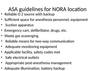
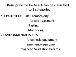
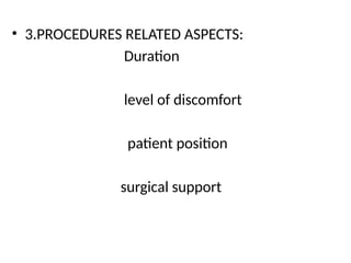
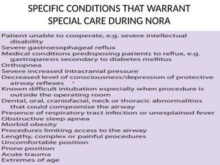
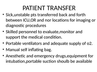
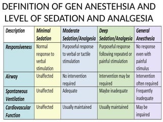
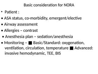
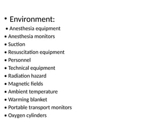
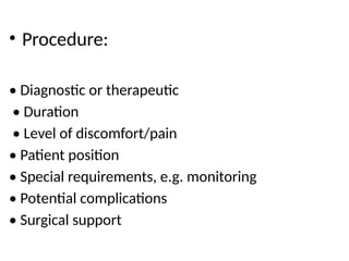
ANAESTHESIA OUTSIDE
OPERATING ROOM
PRESENTER:DR AMIT KUMAR PRADHAN
GUIDE:DR SUBHADARSINI CHOUDHARY
INTRODUCTION
• Non-operating room anesthesia(NORA) refers to all
procedures performed in locations other than the
operating room(OR).
• It includes diverse environmenments and
procedures often associated with challenges not
regularly confronted for procedures performed in
the OR.
Area remote from operating room:
radiology deparment
endoscopic suites
MRI
Dental clinics
cardiac catheterisation Lab
electrophysiology Lab
critical care unit
ASA guidelines for NORA location
• Reliable O 2 source with backup
• Sufficient space for anesthesia personnel, equipment
• Suction apparatus
• Emergency cart, defibrillator, drugs, etc.
• Waste gas scavenging
• Reliable means for two-way communication
• Adequate monitoring equipment
• Applicable facility, safety codes met
• Safe electrical outlets
• Appropriate post-anesthesia management
• Adequate illumination, battery backup
Basic principle for NORA can be classsified
into 3 categories
• 1.PATIENT FACTORS: comorbidity
Airway assesment
Fasting
Monitoring
2.ENVIRONMENTAL ISSUES:
Anesthesia equipment
emergency equipment
magnetic &radiation hazards
• 3.PROCEDURES RELATED ASPECTS:
Duration
level of discomfort
patient position
surgical support
SPECIFIC CONDITIONS THAT WARRANT
SPECIAL CARE DURING NORA
PATIENT TRANSFER
• Sick,unstable pts transferred back and forth
between ICU,OR and nor locations for imaging or
diagnostic procedures
• Skilled personnel to evaluate,monitor and
support the medical condition.
• Portable ventilators and adequate supply of o2.
• Manual self inflating bag.
• Anesthetic and emergency drugs,equipment for
intubation,portable suction shoulb be available
COMPLICATIONS OF NORA
• MINOR:
• -ponv
• -inadequate post op pain control
• Hemodynamic instability
• Minor neurologic complication such as PDPH
• Respiratory complications,cns complications
• Need for opioid reversal
• MAJOR:
-Unintended patient awareness
-Anaphylaxis
-Need for upgrade of care
-serious hemodynamic instability
-respiratory complications
-need for resuscitations
-central and peripheral nervous system injurys
-vascular access
PROBLEMS IN PROVIDING NORA
• 1. equipment might be old,not regularly serviced
and not in standard use as in OT
• 2.monitoring standards may not adequate
• 3.piped gas may not be supplied
• 4.other perssonel may be unaware of problems
facing by anaestesist
• 5. limited space by bulky equipments making access
to patient difficult
• 6.poor environment condition
• Recovery facilities may not be available
• Inadequate ventillation/scavenging causing
pollution
• Problems related to transferring of patients
DEFINITION OF GEN ANESTEHSIA AND
LEVEL OF SEDATION AND ANALGESIA
Basic consideration for NORA
• Patient :
• ASA status, co-morbidity, emergent/elective
• Airway assessment
• Allergies – contrast
• Anesthesia plan – sedation/anesthesia
• Monitoring – Basic/Standard: oxygenation,
■
ventilation, circulation, temperature Advanced:
■
invasive hemodynamic, TEE, BIS
• Environment:
• Anesthesia equipment
• Anesthesia monitors
• Suction
• Resuscitation equipment
• Personnel
• Technical equipment
• Radiation hazard
• Magnetic fields
• Ambient temperature
• Warming blanket
• Portable transport monitors
• Oxygen cylinders
• Procedure:
• Diagnostic or therapeutic
• Duration
• Level of discomfort/pain
• Patient position
• Special requirements, e.g. monitoring
• Potential complications
• Surgical support
ANAESTHESIA FOR IMAGE GUIDED INTERVENTIONS
• DIAGNOSTIC & THERAPEUTIC INTERVENTION:
1. Equipment layout:
- xray tubes and moving c arm create a zone
of inaccesecibility around pt head &limit
placement of anaesthesia macine.
-This neccesiate use of extension on
ventilltor circuits & iv line
- Infusion pump,blood warmers,monitors placed far
away from moving imaging equipoment to prevent
them from knocked down or tangled during c arm
rotation.
- Imaging screens are right angles to
anaesthesiologist making it difficult to track
progress of the case.
- So good communication between both the team is
necessary
• 2.RADIATION EXPOSURE:
-all radiation exposure should follow the ALARA
principle.
-reducing exposure can be by Reducing exposure
time,increased distance from source of
radiation&protective barriers(lead shield) along
with lead glasses and thyroid shield.
• 3.CONTRAST MATERIAL:
XRAY&CT :positive contrast media
MRI:negative contrast media
Reaction to iodinated contrast media:
Mild:nausea,retching,warmth perception,headacheitchy rash,
urticaria
Severe:vomiting,rigors,feeling faint,chest pain,severe
urticaria,bronchospasm,dyspnoea,abd pain,arrythmias,renal
failure
Life threatening:glottic edema/bronchospasm,pulm
oedema,cardiac arrest,seizures/unconsciousness
• TREATMENT: Symptomatic
severe/resistant bronchospasm require t/t
with epinephrine
pt with prev reaction to CM can be given
prednisolone 12 and 2 hrs before procedure.
also diphenhydramine 50 mg immediately before
the procedure.
life threatening lactic acidosis may devlope in
NIDDM pts receiveing metformin& have preexisting
renal dysfunction
hence these pts require extra care
• 4.BLEEDING:
- Occurs in most percutaneous intervention while in some
it is the reason for the procedure( splenic embolisation)
- pts not undergoing anticoagulation for other reason
INR<1.5 and PLT >50000 is required
-if possible warfarin for 5-7 days clopidrogrel and aspirin
for 5 days &fractionated heparin for 12-24 hr should be
stopped.
-NSAIDS 1-2 days prior should be held
• 5.VASCULAR INTERVENTIONAL PROCEDURE:
procedures like Angiography,CT
angiography,arteriography.
thrombolytic therapies with r-TPA,urokinase (CI in
pt with ongoing/recent bleeding,pregnancy,known
allergy to thrombolytic agents,suspected aortic
dissection).
embolisation therapies in cases of
trauma,haemorrahage,vascular
anomalies,fibroids,aneurysm,tumor
• FOR all these vascular anomalies nature of
case,commorbities of patient,intricacies of
procedure determine need of anaesthesiologist.
• Complications to anticipate include bleeding during
thrombolysis,undisired embolisation& vessel
disruption.
• In procedures like venography there is requirement
of low/no sedation.but if the pt is anxious or
moving then we may require some sedation.
• fistulograms,graftograms tunneled haemodialysis
lines called haemodialysis vascular access
procedures.
• These pts are unique as most of them have end
stage renal disease.
• In these pts pre op evaluation is essential with
Special consideration to volume status,Ecg
changes and sr pottasium levels
ANAESTHETIC CONSIDERATION FOR CT MRI
&ECT
• COMPUTED TOMOGRAPHY:
-contrast media introduced in patients who are
sedated or anesthetised by NG tube
-if airway is not secured there is risk of aspiration.
-in anesthetised pt ensure that sides of the scannig
tunnel donot occlude or dislodge the breathing
circuits or monitoring leads during procedure.
• Monitored through a lead glass window,supplemented if
necessary by closed circuit television.
-selection of RA technique and timing of placement of
catheter9 if a catheter based technique is selected) depends
on pts condition as well as space and equipment available.
-recovery room present in radiology suite: safe place forgiving
RA catheter
-if absent: perform the technique before/after the ct procedure
- Be viggilant for complications
MRI(MAGNETIC RESONANCE IMAGING)
- Based on principle that atomic nuclei with odd no of
protons or neutrons have potential to act as magnetic
dipoles.
- Biological tissues have high water content,H1 detection
is the basis of mri.
- Pts are placed within strong magnetic field(0.5-3 tesla)
- Pulses of radiofrequency (RF)energy are applied
• Intermittent release of RF energy from H1 nuclei.
-energy is detected by a series of close fitting
receiving antennae known as coils.
-the RF signals are collected and interpretated by
computer to produce images.
-increased strength( in tesla) is used for better spatial
resolution
Anaesthesia for MRI
• Use of MRI:
• Central nervous system:
-posterior fossa tumors
-head trauma
-cerebral infarct
-dementia
-intracranial infections
• OTHERS:
-spinal cord
-cardiac chambers
-musculoskeletal
system;tendon/muscle/ligament injuries
-intrathoracic disorders
-intraabdominal disorders
• Advantages of MRI
• Noninvasive
• Obtains images in multiple planes simultaneously:
• • Transverse
• • Sagittal
• • Coronal/oblique
• Excellent spatial resolution and soft tissue contrast
• Not affected by bony artefacts
• Does not employ ionizing radiation
• Requires little patient preparation
• May provide intravascular contrast
• Does not in itself produce biologically deleterious effects
• Problems of MRI
• Remoteness of location
• Lack of trained personnel to assist in the event of
emergency
• Minimum consideration given to anesthetic pipeline
gases and suction
• Lack of waste gas exhaust capabilities
• Limitations from the magnetic field of imager
SAFETY ISSUES
Implanted ferromagnetic objects:
IMPLANTED FERROMAGNETIC OBJECTS:
• Includes:
–– Intraocular foreign bodies
–– Scissors
–– Oxygen cylinders
–– Laryngoscopes and stylets
• move in the magnetic field and become dangerous projectiles
• Some objectsmay become inactivated/dislodged :
–– Implanted pacemakers
–– AEDs
–– Cochlear implants
–– Cerebrovascular clips
• These cause hemorrhage/motile injury to
adjacent structures
METAL OBJECTS/ELECTRONIC MONITORS:
• May produce RF waves which interfere with
images generated by MRI
• This can result in generation of degraded and distorted images
• Alternate, specially adapted monitors and
equipment are required
• These should be positioned as far away from the magnet as possible
Thermal injury:
THERMAL INJURY:
• RF energy may be absorbed by tissues/other objects
• This results in localized heating
• Unlikely that body temperature increases by more than
1ºC
• Thermal effects may not be great enough to cause tissue
damage
NOISE POLLUTION:
• Noise generated by the scanner can be as high as 125 dB
• cause temporary/permanent hearing
loss
• Difficult to monitor heart sounds via esophageal/precordial
stethoscope
• Ear is protected with ear plugs
™™
Pregnancy: Pregnant patients in first trimester should not enter
scanner
• MAGNETIC RESONANCE IMAGING:
- A non invasive diagnostic procedure that uses
magnetic properties of atom nuclei
-to produce high resolution, multiplanar cross sectional
images of the body
-strong magnetic field of 0.5 to 3.0 tesla
-so ferromagnetic material should be excluded from the
adea of magnet
-implantable medical devices : pacemakers, vascular
clips,automatic implantable cardioverter-
defibrillators,mechanical heart valves.
-radiofrequency noise;sound >85 db from mri scanners
• Choice of anaesthetic technique depends
upon
-patient comorbidities
-duration
- Practitioner preferences
- Patient requirements
- Deep sedation or
- GA with intubation or supraglottic device
INDICATION OF ANAESTHESIA IN MRI
• -trauma and shock
• -children
• -ventillated pts
• -mental illness
• -pt with severe movement disorders
• -pt whose position is limited by pain
• -pts with learning difficulties
• -claustriophobic patients
• -intraoperative mri for stereotactic neurosurgery
• Preoperative Assessment
• All female patients to have pregnancy test
• Assess all implanted devices:
• Type of orthopedic implant
• Pacemaker & internal defibrillators—MRI contraindicated
• Aneurysms clips, cochlear implants, prosthetic heart valves,
may get dislodged /heat up/cause induction of electric
currents
• Metal workers may have intraocular foreign bodies–screen by
plain radiography before MRI
• Tattoos may heat up in magnetic field due to presence of FeO
• Contraindication of MRI
• Obese patients may not fit into bore of magnet
• Cardiac pacemakers & ICDS as the following may occur:
-Heating of leads
-Inhibiting of pacemaker output of rapid pacing
- Reed switch malfunction
-ICD malfunction
• Pregnancy in first trimester
• Suspected ferromagnetic intraocular FB
• Ferromagnetic vascular clip patients
• Titanium clips are an exception as they are non ferromagnetic
‑
• Hemodynamically unstable patients on infusion pumps as pumps cannot
be taken inside
MONITORING IN MRI SUITES
General principles:
• Must conform to ASA standards
• Important as direct patient visualization in the MRI suite is difficult
• MR compatible monitors used and placed 6–8 feet from the magnet
bore
• non MR compatible
‑ monitors,placed outside the scan room
• Long cables passed through specially
shielded holes in walls
• MR–compatible anesthesia machine with piped anesthetic gases to be
available
• Non MR compatible machines kept outside 50 G
‑ line and bolted onto
floor
• All gas cylinders to be MR compatible
Specific monitors
abo avoid burns
Capnograph—delay in obtaining signal as
sampling tube will be longer
• NIBP ;connections of the cuff
and hose are plastic
• Temperature monitoring difficult unless
probes with RF filters are used
• ECG:
–– Cables shielded and use specific electrodes
–– ST–T changes similar to pericarditis due to magnetic field
–– completely distorted due to the static magnetic field
• Padding to be placed between patients skin and monitor cables to prevent burns
Pulse oximeter with fibreoptic cables to avoid burns
• Sedation with benzodiazepines or with
inhahalational agents
• Small infants:feed wrap and scan
• Oral chloral hydrate : 80-100 mg/kg 30-60 min
before procedure
• rectally administered barbiturates
• General anesthesia with propofol ketamine or
inhaled anestetics
ANAESTHETIC MANAGEMENT
• CHOICE OF ANESTEHSIA:
-GA WITH ETT/LMA COMMONLY USED
-CHILDREN BETWEEN 3-7 YEAR MAY REQUIRE SEDATION
-YOUNGER CHILDREN SLEEP DEEPLY AFTER FEED.
SITE OF INDUCTION:
-best induced outside mri room
-pt transferred to docking table and shifted to non
ferromagnetic trolley
AIRWAY MANAGEMENT
• MR LMA: most commonly used airway adjunct
-pilot ballon taped away from scan site
-pts with poor gag reflex/pregnant pt require
intubation
-preformed ett is used with pilot ballon taped away
from scan site
-flexometallic tube not used
-plastic laryngoscope with lithium battery and
aluminium spares are used
-airways to be clear as obstruction may cause
increased resp movement &image artifact
Maintainence
• Easier with sevofluorane
• -TIVA with propofol and ramifentanyl
• -very long circuits
• Contrast used is gadolinium DTPA;very low
incidence of anaphylaxis
• AWAKENING: patient awakened once shifted back
to induction area with conventional monitoring.
• SEDATION IN MRI:
• ADULTS:
• oral bzds most commonly used
-iv sedation bolus midazolam/low dose propofol
With ramifentanyl infusion
-monitor pulse oximetry,ETCO2 if possible
-short MR sequences improves pt compliance
• CHILDREN:
<3year sleep deeply after feed
->7 year are compliant without sedation
-chloral hydrate,BZDs and low dose propofol
infusion used
-conscious sedation doesnot ensure compliance due
to noise
-supplemental o2 and adequate monitoring
mandatory.
CASE DISCUSSION
• Management of a 6month child with
hydrocephalus for MRI:
*pre procedure evaluation:
-Similar to that requiring surger
- Special challenges relating to radiology site
- Physian may not be familiar with the
anaesthetic implication of the disease
• -key is to educating physian when an anestetic
consultation is appropiate
-when taking history helpful to focus on those risk
factors specific to remote site anaesthesia.
-example: history of difficult airway or difficult
airway during examination,
difficult iv access,
history of anaesthesia complication
MANAGEMENT OF INFANTS AND CHILD IN MRI ROOM
• Different techniques for children and infants in mri
room :
-Sedation( chloral hydrate or pentobarbital)
-propofol infusion
-inhalational agent administered by lma or ett
-extreme young infants can be sometimes scanned
after fed and swaddled
• Scan >60 min in length can outlast the sedation thus
necessiating additional dosing
-multiple changes of position or coil can also stimulate
the pt and cause additional drug to be
administered.
-often children<2 yr presenting for short scans(<60
min) can be successfully sedated with oral
medication, which avoid the need for invasive
airway management
• In our case we can go for inhalation agent along
with sedation or if the procedure will take more
time then we will intubate the patient beforehand
only.
Concern with hydro cephalus
• Hydrocephalus: imbalance between csf formation
and absorption resulting in execessive
accumulation of csf in ventricles
• Abnormal csf accumulation in ventricles leads to
ventricular dilatation and increased icp.
• CHALLENGES ARE:-abnormally large head making
difficult to anticipate BMV and intubation
• Hence pre op assement is essential including
history taking,c/f,physical examination and most
imp. Difficult airway assesment.
case presentation
• In our case the main problem we foresaw were difficult
BMV and intubation due to gross macrocephalus,
positioning of the baby for intubation and risk of
hypothermia
• We will keep the difficult aircart ready
• PATIENT POSITION: will keep ½ folded blanket below
body of the child where as head directly placed over
the table so the body had come to the level of the head
which will facilitate easy intubation.
• after shifting thre pt to OR basic monitors like ecg,
oximeter,temp probe,bp cuff are attached and
baseline vitals measured.
• Secure proper iv access(24g)
• Inj glycopyrolate 0.05 mgiv given(expected wt 6
kg)
• Induction with sevofluorane carreied out with
preserving spont. Respiration.
• After checkin effective BMV,inj ketamine 10 mg given.
• Inj atracurarium 3mg given
• after performing BMV for 3 min DL or video
laryngoscopy( if avail) with miller blade 1 and
intubation done with 4 size uncuffed ett and fixed at 9
cm and b/l air entry checked.
• GA was maintained with o2,nitrous oxide and
sevoflurane along with intermittent maintainance
dose of atracurarium
• After finishing the procedureinhalation agent were
cuttoff.
• after checking for spont respiration it was reversed
with neostigmine 0.4 mg and glycopyrolate 0.05 mg.
• Patient was extubated and put on a facemask at 4 l/
min.
• Upon followup next day pt was dischaged as he was
fine.
THANK YOU
Reference :
MILLERS ANAESTHESIA
MORGAN &MIKHAILS
ANAESTHESIOLOGY

ANAESTHESIA OUTSIDE OPERATING ROOM 1 .pptx

  • 1.
    ANAESTHESIA OUTSIDE OPERATING ROOM PRESENTER:DRAMIT KUMAR PRADHAN GUIDE:DR SUBHADARSINI CHOUDHARY
  • 2.
    INTRODUCTION • Non-operating roomanesthesia(NORA) refers to all procedures performed in locations other than the operating room(OR). • It includes diverse environmenments and procedures often associated with challenges not regularly confronted for procedures performed in the OR.
  • 3.
    Area remote fromoperating room: radiology deparment endoscopic suites MRI Dental clinics cardiac catheterisation Lab electrophysiology Lab critical care unit
  • 4.
    ASA guidelines forNORA location • Reliable O 2 source with backup • Sufficient space for anesthesia personnel, equipment • Suction apparatus • Emergency cart, defibrillator, drugs, etc. • Waste gas scavenging • Reliable means for two-way communication • Adequate monitoring equipment • Applicable facility, safety codes met • Safe electrical outlets • Appropriate post-anesthesia management • Adequate illumination, battery backup
  • 5.
    Basic principle forNORA can be classsified into 3 categories • 1.PATIENT FACTORS: comorbidity Airway assesment Fasting Monitoring 2.ENVIRONMENTAL ISSUES: Anesthesia equipment emergency equipment magnetic &radiation hazards
  • 6.
    • 3.PROCEDURES RELATEDASPECTS: Duration level of discomfort patient position surgical support
  • 7.
    SPECIFIC CONDITIONS THATWARRANT SPECIAL CARE DURING NORA
  • 8.
    PATIENT TRANSFER • Sick,unstablepts transferred back and forth between ICU,OR and nor locations for imaging or diagnostic procedures • Skilled personnel to evaluate,monitor and support the medical condition. • Portable ventilators and adequate supply of o2. • Manual self inflating bag. • Anesthetic and emergency drugs,equipment for intubation,portable suction shoulb be available
  • 9.
    COMPLICATIONS OF NORA •MINOR: • -ponv • -inadequate post op pain control • Hemodynamic instability • Minor neurologic complication such as PDPH • Respiratory complications,cns complications • Need for opioid reversal
  • 10.
    • MAJOR: -Unintended patientawareness -Anaphylaxis -Need for upgrade of care -serious hemodynamic instability -respiratory complications -need for resuscitations -central and peripheral nervous system injurys -vascular access
  • 11.
    PROBLEMS IN PROVIDINGNORA • 1. equipment might be old,not regularly serviced and not in standard use as in OT • 2.monitoring standards may not adequate • 3.piped gas may not be supplied • 4.other perssonel may be unaware of problems facing by anaestesist • 5. limited space by bulky equipments making access to patient difficult • 6.poor environment condition
  • 12.
    • Recovery facilitiesmay not be available • Inadequate ventillation/scavenging causing pollution • Problems related to transferring of patients
  • 13.
    DEFINITION OF GENANESTEHSIA AND LEVEL OF SEDATION AND ANALGESIA
  • 14.
    Basic consideration forNORA • Patient : • ASA status, co-morbidity, emergent/elective • Airway assessment • Allergies – contrast • Anesthesia plan – sedation/anesthesia • Monitoring – Basic/Standard: oxygenation, ■ ventilation, circulation, temperature Advanced: ■ invasive hemodynamic, TEE, BIS
  • 15.
    • Environment: • Anesthesiaequipment • Anesthesia monitors • Suction • Resuscitation equipment • Personnel • Technical equipment • Radiation hazard • Magnetic fields • Ambient temperature • Warming blanket • Portable transport monitors • Oxygen cylinders
  • 16.
    • Procedure: • Diagnosticor therapeutic • Duration • Level of discomfort/pain • Patient position • Special requirements, e.g. monitoring • Potential complications • Surgical support
  • 17.
    ANAESTHESIA FOR IMAGEGUIDED INTERVENTIONS • DIAGNOSTIC & THERAPEUTIC INTERVENTION: 1. Equipment layout: - xray tubes and moving c arm create a zone of inaccesecibility around pt head &limit placement of anaesthesia macine. -This neccesiate use of extension on ventilltor circuits & iv line
  • 18.
    - Infusion pump,bloodwarmers,monitors placed far away from moving imaging equipoment to prevent them from knocked down or tangled during c arm rotation. - Imaging screens are right angles to anaesthesiologist making it difficult to track progress of the case. - So good communication between both the team is necessary
  • 19.
    • 2.RADIATION EXPOSURE: -allradiation exposure should follow the ALARA principle. -reducing exposure can be by Reducing exposure time,increased distance from source of radiation&protective barriers(lead shield) along with lead glasses and thyroid shield.
  • 20.
    • 3.CONTRAST MATERIAL: XRAY&CT:positive contrast media MRI:negative contrast media Reaction to iodinated contrast media: Mild:nausea,retching,warmth perception,headacheitchy rash, urticaria Severe:vomiting,rigors,feeling faint,chest pain,severe urticaria,bronchospasm,dyspnoea,abd pain,arrythmias,renal failure Life threatening:glottic edema/bronchospasm,pulm oedema,cardiac arrest,seizures/unconsciousness
  • 21.
    • TREATMENT: Symptomatic severe/resistantbronchospasm require t/t with epinephrine pt with prev reaction to CM can be given prednisolone 12 and 2 hrs before procedure. also diphenhydramine 50 mg immediately before the procedure. life threatening lactic acidosis may devlope in NIDDM pts receiveing metformin& have preexisting renal dysfunction hence these pts require extra care
  • 22.
    • 4.BLEEDING: - Occursin most percutaneous intervention while in some it is the reason for the procedure( splenic embolisation) - pts not undergoing anticoagulation for other reason INR<1.5 and PLT >50000 is required -if possible warfarin for 5-7 days clopidrogrel and aspirin for 5 days &fractionated heparin for 12-24 hr should be stopped. -NSAIDS 1-2 days prior should be held
  • 23.
    • 5.VASCULAR INTERVENTIONALPROCEDURE: procedures like Angiography,CT angiography,arteriography. thrombolytic therapies with r-TPA,urokinase (CI in pt with ongoing/recent bleeding,pregnancy,known allergy to thrombolytic agents,suspected aortic dissection). embolisation therapies in cases of trauma,haemorrahage,vascular anomalies,fibroids,aneurysm,tumor
  • 24.
    • FOR allthese vascular anomalies nature of case,commorbities of patient,intricacies of procedure determine need of anaesthesiologist. • Complications to anticipate include bleeding during thrombolysis,undisired embolisation& vessel disruption. • In procedures like venography there is requirement of low/no sedation.but if the pt is anxious or moving then we may require some sedation.
  • 25.
    • fistulograms,graftograms tunneledhaemodialysis lines called haemodialysis vascular access procedures. • These pts are unique as most of them have end stage renal disease. • In these pts pre op evaluation is essential with Special consideration to volume status,Ecg changes and sr pottasium levels
  • 26.
    ANAESTHETIC CONSIDERATION FORCT MRI &ECT • COMPUTED TOMOGRAPHY: -contrast media introduced in patients who are sedated or anesthetised by NG tube -if airway is not secured there is risk of aspiration. -in anesthetised pt ensure that sides of the scannig tunnel donot occlude or dislodge the breathing circuits or monitoring leads during procedure.
  • 27.
    • Monitored througha lead glass window,supplemented if necessary by closed circuit television. -selection of RA technique and timing of placement of catheter9 if a catheter based technique is selected) depends on pts condition as well as space and equipment available. -recovery room present in radiology suite: safe place forgiving RA catheter -if absent: perform the technique before/after the ct procedure - Be viggilant for complications
  • 28.
    MRI(MAGNETIC RESONANCE IMAGING) -Based on principle that atomic nuclei with odd no of protons or neutrons have potential to act as magnetic dipoles. - Biological tissues have high water content,H1 detection is the basis of mri. - Pts are placed within strong magnetic field(0.5-3 tesla) - Pulses of radiofrequency (RF)energy are applied
  • 29.
    • Intermittent releaseof RF energy from H1 nuclei. -energy is detected by a series of close fitting receiving antennae known as coils. -the RF signals are collected and interpretated by computer to produce images. -increased strength( in tesla) is used for better spatial resolution
  • 30.
    Anaesthesia for MRI •Use of MRI: • Central nervous system: -posterior fossa tumors -head trauma -cerebral infarct -dementia -intracranial infections
  • 31.
    • OTHERS: -spinal cord -cardiacchambers -musculoskeletal system;tendon/muscle/ligament injuries -intrathoracic disorders -intraabdominal disorders
  • 32.
    • Advantages ofMRI • Noninvasive • Obtains images in multiple planes simultaneously: • • Transverse • • Sagittal • • Coronal/oblique • Excellent spatial resolution and soft tissue contrast • Not affected by bony artefacts • Does not employ ionizing radiation • Requires little patient preparation • May provide intravascular contrast • Does not in itself produce biologically deleterious effects
  • 33.
    • Problems ofMRI • Remoteness of location • Lack of trained personnel to assist in the event of emergency • Minimum consideration given to anesthetic pipeline gases and suction • Lack of waste gas exhaust capabilities • Limitations from the magnetic field of imager
  • 34.
    SAFETY ISSUES Implanted ferromagneticobjects: IMPLANTED FERROMAGNETIC OBJECTS: • Includes: –– Intraocular foreign bodies –– Scissors –– Oxygen cylinders –– Laryngoscopes and stylets • move in the magnetic field and become dangerous projectiles • Some objectsmay become inactivated/dislodged : –– Implanted pacemakers –– AEDs –– Cochlear implants –– Cerebrovascular clips • These cause hemorrhage/motile injury to adjacent structures
  • 35.
    METAL OBJECTS/ELECTRONIC MONITORS: •May produce RF waves which interfere with images generated by MRI • This can result in generation of degraded and distorted images • Alternate, specially adapted monitors and equipment are required • These should be positioned as far away from the magnet as possible
  • 36.
    Thermal injury: THERMAL INJURY: •RF energy may be absorbed by tissues/other objects • This results in localized heating • Unlikely that body temperature increases by more than 1ºC • Thermal effects may not be great enough to cause tissue damage
  • 37.
    NOISE POLLUTION: • Noisegenerated by the scanner can be as high as 125 dB • cause temporary/permanent hearing loss • Difficult to monitor heart sounds via esophageal/precordial stethoscope • Ear is protected with ear plugs ™™ Pregnancy: Pregnant patients in first trimester should not enter scanner
  • 39.
    • MAGNETIC RESONANCEIMAGING: - A non invasive diagnostic procedure that uses magnetic properties of atom nuclei -to produce high resolution, multiplanar cross sectional images of the body -strong magnetic field of 0.5 to 3.0 tesla -so ferromagnetic material should be excluded from the adea of magnet -implantable medical devices : pacemakers, vascular clips,automatic implantable cardioverter- defibrillators,mechanical heart valves. -radiofrequency noise;sound >85 db from mri scanners
  • 40.
    • Choice ofanaesthetic technique depends upon -patient comorbidities -duration - Practitioner preferences - Patient requirements - Deep sedation or - GA with intubation or supraglottic device
  • 41.
    INDICATION OF ANAESTHESIAIN MRI • -trauma and shock • -children • -ventillated pts • -mental illness • -pt with severe movement disorders • -pt whose position is limited by pain • -pts with learning difficulties • -claustriophobic patients • -intraoperative mri for stereotactic neurosurgery
  • 43.
    • Preoperative Assessment •All female patients to have pregnancy test • Assess all implanted devices: • Type of orthopedic implant • Pacemaker & internal defibrillators—MRI contraindicated • Aneurysms clips, cochlear implants, prosthetic heart valves, may get dislodged /heat up/cause induction of electric currents • Metal workers may have intraocular foreign bodies–screen by plain radiography before MRI • Tattoos may heat up in magnetic field due to presence of FeO
  • 44.
    • Contraindication ofMRI • Obese patients may not fit into bore of magnet • Cardiac pacemakers & ICDS as the following may occur: -Heating of leads -Inhibiting of pacemaker output of rapid pacing - Reed switch malfunction -ICD malfunction • Pregnancy in first trimester • Suspected ferromagnetic intraocular FB • Ferromagnetic vascular clip patients • Titanium clips are an exception as they are non ferromagnetic ‑ • Hemodynamically unstable patients on infusion pumps as pumps cannot be taken inside
  • 45.
    MONITORING IN MRISUITES General principles: • Must conform to ASA standards • Important as direct patient visualization in the MRI suite is difficult • MR compatible monitors used and placed 6–8 feet from the magnet bore • non MR compatible ‑ monitors,placed outside the scan room • Long cables passed through specially shielded holes in walls • MR–compatible anesthesia machine with piped anesthetic gases to be available • Non MR compatible machines kept outside 50 G ‑ line and bolted onto floor • All gas cylinders to be MR compatible
  • 46.
    Specific monitors abo avoidburns Capnograph—delay in obtaining signal as sampling tube will be longer • NIBP ;connections of the cuff and hose are plastic • Temperature monitoring difficult unless probes with RF filters are used • ECG: –– Cables shielded and use specific electrodes –– ST–T changes similar to pericarditis due to magnetic field –– completely distorted due to the static magnetic field • Padding to be placed between patients skin and monitor cables to prevent burns Pulse oximeter with fibreoptic cables to avoid burns
  • 47.
    • Sedation withbenzodiazepines or with inhahalational agents • Small infants:feed wrap and scan • Oral chloral hydrate : 80-100 mg/kg 30-60 min before procedure • rectally administered barbiturates • General anesthesia with propofol ketamine or inhaled anestetics
  • 48.
    ANAESTHETIC MANAGEMENT • CHOICEOF ANESTEHSIA: -GA WITH ETT/LMA COMMONLY USED -CHILDREN BETWEEN 3-7 YEAR MAY REQUIRE SEDATION -YOUNGER CHILDREN SLEEP DEEPLY AFTER FEED. SITE OF INDUCTION: -best induced outside mri room -pt transferred to docking table and shifted to non ferromagnetic trolley
  • 49.
    AIRWAY MANAGEMENT • MRLMA: most commonly used airway adjunct -pilot ballon taped away from scan site -pts with poor gag reflex/pregnant pt require intubation -preformed ett is used with pilot ballon taped away from scan site -flexometallic tube not used -plastic laryngoscope with lithium battery and aluminium spares are used -airways to be clear as obstruction may cause increased resp movement &image artifact
  • 50.
    Maintainence • Easier withsevofluorane • -TIVA with propofol and ramifentanyl • -very long circuits • Contrast used is gadolinium DTPA;very low incidence of anaphylaxis • AWAKENING: patient awakened once shifted back to induction area with conventional monitoring.
  • 51.
    • SEDATION INMRI: • ADULTS: • oral bzds most commonly used -iv sedation bolus midazolam/low dose propofol With ramifentanyl infusion -monitor pulse oximetry,ETCO2 if possible -short MR sequences improves pt compliance
  • 52.
    • CHILDREN: <3year sleepdeeply after feed ->7 year are compliant without sedation -chloral hydrate,BZDs and low dose propofol infusion used -conscious sedation doesnot ensure compliance due to noise -supplemental o2 and adequate monitoring mandatory.
  • 53.
    CASE DISCUSSION • Managementof a 6month child with hydrocephalus for MRI: *pre procedure evaluation: -Similar to that requiring surger - Special challenges relating to radiology site - Physian may not be familiar with the anaesthetic implication of the disease
  • 54.
    • -key isto educating physian when an anestetic consultation is appropiate -when taking history helpful to focus on those risk factors specific to remote site anaesthesia. -example: history of difficult airway or difficult airway during examination, difficult iv access, history of anaesthesia complication
  • 55.
    MANAGEMENT OF INFANTSAND CHILD IN MRI ROOM • Different techniques for children and infants in mri room : -Sedation( chloral hydrate or pentobarbital) -propofol infusion -inhalational agent administered by lma or ett -extreme young infants can be sometimes scanned after fed and swaddled
  • 56.
    • Scan >60min in length can outlast the sedation thus necessiating additional dosing -multiple changes of position or coil can also stimulate the pt and cause additional drug to be administered. -often children<2 yr presenting for short scans(<60 min) can be successfully sedated with oral medication, which avoid the need for invasive airway management
  • 57.
    • In ourcase we can go for inhalation agent along with sedation or if the procedure will take more time then we will intubate the patient beforehand only.
  • 58.
    Concern with hydrocephalus • Hydrocephalus: imbalance between csf formation and absorption resulting in execessive accumulation of csf in ventricles • Abnormal csf accumulation in ventricles leads to ventricular dilatation and increased icp. • CHALLENGES ARE:-abnormally large head making difficult to anticipate BMV and intubation • Hence pre op assement is essential including history taking,c/f,physical examination and most imp. Difficult airway assesment.
  • 59.
    case presentation • Inour case the main problem we foresaw were difficult BMV and intubation due to gross macrocephalus, positioning of the baby for intubation and risk of hypothermia • We will keep the difficult aircart ready • PATIENT POSITION: will keep ½ folded blanket below body of the child where as head directly placed over the table so the body had come to the level of the head which will facilitate easy intubation.
  • 60.
    • after shiftingthre pt to OR basic monitors like ecg, oximeter,temp probe,bp cuff are attached and baseline vitals measured. • Secure proper iv access(24g) • Inj glycopyrolate 0.05 mgiv given(expected wt 6 kg) • Induction with sevofluorane carreied out with preserving spont. Respiration.
  • 61.
    • After checkineffective BMV,inj ketamine 10 mg given. • Inj atracurarium 3mg given • after performing BMV for 3 min DL or video laryngoscopy( if avail) with miller blade 1 and intubation done with 4 size uncuffed ett and fixed at 9 cm and b/l air entry checked. • GA was maintained with o2,nitrous oxide and sevoflurane along with intermittent maintainance dose of atracurarium
  • 62.
    • After finishingthe procedureinhalation agent were cuttoff. • after checking for spont respiration it was reversed with neostigmine 0.4 mg and glycopyrolate 0.05 mg. • Patient was extubated and put on a facemask at 4 l/ min. • Upon followup next day pt was dischaged as he was fine.
  • 63.
    THANK YOU Reference : MILLERSANAESTHESIA MORGAN &MIKHAILS ANAESTHESIOLOGY