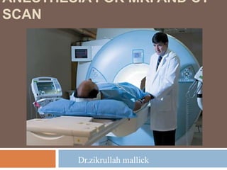
ANESTHESIA FOR MRI AND CT SCANs suite room
•Download as PPTX, PDF•
0 likes•8 views

ANESTHESIA FOR MRI AND CT SCANs .pptx

Recommended
Recommended
More Related Content
Similar to ANESTHESIA FOR MRI AND CT SCANs suite room
Similar to ANESTHESIA FOR MRI AND CT SCANs suite room (20)
More from ZIKRULLAH MALLICK
More from ZIKRULLAH MALLICK (20)
Recently uploaded
Recently uploaded (20)
ANESTHESIA FOR MRI AND CT SCANs suite room
- 1. ANESTHESIA FOR MRI AND CT SCAN Dr.zikrullah mallick
- 2. HEADINGS Introduction. Radiological Suits requiring Sedation/Anesthesia Indications for sedation/Anaesthesia. Problems in Radiological suite. Types of anaesthesia. Anaesthesia for CT. Anaesthesia for MRI. Practice guidelines for sedation and anaesthesia.
- 3. INTRODUCTION Anesthesiologists are increasingly being asked to provide anesthetic care in locations outside of the OT. Key to efficient and safe remote anesthetic depends on open communication between the anesthesiologist and non-operating room personnel. Because remote locations have different safety concerns, such as radiation and powerful magnetic fields
- 4. Radiological Suits requiring Sedation/Anesthesia Magnetic Resonance imaging(MRI) Computed Tomography Interventional Neuroradiology Ultrasound guided percut. tissue Ablation PET Scan/SPECT Scan Endoscopy suites Cardiac Angiography Psychiatric unit for electroconvulsive therapy Renal unit for lithotripsy
- 5. PROCEDURES Plain CT Contrast CT MRI MRI Angiography Functional MRI
- 6. Functional MRI fMRI is the use of MRI to measure the haemodynamic response related to neural activity in the brain
- 7. INDICATIONS FOR SEDATION/ANESTHESIA Infants or uncooperative children Children or adults with psychological problems Children or adults with Movement disorder Intubated critically ill patients
- 8. PROBLEMS IN RADIOLOGY SUITE Patient Anesthetist Environme nt Procedure
- 9. PROBLEM FOR PATIENTS Too cold/hot enviornment Immobility Noise Anxiety Claustrophobia
- 10. ENVIORNMENTAL PROBLEMS Dark/Dim light Inadequate Space Untrained staff Cylinder Supply Non availability Drug Monitors Equipment Lack of Direct observation
- 11. CHALLENGES FOR ANESTHESIST Developmental delay Epilepsy Malignancy Psychiatric Patient CNS disease Cardiac Respiratory Acute trauma with unstable Cardiovascular, Respiratory or Neurological function
- 12. CHALLENGES OF PROCEDURE Reaction to iodinated contrast media Patient positioning Limited access to patient/airway Exclusion of ferromagnetic objects in MRI Radiation hazard to the Anesthesiologist
- 13. TYPES OF ANESTHESIA Monitoring only General Anesthesia Using ET Tube and IPPV Using LMA Conscious Sedation TIVA
- 14. AIMS OF THE ANAESTHETIST Safety of the patient is the overiding goal of anaesthesia in remote locations and the standard of care should not differ from that offered in the operating theatre. Rapid recovery from anaestheisa or sedation is beneficial. Anesthetic implications of patient’s medical conditions do not vary with location
- 15. ANAESTHESIA FOR CT Anaesthetist can remain in the room wearing X-ray protection or view the patient and monitors from the control room The CT scanner does not interfere with monitoring equipment. The scans are short and can be interrupted The patient couch moves during examination Temporarily interruption of ventilation to improve image quality – immediately re-ventilate Patient positioning
- 16. ANESTHESIA FOR MRI MRI has superceded CT for the examination of CNS and many orthopaedic conditions MRI uses a static magnetic field which is permanently on and super-imposed rapidly changing magnetic fields and radio-frequency currents Low ionising radiation is used and there are no known ill effects Everyone must be screened before entering magnetic area and all ferro-magnetic items removed MRI can last over an hour and an individual scan can last upto 20 minutes
- 17. UNIQUE PROBLEMS OF MRI High magnetic field which is always on The bore of the magnet is narrow, noisy and claustrophobic. Access to the patient is difficult so airway must be secured Monitoring equipment can introduce stray radiofrequency current causing degradation of the image
- 18. MONITORING CONSIDERATIONS ECG Rapidly changing magnetic fields produce artifact, ST and T wave abnormality…may mimic arrhythmia If ECG wires are in loop, the magnetic field may heat the wires and leads, thus leading to thermal injury (antenna coupling effect) PULSE OXIMETRY Like ECG wires, antenna effect may produce thermal injury CAPNOGRAPHY BLOOD PRESSURE
- 19. Equipment for MRI Specialized MRI compatible equipment within scan room or conventional equipment outside scanner magnetic field Non magnetic tipping trolley Piped gases, scavenging & suction in both induction area & control room Respiratory gas side stream analyzer with capnograph with extended sampling line MRI compatible pulse oximeter- Fibreoptic patient probe & shielded cable ECG with MRI compatible carbon fibre leads & electrodes
- 20. SOLUTIONS… MRI compatible anaesthetic equipment or non-MRI compatible anaesthetic equipment kept outside the magnetic field Monitoring during MRI To minimize interference, use of Faraday cage and low band filters Capnography:- for monitoring of adequacy of ventilation and has a disconnection alarm Blood pressure-automated blood pressure recording can be performed using extended cable and nylon threads on cuff attachment. Invasive blood pressure measurements need long extension tubing
- 21. PROBLEMS FOR ANESTHESIST Unfamiliar/outdated Equipments Isolated - support services No piped gases, no suction Moving away from patient Difficult to resuscitate
- 22. The particular goals to consider when sedating patients Guard the patient’s safety and welfare Minimize physical discomfort and pain Control anxiety, minimize psychological trauma and maximize the potential for amnesia Control movement to allow safe completion of the procedure Return the patient to a state in which safe discharge from medical supervision is possible.
- 23. Practice Guidelines for Sedation and Anesthesia Patient must be evaluated before the procedure by qualified personnel to ensure that patients are not compromised by coexisting medical conditions Appropriate NPO status Informed consent During the procedure, the level of consciousness, ventilation, oxygenation, and hemodynamics are to be evaluated with standard monitors by a designated individual who should be trained to recognize complications of analgesia and sedation
- 24. Practice Guidelines for Sedation and Anesthesia At least one individual trained in basic life support skills should be present continuously when moderate or deep sedation is used Supplemental oxygen should be used for moderate and deep sedation, and emergency equipment, including pharmacologic antagonists, should be available Adequate recovery care should be provided, with the patient observed in an adequately staffed and equipped recovery area
- 25. MRI ZONES
- 26. PRACTICE ADVISORY ON ANESTHETIC CARE I. Education 1. MRI education for magnet hazards 2. MRI education for monitoring limitations 3. MRI education for long-term health hazards II. Screening of Anesthesia Care Providers and Ancillary Support Personnel 4. Mandatory screening of all personnel entering zone III or IV
- 27. III. Patient Screening 5. Patient-related risks for adverse outcomes related to MRI 6. Equipment-related risks for adverse outcomes related to MRI IV. Preparation 7. Planning for the anesthetic care of the patient for the scan 8. Planning for rapidly summoning additional personnel in the event of an emergency
- 28. V. Patient Management during MRI 9. Monitoring during MRI 10. Anesthetic care during MRI 11. Airway management during MRI VI. Management of Emergencies 12. Medical emergencies 13. Environmental emergencies
- 29. VII. Post procedure Care 14. Post procedure care consistent with that provided for other areas of the institution
- 30. Sedating agent Most children younger than the age of 5 years and many as old as age 11 require sedation or general anesthesia to tolerate MRI Oral sedation techniques, if appropriately administered, have a success rate of 93% Oral chloral hydrate is a popular agent ( 25-50 mg/kg for infants younger than 4 months, 50 mg/kg for older children)
- 31. Benzodiazepines such as midazolam administered either orally (0.25 to 0.75 mg/kg) or intravenously (0.05 to 0.15 mg/kg) are also commonly used for sedation Deep sedation with propofol, oxygen administration via nasal cannula, and end-tidal carbon dioxide monitoring is a successful technique
- 32. Children are initially sedated with incremental propofol boluses up to 3 mg/kg with or without midazolam, 0.03 to 0.05 mg/kg, and then maintained with an infusion rate of propofol, 1 to 3 mg/kg/hr, with supplemental boluses of 1 mg/kg for movement In the case of an emergency , the MRI technicians should be notified, the scan sequence stopped, and the patient rapidly removed
- 33. Anaphylaxis to iodinated dyes is possible. All the drugs for management of anaphylaxis should always be immediately available Resuscitation attempts should take place outside the scanner because equipment such as laryngoscopes, oxygen cylinders, and cardiac defibrillators cannot be taken close to the magnet
- 35. SUMMARY These require sedation/Anaesthesia :: o Infants and uncooperative children. o People with movement disorders. o People with psychological disorders. o Intubated critically ill patients. MRI compatible equipment's are required. Propofol, Midazolam and Thiopentone are commonly used.