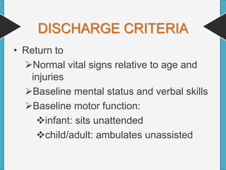
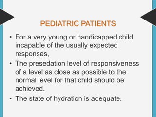
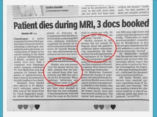
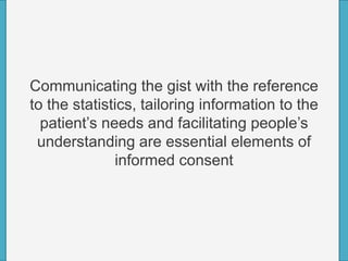
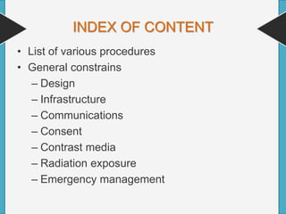
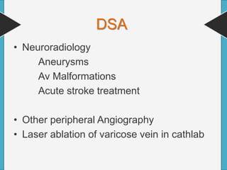
Use of radiological support involves nearly
every medical specialty ,generates volume
and revenue equivalent to that of OT ,and
are as demanding of anesthesiologist as the
most advanced surgical OT procedures.
They require the same attention to
operational efficiency ,planning and
consistency with which the OT is managed
Intrinsic ,common and unique characteristics
of such cases that imposes unusual
constrains on anesthesiologists in the out of
OT arena.
INDEX OF CONTENT
• List of various procedures
• General constrains
– Design
– Infrastructure
– Communications
– Consent
– Contrast media
– Radiation exposure
– Emergency management
– Post procedure care
– Documentation & discharge
• Specific concerns
• Concept of Sedation
• Drugs needed for sedation
• Concerns of isolated radiological centers
• Pediatric pts.
• Medico legal issues.
• USG guided procedures ( Biopsy, pleural
tapping etc.)
• Myelography
• IVP
• CT scan
• MRI
• Endoscopic procedures
• Cath lab procedures
• DSA(neuroradiology, peripheral angiography)
List of procedures
GENERAL CONSTRAINTS
• Location=the procedure never takes place
in a typical operation theater.
• Operator=for the most part ,the operator
performing the procedure is not a surgeon.
• Relative Novelty=the procedure and
technologies used may be novel in one or
other way.
EQUIPMENT LAYOUT
• Layout of radiological suit is made as per
the need of radiologist, so anaesthesia
equipments gets minimum space or non
reachable corner of the room.
ANAESTHESIA EQUIPMENTS
• Anaesthesia workstation is must(or redily
available) for every radiological unit.
• MRI compatible workstation is costly but
mandatory.
• Extra long tubeings and extensions are
very important.
• Syringe pump and defibrillator is must
• lack of equipment and storage
• Many procedures are carried out in
darkened room
• Should be able to visualize the flow meter
and to check accurate flow.
SOAPME
• Suction
• Oxygen
• Airways
• Pharmacy (all drugs)
• Monitors
• Equipment (other equipment's)
MONITORS
• Pulse oximeter
• NIBP
• ECG
• ETCO2
Nasal cannula for etco2 monitor is very
helpful
STAFF
• Number of staff is less and Staff-trained
only in their specialty.
• Sole responsibility of anesthesiologist to
check and ensure safety
• The indifferent reflexes shown by the non
operating room staff in emergency
situations.
• Unlike surgeons-most medical providers
have minimal knowledge about
anaesthesia, less familiar with the skill set
of anaesthesiologist lack experience with
many relatively rare but serious
complications.
PLANNING OF THE PROCEDURE
Schedule for the procedures may not be in
place the cases schedule is sporadic making
anaesthesia services less productive.
• Beware the situation where the
anesthesiologist is called after the
intervention has started and the pt. is
found to be uncooperative.
• Without a prior plan or airway assessment
the situation is hazardous.-if situation
allows, it is better to abort the procedure
and reschedule it.
PRE OP ASSESSMENT
• The reason for which they require the
intervention.
• Associated co morbidities
• Fasting status
• Quick airway assessment
• Presence of denture
DURATION
• Duration of procedure is difficult to predict
• They may finish very abruptly
COMMUNICATION
• Communication with team is extremely
important
• Confirm the communication with other
departments also.
CONSENT FOR ANAESTHESIA
• Detailed description of current status
should be discussed.
• Plan for MAC,Sedation or GA should be
discussed properly.
• Must discuss about postop outcome and
admission or discharge criteria.
Extremely important to explain the current
condition and likely condition post
procedural sedation, like
pt. is rowdy due to CNS bleed and we give
sedation, that should not confuse relative
that pt. is under the influence of
anaesthesia.
POSITIONING
• Generally the staff and other assistants
are less familiar and equipped to keep the
pt. in ‘prone ‘ or ‘lateral’ position.
OUR AIMS
• Guard the patient’s safety.
• Minimize physical discomfort and pain
• control anxiety,
• minimize psychological traumas
• maximize the potential for amnesia
• Control movement of pt. during procedure
• Safe discharge
PREPROCEDURAL EVALUATION OF
PROCEDURE ROOM.
• Many procedure suits have beds with low wt.
limits and less mobility
• Head up and head low is not possible most of
the time.
• Anticoagulation status is often a issue.
• Contrast and renal status should be planned
before.
• Bleeding is generally occult
• Many pt. do not tolerate supine position for
long
Contrast media
Commonly used iodinated contrast agents
• One of the primary differences between
ionic and non-ionic contrast media is that
an ionic compound dissociates or
dissolves into charged particles when it
enters into blood.
• Ionic media breakdown into cations and
anions.
• For every three iodine molecules present
in an ionic media, one cation and one
anion are produced when it enters blood.
• Ionic contrast media are generally referred
to as 3:2 compounds.
REACTION TO CONTRAST MEDIA
• Although most reactions occur in the first
hour after administration, and many occur
within the first 5 minutes, there are rare
instances of late adverse reactions that
occur between 1 hour and 1 week after
injection of iodinated contrast agents.
HIGH RISK FOR CONTRAST
REACTION
• Previous reactions to iodinated contrast
agents
• All severe allergies and reactions (to
medications and food)
• History of asthma, bronchospasm, or
atopy
• History of cardiac or renal disease
• Especially those aged >60 y or <5 y
EXTRAVASATION
• Intravenous access should be evaluated
before the administration of contrast
media.
• Use of a 20-gauge or larger catheter in an
antecubital or other large forearm vein is
recommended for flow rates of at least 3
mL/sec, with flow rates no greater than 1.5
mL/sec for peripherally placed or 22-
gauge catheters.
• The use of deep brachial intravenous
catheters should be avoided because of
the markedly higher relative risk of
extravasation .
PROPHYLAXIS AGAINST
REACTION
• Contrast Media Safety Committee of
ESUR considers the use of premedication
with steroids and antihistaminic , although
evidence of its effectiveness is limited in
patients with previous moderate or severe
acute reactions.
• Aspiration of high-osmolar water-soluble
contrast agents can lead to severe
pulmonary edema. Therefore, iso-osmolar
or low-osmolar agents should be used in
patients at increased risk for aspiration.
Oral Contrast
• A small amount (approximately 1%–2%) of
ingested iodinated contrast material
normally is absorbed. This absorption
theoretically can cause dose-independent
anaphylactoid reactions
PREVENTION FOR ARF
• Alkalization of the urine with sodium
bicarbonate is also useful.
• Bicarbonate should be infused at a rate
3ml/kg/hr. for 1 hr. before injection of
contrast agent, followed by 1ml/kg/hr. for 6
hrs.
• Mucomist is relatively inexpensive and
may be useful.
RADIATION EXPOSURE
• Occupational exposure to radiation comes
primarily from x rays (ionizing
radiation)scattered by the pt. and
surrounding equipments.
• A distance of 3 feet from the pt. is
recommended to minimize physiological
damage from occupational hazards.
• A distance of 6 feet from the pt. provides
the same protection as 2.5 mm lead.
EMERGENCY MANAGEMENT
• Emergency managemet is difficult due to
lack of equipments and drugs
• Call for help as early as possible.
• Stabilizing the pt. and shifting to ICU
ASAP is extremely important for better
outcome.
• Stopping the magnetic field and shifting
the pt. outside the magnet is primary thing
in MRI.
SPECIFIC ISSUES
CT SCAN
• CT scan
• CT guided biopsy
• CT guided therapeutic
intervention
• CT guided pain
management procedures
• Short procedure lasting from few seconds
to few minutes only.
• Radiation hazards are more for
anaesthesiologist
• High speed contrast injection.
MRI
• MRI
• MRI guided interventions
– Biopsy
– tumor ablation using cytoablative technology
for which repeted breath holds, long time in
one position, repeted heating and freezing of
tissue is needed ..so GA may be needed.
• Movement of head not allowed in case of
MRI Brain.
• Lack of MRI compatible monitors
• Magnetic field
• Pacemaker,
• Implanted defibrillators,
• Cochlear implants,
• Pumps,
• Nerve stimulators,
• Aneurysm clips,
• Metal fragments
• Bullets
SHOULD NOT BE SCANNED
• Procedure is as long as 15 min to 45 min
• Loud noise
• Pt. not accessible
• Claustrophobic environment
• Low temperature of MRI suit.
CATH LAB
• Pacemaker insertion
• Device insertion for ASD VSD
• Balloon valvoplasty & valve replacement.
• Catheter ablation technique for
arrhythmias
• Implantable cardioverter-defibrilators
• Percutaneous ventricular assist devices.
• Mostly requirement is MAC
• But for many procedure like devise
insertion in teenage group requires deep
sedation or GA
• Many procedure are with the use of TEE
also.
• Hemodynamic support, ventilation support
and emergency management is vital here.
DSA
• Neuroradiology
Aneurysms
Av Malformations
Acute stroke treatment
• Other peripheral Angiography
• Laser ablation of varicose vein in cathlab
Conscious sedation
Vital Signs
stable and
normal
All protective
Reflexes intact
Child’s pain
Threshold
Should be
Increased
Amnesia
Should occur
The patient’s
Mood should
be altered
Patient should
be conscious,
respond to
verbal stimuli
Patient should
be cooperative
Objectives of
conscious
sedation
CHOICE OF ANAESTHESIA
• Monitored anaesthesia care
• Sedation
• Regional anesthesia
• General anaesthesia
MIDAZOLAM
• Minimal sedation, anxiolytic and amnesia
• Combine with fentanyl for deeper sedation
& analgesia
• Onset: 2-5 minutes
• Duration: 30-60 minutes
• Dose: 0.05-0.1 mg/kg over 1-2min
• then q 2-5 min
FENTANYL
• Short acting Opioid.
• Adult dose 1-2 mcg/kg slow IV push (over 1-2
min); may repeat dose after 30 min.
• Onset of action 1-2 min, and duration 30-60
min.
• Does not stimulate histamine release.
• May cause chest Wall rigidity, apnea,
respiratory depression, or hypotension.
• Action reversed by naloxone.
Midazolam + Fentanyl
Dose Midazolam Fentanyl
Initial 0.02 mg/kg 0.5 mcg/kg
Subsequent
q 2 min
0.25 mg/kg 0.25 mcg/kg
WAIT
KETAMINE HYDROCHLORIDE
Indications :
• Shocked patient
• Pediatric anesthesia
• Analgesia And sedation.
SHORT TERM SIDE EFFECTS OF
KETAMINE ARE:
• Increase in heart
rate
• Slurred speech
• Confusion,
disorientation
• Out-of-body
experience
• Nausea
• Sedation
• Hypertension
• Euphoria
• Salivation
• Delirium
• Raised ICT
DEXMEDETOMIDINE
• For procedural sedation in pediatric and
adult patients
• An alpha2-adrenergic agonist that
provides sedation, anxiolysis, hypnosis,
analgesia, and sympatholysis
• provides little to no respiratory depression
• patients are able to follow commands and
respond to verbal and tactile stimulus but
fall quickly asleep when not stimulated
• provide some pain relief, like ketamine, but
not to the same degree (use of other
analgesics necessary for the more painful
procedures)
• Minimal cardiovascular effects are seen
and include mild bradycardia and a
decrease in systemic vascular resistance
PROPOFOL
• It is alkylphenol derivative.
• Adult dose 0.5-1 mg/kg IV loading dose;
may repeat by 0.5-mg increments q3-
5min.
• Onset of action <1 min, and duration 3-10
min.
• Provides rapid onset and recovery phase,
and brief duration of action.
• No analgesia.
• Has anticonvulsant properties.
• Can rapidly cause deep sedation.
• Causes cardiovascular depression and
hypotension.
Ketofol (1 ketamine; 1 propofol)
• Draw up 10ml of Propofol in a 20cc
syringe.
– Propofol comes 10mg/ml.
• Discard 2cc from a 10cc saline flush.
Drawl up 2cc of Ketamine.
– Ketamine 50mg/ml (adjust the dose if you use
a different concentration)
– You now have 10mg/ml
• Inject the Ketamine in the saline flush into
a 20cc syringe of Propofol.
• Dose at O.5 mg/kg IV Ketofol, then redose
as needed
ETOMIDATE
• Etomidate is given IV over 30 to 60
seconds in doses of 0.1 to 0.15 mg/kg
• Maintains cardiovascular stability
• Has no analgesic properties
• Etomidate causes pain during injection
into peripheral veins
• Potential side effects of etomidate include
myoclonus (most frequently), respiratory
depression, adrenal suppression, and
nausea and Vomiting
• When given by continuous infusion,
causes adrenal insufficiency.
TIVA
• Careful about airway
• Chin lift, jaw thrust, or oral airway/nasal
airway or LMA may be needed.
GA
• Rarely needed.
• But may be needed for very uncooperative
pt. who does not tolerate sedation.
• pt. with endotracheal intubation
POST PROCEDURE CARE
• Recovery room with monitors & staff is
must for all the procedures.
• Isolated centers outside hospital campus
has more problems related to this.
• Communication with ICU prior to the
procedure is need in case of shifting to
ICU.
• Transfer of the pt. to a standard recovery
room with the monitors ,oxygen along with
the anesthesiologist.
• Transfer from isolated centers should be
done in well equipped ambulance only.
DISCHARGE CRITERIA
• Return to
Normal vital signs relative to age and
injuries
Baseline mental status and verbal skills
Baseline motor function:
infant: sits unattended
child/adult: ambulates unassisted
• Tolerating oral fluids
• Pain controlled with oral analgesia
• Discharged to responsible adult who
understands and can comply with
discharge instructions
PEDIATRIC PATIENTS
• For a very young or handicapped child
incapable of the usually expected
responses,
• The presedation level of responsiveness
of a level as close as possible to the
normal level for that child should be
achieved.
• The state of hydration is adequate.
Communicating the gist with the reference
to the statistics, tailoring information to the
patient’s needs and facilitating people’s
understanding are essential elements of
informed consent
Anaesthesia for radiological procedures final

Anaesthesia for radiological procedures final

  • 2.
    Use of radiologicalsupport involves nearly every medical specialty ,generates volume and revenue equivalent to that of OT ,and are as demanding of anesthesiologist as the most advanced surgical OT procedures.
  • 3.
    They require thesame attention to operational efficiency ,planning and consistency with which the OT is managed
  • 4.
    Intrinsic ,common andunique characteristics of such cases that imposes unusual constrains on anesthesiologists in the out of OT arena.
  • 5.
    INDEX OF CONTENT •List of various procedures • General constrains – Design – Infrastructure – Communications – Consent – Contrast media – Radiation exposure – Emergency management
  • 6.
    – Post procedurecare – Documentation & discharge • Specific concerns • Concept of Sedation • Drugs needed for sedation • Concerns of isolated radiological centers • Pediatric pts. • Medico legal issues.
  • 7.
    • USG guidedprocedures ( Biopsy, pleural tapping etc.) • Myelography • IVP • CT scan • MRI • Endoscopic procedures • Cath lab procedures • DSA(neuroradiology, peripheral angiography) List of procedures
  • 8.
    GENERAL CONSTRAINTS • Location=theprocedure never takes place in a typical operation theater. • Operator=for the most part ,the operator performing the procedure is not a surgeon. • Relative Novelty=the procedure and technologies used may be novel in one or other way.
  • 9.
    EQUIPMENT LAYOUT • Layoutof radiological suit is made as per the need of radiologist, so anaesthesia equipments gets minimum space or non reachable corner of the room.
  • 10.
    ANAESTHESIA EQUIPMENTS • Anaesthesiaworkstation is must(or redily available) for every radiological unit. • MRI compatible workstation is costly but mandatory. • Extra long tubeings and extensions are very important. • Syringe pump and defibrillator is must
  • 11.
    • lack ofequipment and storage • Many procedures are carried out in darkened room • Should be able to visualize the flow meter and to check accurate flow.
  • 12.
    SOAPME • Suction • Oxygen •Airways • Pharmacy (all drugs) • Monitors • Equipment (other equipment's)
  • 13.
    MONITORS • Pulse oximeter •NIBP • ECG • ETCO2 Nasal cannula for etco2 monitor is very helpful
  • 14.
    STAFF • Number ofstaff is less and Staff-trained only in their specialty. • Sole responsibility of anesthesiologist to check and ensure safety • The indifferent reflexes shown by the non operating room staff in emergency situations.
  • 15.
    • Unlike surgeons-mostmedical providers have minimal knowledge about anaesthesia, less familiar with the skill set of anaesthesiologist lack experience with many relatively rare but serious complications.
  • 16.
    PLANNING OF THEPROCEDURE Schedule for the procedures may not be in place the cases schedule is sporadic making anaesthesia services less productive.
  • 17.
    • Beware thesituation where the anesthesiologist is called after the intervention has started and the pt. is found to be uncooperative. • Without a prior plan or airway assessment the situation is hazardous.-if situation allows, it is better to abort the procedure and reschedule it.
  • 18.
    PRE OP ASSESSMENT •The reason for which they require the intervention. • Associated co morbidities • Fasting status • Quick airway assessment • Presence of denture
  • 20.
    DURATION • Duration ofprocedure is difficult to predict • They may finish very abruptly
  • 21.
    COMMUNICATION • Communication withteam is extremely important • Confirm the communication with other departments also.
  • 22.
    CONSENT FOR ANAESTHESIA •Detailed description of current status should be discussed. • Plan for MAC,Sedation or GA should be discussed properly. • Must discuss about postop outcome and admission or discharge criteria.
  • 23.
    Extremely important toexplain the current condition and likely condition post procedural sedation, like pt. is rowdy due to CNS bleed and we give sedation, that should not confuse relative that pt. is under the influence of anaesthesia.
  • 24.
    POSITIONING • Generally thestaff and other assistants are less familiar and equipped to keep the pt. in ‘prone ‘ or ‘lateral’ position.
  • 25.
    OUR AIMS • Guardthe patient’s safety. • Minimize physical discomfort and pain • control anxiety, • minimize psychological traumas • maximize the potential for amnesia • Control movement of pt. during procedure • Safe discharge
  • 26.
    PREPROCEDURAL EVALUATION OF PROCEDUREROOM. • Many procedure suits have beds with low wt. limits and less mobility • Head up and head low is not possible most of the time. • Anticoagulation status is often a issue. • Contrast and renal status should be planned before. • Bleeding is generally occult • Many pt. do not tolerate supine position for long
  • 27.
  • 28.
    Commonly used iodinatedcontrast agents
  • 29.
    • One ofthe primary differences between ionic and non-ionic contrast media is that an ionic compound dissociates or dissolves into charged particles when it enters into blood. • Ionic media breakdown into cations and anions.
  • 30.
    • For everythree iodine molecules present in an ionic media, one cation and one anion are produced when it enters blood. • Ionic contrast media are generally referred to as 3:2 compounds.
  • 31.
    REACTION TO CONTRASTMEDIA • Although most reactions occur in the first hour after administration, and many occur within the first 5 minutes, there are rare instances of late adverse reactions that occur between 1 hour and 1 week after injection of iodinated contrast agents.
  • 32.
    HIGH RISK FORCONTRAST REACTION • Previous reactions to iodinated contrast agents • All severe allergies and reactions (to medications and food) • History of asthma, bronchospasm, or atopy • History of cardiac or renal disease • Especially those aged >60 y or <5 y
  • 35.
    EXTRAVASATION • Intravenous accessshould be evaluated before the administration of contrast media.
  • 36.
    • Use ofa 20-gauge or larger catheter in an antecubital or other large forearm vein is recommended for flow rates of at least 3 mL/sec, with flow rates no greater than 1.5 mL/sec for peripherally placed or 22- gauge catheters. • The use of deep brachial intravenous catheters should be avoided because of the markedly higher relative risk of extravasation .
  • 37.
    PROPHYLAXIS AGAINST REACTION • ContrastMedia Safety Committee of ESUR considers the use of premedication with steroids and antihistaminic , although evidence of its effectiveness is limited in patients with previous moderate or severe acute reactions.
  • 38.
    • Aspiration ofhigh-osmolar water-soluble contrast agents can lead to severe pulmonary edema. Therefore, iso-osmolar or low-osmolar agents should be used in patients at increased risk for aspiration. Oral Contrast
  • 39.
    • A smallamount (approximately 1%–2%) of ingested iodinated contrast material normally is absorbed. This absorption theoretically can cause dose-independent anaphylactoid reactions
  • 40.
    PREVENTION FOR ARF •Alkalization of the urine with sodium bicarbonate is also useful. • Bicarbonate should be infused at a rate 3ml/kg/hr. for 1 hr. before injection of contrast agent, followed by 1ml/kg/hr. for 6 hrs. • Mucomist is relatively inexpensive and may be useful.
  • 41.
    RADIATION EXPOSURE • Occupationalexposure to radiation comes primarily from x rays (ionizing radiation)scattered by the pt. and surrounding equipments.
  • 42.
    • A distanceof 3 feet from the pt. is recommended to minimize physiological damage from occupational hazards. • A distance of 6 feet from the pt. provides the same protection as 2.5 mm lead.
  • 43.
    EMERGENCY MANAGEMENT • Emergencymanagemet is difficult due to lack of equipments and drugs • Call for help as early as possible. • Stabilizing the pt. and shifting to ICU ASAP is extremely important for better outcome. • Stopping the magnetic field and shifting the pt. outside the magnet is primary thing in MRI.
  • 44.
  • 45.
    CT SCAN • CTscan • CT guided biopsy • CT guided therapeutic intervention • CT guided pain management procedures
  • 46.
    • Short procedurelasting from few seconds to few minutes only. • Radiation hazards are more for anaesthesiologist • High speed contrast injection.
  • 47.
    MRI • MRI • MRIguided interventions – Biopsy – tumor ablation using cytoablative technology for which repeted breath holds, long time in one position, repeted heating and freezing of tissue is needed ..so GA may be needed.
  • 48.
    • Movement ofhead not allowed in case of MRI Brain. • Lack of MRI compatible monitors • Magnetic field
  • 49.
    • Pacemaker, • Implanteddefibrillators, • Cochlear implants, • Pumps, • Nerve stimulators, • Aneurysm clips, • Metal fragments • Bullets SHOULD NOT BE SCANNED
  • 50.
    • Procedure isas long as 15 min to 45 min • Loud noise • Pt. not accessible • Claustrophobic environment • Low temperature of MRI suit.
  • 51.
    CATH LAB • Pacemakerinsertion • Device insertion for ASD VSD • Balloon valvoplasty & valve replacement. • Catheter ablation technique for arrhythmias • Implantable cardioverter-defibrilators • Percutaneous ventricular assist devices.
  • 52.
    • Mostly requirementis MAC • But for many procedure like devise insertion in teenage group requires deep sedation or GA • Many procedure are with the use of TEE also.
  • 53.
    • Hemodynamic support,ventilation support and emergency management is vital here.
  • 54.
    DSA • Neuroradiology Aneurysms Av Malformations Acutestroke treatment • Other peripheral Angiography • Laser ablation of varicose vein in cathlab
  • 55.
  • 57.
    Vital Signs stable and normal Allprotective Reflexes intact Child’s pain Threshold Should be Increased Amnesia Should occur The patient’s Mood should be altered Patient should be conscious, respond to verbal stimuli Patient should be cooperative Objectives of conscious sedation
  • 59.
    CHOICE OF ANAESTHESIA •Monitored anaesthesia care • Sedation • Regional anesthesia • General anaesthesia
  • 60.
    MIDAZOLAM • Minimal sedation,anxiolytic and amnesia • Combine with fentanyl for deeper sedation & analgesia • Onset: 2-5 minutes • Duration: 30-60 minutes • Dose: 0.05-0.1 mg/kg over 1-2min • then q 2-5 min
  • 61.
    FENTANYL • Short actingOpioid. • Adult dose 1-2 mcg/kg slow IV push (over 1-2 min); may repeat dose after 30 min. • Onset of action 1-2 min, and duration 30-60 min. • Does not stimulate histamine release. • May cause chest Wall rigidity, apnea, respiratory depression, or hypotension. • Action reversed by naloxone.
  • 62.
    Midazolam + Fentanyl DoseMidazolam Fentanyl Initial 0.02 mg/kg 0.5 mcg/kg Subsequent q 2 min 0.25 mg/kg 0.25 mcg/kg WAIT
  • 63.
    KETAMINE HYDROCHLORIDE Indications : •Shocked patient • Pediatric anesthesia • Analgesia And sedation.
  • 64.
    SHORT TERM SIDEEFFECTS OF KETAMINE ARE: • Increase in heart rate • Slurred speech • Confusion, disorientation • Out-of-body experience • Nausea • Sedation • Hypertension • Euphoria • Salivation • Delirium • Raised ICT
  • 65.
    DEXMEDETOMIDINE • For proceduralsedation in pediatric and adult patients • An alpha2-adrenergic agonist that provides sedation, anxiolysis, hypnosis, analgesia, and sympatholysis
  • 66.
    • provides littleto no respiratory depression • patients are able to follow commands and respond to verbal and tactile stimulus but fall quickly asleep when not stimulated
  • 67.
    • provide somepain relief, like ketamine, but not to the same degree (use of other analgesics necessary for the more painful procedures) • Minimal cardiovascular effects are seen and include mild bradycardia and a decrease in systemic vascular resistance
  • 68.
    PROPOFOL • It isalkylphenol derivative. • Adult dose 0.5-1 mg/kg IV loading dose; may repeat by 0.5-mg increments q3- 5min. • Onset of action <1 min, and duration 3-10 min. • Provides rapid onset and recovery phase, and brief duration of action.
  • 69.
    • No analgesia. •Has anticonvulsant properties. • Can rapidly cause deep sedation. • Causes cardiovascular depression and hypotension.
  • 70.
    Ketofol (1 ketamine;1 propofol) • Draw up 10ml of Propofol in a 20cc syringe. – Propofol comes 10mg/ml. • Discard 2cc from a 10cc saline flush. Drawl up 2cc of Ketamine. – Ketamine 50mg/ml (adjust the dose if you use a different concentration) – You now have 10mg/ml
  • 71.
    • Inject theKetamine in the saline flush into a 20cc syringe of Propofol. • Dose at O.5 mg/kg IV Ketofol, then redose as needed
  • 72.
    ETOMIDATE • Etomidate isgiven IV over 30 to 60 seconds in doses of 0.1 to 0.15 mg/kg • Maintains cardiovascular stability • Has no analgesic properties • Etomidate causes pain during injection into peripheral veins
  • 73.
    • Potential sideeffects of etomidate include myoclonus (most frequently), respiratory depression, adrenal suppression, and nausea and Vomiting • When given by continuous infusion, causes adrenal insufficiency.
  • 74.
    TIVA • Careful aboutairway • Chin lift, jaw thrust, or oral airway/nasal airway or LMA may be needed.
  • 75.
    GA • Rarely needed. •But may be needed for very uncooperative pt. who does not tolerate sedation. • pt. with endotracheal intubation
  • 76.
    POST PROCEDURE CARE •Recovery room with monitors & staff is must for all the procedures. • Isolated centers outside hospital campus has more problems related to this. • Communication with ICU prior to the procedure is need in case of shifting to ICU.
  • 77.
    • Transfer ofthe pt. to a standard recovery room with the monitors ,oxygen along with the anesthesiologist. • Transfer from isolated centers should be done in well equipped ambulance only.
  • 78.
    DISCHARGE CRITERIA • Returnto Normal vital signs relative to age and injuries Baseline mental status and verbal skills Baseline motor function: infant: sits unattended child/adult: ambulates unassisted
  • 79.
    • Tolerating oralfluids • Pain controlled with oral analgesia • Discharged to responsible adult who understands and can comply with discharge instructions
  • 80.
    PEDIATRIC PATIENTS • Fora very young or handicapped child incapable of the usually expected responses, • The presedation level of responsiveness of a level as close as possible to the normal level for that child should be achieved. • The state of hydration is adequate.
  • 82.
    Communicating the gistwith the reference to the statistics, tailoring information to the patient’s needs and facilitating people’s understanding are essential elements of informed consent