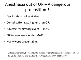
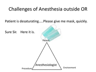
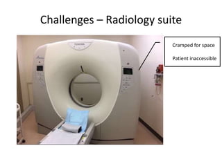
Anaesthesia & sedation in offsite
locations:
How to maintain quality & avoid
complications
Dr Sandeep Kundra
Associate Professor,
Department of Anesthesia,
DMC & H, Ludhiana.
What’s there in the name
- Non operating room anesthesia.
(NORA)
- Anesthesia at remote location.
- Outpatient anesthesia.
- Office based anesthesia.
That which we call a Rose by any other name would smell as sweet.
- William Shakespeare
Common location outside OR
• Radiology. (CT and MRI)
• Interventional Radiology suites.
• Endoscopy suite.
• Cardiac catheterization Lab.
• Nuclear medicine. (PET-CT, PET-MRI)
• Radiation therapy.
• Electroconvulsive therapy.
• Dentistry.
Anesthesia outside OR : Goals.
• Ensure safety.
• Minimize pain and discomfort.
• Control patient movement.
• Minimize psychological discomfort and anxiety.
• Careful monitoring and recording of vital signs.
• Develop efficient and cost effective system.
Anesthesia out of OR – A dangerous
proposition!!!
• Exact data – not available.
• Complication rate higher than OR.
• Adverse respiratory event – 44 %.
• 50 % cases were under MAC.
• Many were preventable.
MetznerJ, Posner KL, Domino KB. The risk and safety of anesthesia at remote locations:
the US closed claims analysis. Curr Opin Anaesthesiol 2009; 22:502–508.
Challenges of Anesthesia outside OR
Patient is desaturating…..Please give me mask, quickly.
Sure Sir. Here it is.
Patient
Procedure Environment
Anesthesiologist
Challenges - Patient
• Extremes of age.
• Sick patients offered procedures, otherwise unfit.
• Proceduralist unaware of general condition.
• Little time for cross consultation and optimization.
• Lack of patient awareness.
Challenges - Procedure
Sisterrrrrr…………. I asked for Laryngoscope,
not this endoscope!!!!!
• Staff not acquainted with anesthetic management.
• In case of any problem, additional help not available.
• Procedure details may not be known to anesthetist.
• Limited airway rescue devices.
Challenge - Environment
• Cramped for space.
• Limited access to patient.
• Poor Illumination.
• Often Old, unfamiliar equipment, not serviced.
• Emergency drugs and resuscitation equipment not
readily available.
Simile with surgical strike
Challenges – Environment
CT
• Radiation hazard.
• Takes little time.
• Oral contrast administered requires deviation from
NPO guidelines.
• Look out for Contrast reactions.
• Breath holding may be required for chest CT.
Challenges – Environment
MRI
1 Tesla = 10,000 Gauss
Earth’s magnetic field = 0.5 G
3 T MRI = 15,000 times earth’s magnetic field
MRI - Challenges
MRI Challenges
MRI suite
MRI safe equipment
Ensure patient has
no ferromagnetic
implants
Anesthetist has no
metal objects
Absolute immobility
needed
Young children
Claustrophobic
Mentally challenged
Auditory protection
Ferromagnetic Objects
• ICD/ Pacemakers
• Vascular clips
• Ortho Implants
• Insulin pump
• Dentures
• Metallic Heart valve
• Pellets, bullets.
• Magnetic strips including credit cards and ID badges.
MRI zones
There is a yellow line
within the MRI room
which cannot be crossed
with any ferromagnetic
materials.
Interventional Radiology
• Liver biopsy, abscess drainage, TIPPS, embolization.
• Unfit for surgery are offered interventions so many
patients are high risk patients.
• Sclero-therapeutic agents:
- Ethanol - Bleomycin
- Polyvinyl alcohol - sodium tetradecyl sulfate
- Ethylene vinyl alcohal - cyanoacrylate glue
Interventional Radiology : Cerebral
angiography
• Procedure requirements:
- Patient immobility
- Controlled ventilation with
intermittent breath holding
- Meticulous BP control.
Nuclear medicine : PET
• Applications for diagnosis and response to treatment.
• Sedation required during warming phase, 30 min to 1
hour after radioisotope injection.
• High risk of exposure to anesthesia personnel, remote
monitoring can be set up.
• Commonly require long periods of sedation.
External beam radiation therapy
• Complete patient immobility.
• Daily treatments are common.
• Remote monitoring is necessary.
• Children commonly require monitored transport to a
radiation centre.
Cardiac Cath Lab
• PTCA for unstable patients.
• Minimize effects of anesthetic drugs on CVS.
• May need resuscitation during procedure.
• Multiple infusions.
• Device closure for ASD/VSD – GA.
Upper GI endoscopy
GI endoscopy
Shared airway
Risk of aspiration
Bleeding
Copious bowel
prep agents
Hemodynamic
instability esp in
elderly
Limited CVS
reserve &
Dehydration
Vagal response
from distended
bowel
Risk of Apnea, Laryngospasm, Bronchospasm and Airway
obstruction.
Bronchoscopy
• Shared airway.
• Flexible vs Rigid bronchoscopy
• Ventilatory strategies:
- Apnoeic oxygenation
- Spontaneous assisted ventilation
- Controlled ventilation
- Manual jet ventilation
- High frequency jet ventilation.
ECT
• Electrical impulse induced GTC seizues.
• Tonic phase, 5 – 10 sec, Clonic phase 30 - 60 sec.
• Isolated Arm Technique.
• Initial parasympathetic discharge
• Prominent sympathetic response, chances of ST-T
changes.
Dentistry
• Reserved for Complicated
or prolonged cases, uncooperative patients.
• Shared airway - Tracheal intubation, often nasally.
• Postoperative complications:
- Bleeding,
- Airway obstruction
- Laryngeal spasm.
Ensuring safety : Onus is on
Anesthesiologist
• Personnel for NORA.
• Equipments.
• Monitoring.
• Precautions.
• Post procedural care.
Patient
safety
Constraints
Patient
Comfort
Quality
Assurance
Proceduralist
requirements
Who can administer sedation.
• American Society of Anesthesiologists. (ASA)
• American Society for gastro-instestinal endoscopy.
(ASGE)
• Guidelines by American College of Chest Physicians.
• Guidelines by Interventional radiologists
Who can administer sedation
• Non-anesthetists – Well, Yes They can !!
• Person sedating - should not perform procedure.
• Skilled in resuscitation and airway management.
• Nurse administered propofol sedation. (NAPS)
• Adequate Monitoring is must.
• Deep sedation – anesthetist help should be sought.
Sedation continuum
Equipment checklist - SOAPME
S (Suction) Reliable with appropriate size catheters
O ( Oxygen) Reliable source
At least one spare E-type cylinder
A ( Airway) Appropriate sized airway equipment
- Face mask
- Oral and nasal airway
- ETT & Stylets
- Bag-valve-mask or equivalent device
P ( Pharmacy) Emergency Resuscitation drugs
M ( Monitors) As per ASA standards
E ( Equipment) - Defibrillator with pads
- Gas Scavenging if volatile being used
- Safe electrical Outlets (earthed)
- Adequate Lighting ( Torch/ Batteries)
- Means of Communication to main OT
Monitoring requirements
As per ASA minimum monitoring standards in OR
1. Pulse oxymeter
2. NIBP
3. ECG
4. End tidal CO2
5. Temperature
Easier said than done !!!
MRI – monitoring difficulties
• ECG - Voltage induced by blood flow in the aorta
produces T- and ST-wave abnormalities
ECG electrodes using carbon graphite and coaxialised
cables to avoid any coils.
• BLOOD PRESSURE – pneumatically operated no
interference.
Invasive BP – transducers need to be kept outside the
Gauss line
MRI – monitoring difficulties
• VENTILATION
– difficult to visualize pt.
– capnography (signal delay long tubing)
– spontaneously/controlled breathing bag movements.
• OXYGENATION – MR specific oxymeters with
fibreoptic cables which don’t heat and cant be looped.
• TEMPERATURE – Cold room predisposes to hypothermia
MRI compatible equipment
Anesthesia workstation Aluminum cylinder
Laryngoscope handle and blades Infusion pumps
What to do if MRI safe workstation not
available.
Patient preparation
• Detailed PAC & relevant investigations.
• Review previous procedures and sedation.
• Ensure NPO status as per ASA guidelines.
• Airway examination is of utmost importance.
• Informed consent.
• Patient information and Education.
Technique of Anesthesia
• Discuss procedure details with interventionist.
• Benzodiazepine sedation.
• Bzp/Opioid combination.
• Ketamine.
• Propofol ± LMA/ ETT.
• Dexmedetomidine.
Ensuring safety despite constraints
• Acquaintance with the place.
• Check anesthesia machine and monitors.
• Ensure adequate drugs and resuscitation equipment.
• Enough Light and space.
• Have a trained assistant.
Always have a back up plan and have someone on
call.
Sedation - Contraindications
• Full stomach.
• Airway problems – actual/potential obstruction.
• Apneic spells.
• Respiratory distress- SpO₂ <94% with room air,
respiratory failure, inability to cough and cry.
• Raised ICT – drowsiness, headache & vomiting.
Special Conditions
• Burn ICU – difficult airway and IV access.
• Pediatric patients – More prone to obstruction.
• Screen for syndromes and typical facies.
• Obese patients : Difficult airway.
• H/O OSA – challenges.
Anesthetist called in the midst of
procedure
• Hazardous condition.
• No prior planning.
• Sub-optimal monitoring.
• Unsure of the drugs, already administered.
• If possible, postpone the procedure.
Post procedure care
• Ensure proper documentation.
• Transfer to an area where monitoring continues.
• Careful transfer and hand over.
• Clear instructions to the holding area.
• Must fulfill Discharge criteria before shifting back.
Thank you
Challenges – Radiology suite
Cramped for space
Patient inaccessible

Anesthesia at Remote locations

  • 1.
    Anaesthesia & sedationin offsite locations: How to maintain quality & avoid complications Dr Sandeep Kundra Associate Professor, Department of Anesthesia, DMC & H, Ludhiana.
  • 2.
    What’s there inthe name - Non operating room anesthesia. (NORA) - Anesthesia at remote location. - Outpatient anesthesia. - Office based anesthesia. That which we call a Rose by any other name would smell as sweet. - William Shakespeare
  • 3.
    Common location outsideOR • Radiology. (CT and MRI) • Interventional Radiology suites. • Endoscopy suite. • Cardiac catheterization Lab. • Nuclear medicine. (PET-CT, PET-MRI) • Radiation therapy. • Electroconvulsive therapy. • Dentistry.
  • 4.
    Anesthesia outside OR: Goals. • Ensure safety. • Minimize pain and discomfort. • Control patient movement. • Minimize psychological discomfort and anxiety. • Careful monitoring and recording of vital signs. • Develop efficient and cost effective system.
  • 5.
    Anesthesia out ofOR – A dangerous proposition!!! • Exact data – not available. • Complication rate higher than OR. • Adverse respiratory event – 44 %. • 50 % cases were under MAC. • Many were preventable. MetznerJ, Posner KL, Domino KB. The risk and safety of anesthesia at remote locations: the US closed claims analysis. Curr Opin Anaesthesiol 2009; 22:502–508.
  • 6.
    Challenges of Anesthesiaoutside OR Patient is desaturating…..Please give me mask, quickly. Sure Sir. Here it is. Patient Procedure Environment Anesthesiologist
  • 7.
    Challenges - Patient •Extremes of age. • Sick patients offered procedures, otherwise unfit. • Proceduralist unaware of general condition. • Little time for cross consultation and optimization. • Lack of patient awareness.
  • 8.
    Challenges - Procedure Sisterrrrrr………….I asked for Laryngoscope, not this endoscope!!!!! • Staff not acquainted with anesthetic management. • In case of any problem, additional help not available. • Procedure details may not be known to anesthetist. • Limited airway rescue devices.
  • 9.
    Challenge - Environment •Cramped for space. • Limited access to patient. • Poor Illumination. • Often Old, unfamiliar equipment, not serviced. • Emergency drugs and resuscitation equipment not readily available.
  • 10.
  • 11.
    Challenges – Environment CT •Radiation hazard. • Takes little time. • Oral contrast administered requires deviation from NPO guidelines. • Look out for Contrast reactions. • Breath holding may be required for chest CT.
  • 12.
    Challenges – Environment MRI 1Tesla = 10,000 Gauss Earth’s magnetic field = 0.5 G 3 T MRI = 15,000 times earth’s magnetic field
  • 13.
  • 14.
    MRI Challenges MRI suite MRIsafe equipment Ensure patient has no ferromagnetic implants Anesthetist has no metal objects Absolute immobility needed Young children Claustrophobic Mentally challenged Auditory protection
  • 15.
    Ferromagnetic Objects • ICD/Pacemakers • Vascular clips • Ortho Implants • Insulin pump • Dentures • Metallic Heart valve • Pellets, bullets. • Magnetic strips including credit cards and ID badges.
  • 16.
    MRI zones There isa yellow line within the MRI room which cannot be crossed with any ferromagnetic materials.
  • 17.
    Interventional Radiology • Liverbiopsy, abscess drainage, TIPPS, embolization. • Unfit for surgery are offered interventions so many patients are high risk patients. • Sclero-therapeutic agents: - Ethanol - Bleomycin - Polyvinyl alcohol - sodium tetradecyl sulfate - Ethylene vinyl alcohal - cyanoacrylate glue
  • 18.
    Interventional Radiology :Cerebral angiography • Procedure requirements: - Patient immobility - Controlled ventilation with intermittent breath holding - Meticulous BP control.
  • 19.
    Nuclear medicine :PET • Applications for diagnosis and response to treatment. • Sedation required during warming phase, 30 min to 1 hour after radioisotope injection. • High risk of exposure to anesthesia personnel, remote monitoring can be set up. • Commonly require long periods of sedation.
  • 20.
    External beam radiationtherapy • Complete patient immobility. • Daily treatments are common. • Remote monitoring is necessary. • Children commonly require monitored transport to a radiation centre.
  • 21.
    Cardiac Cath Lab •PTCA for unstable patients. • Minimize effects of anesthetic drugs on CVS. • May need resuscitation during procedure. • Multiple infusions. • Device closure for ASD/VSD – GA.
  • 22.
    Upper GI endoscopy GIendoscopy Shared airway Risk of aspiration Bleeding Copious bowel prep agents Hemodynamic instability esp in elderly Limited CVS reserve & Dehydration Vagal response from distended bowel Risk of Apnea, Laryngospasm, Bronchospasm and Airway obstruction.
  • 23.
    Bronchoscopy • Shared airway. •Flexible vs Rigid bronchoscopy • Ventilatory strategies: - Apnoeic oxygenation - Spontaneous assisted ventilation - Controlled ventilation - Manual jet ventilation - High frequency jet ventilation.
  • 24.
    ECT • Electrical impulseinduced GTC seizues. • Tonic phase, 5 – 10 sec, Clonic phase 30 - 60 sec. • Isolated Arm Technique. • Initial parasympathetic discharge • Prominent sympathetic response, chances of ST-T changes.
  • 25.
    Dentistry • Reserved forComplicated or prolonged cases, uncooperative patients. • Shared airway - Tracheal intubation, often nasally. • Postoperative complications: - Bleeding, - Airway obstruction - Laryngeal spasm.
  • 26.
    Ensuring safety :Onus is on Anesthesiologist • Personnel for NORA. • Equipments. • Monitoring. • Precautions. • Post procedural care. Patient safety Constraints Patient Comfort Quality Assurance Proceduralist requirements
  • 27.
    Who can administersedation. • American Society of Anesthesiologists. (ASA) • American Society for gastro-instestinal endoscopy. (ASGE) • Guidelines by American College of Chest Physicians. • Guidelines by Interventional radiologists
  • 28.
    Who can administersedation • Non-anesthetists – Well, Yes They can !! • Person sedating - should not perform procedure. • Skilled in resuscitation and airway management. • Nurse administered propofol sedation. (NAPS) • Adequate Monitoring is must. • Deep sedation – anesthetist help should be sought.
  • 29.
  • 30.
    Equipment checklist -SOAPME S (Suction) Reliable with appropriate size catheters O ( Oxygen) Reliable source At least one spare E-type cylinder A ( Airway) Appropriate sized airway equipment - Face mask - Oral and nasal airway - ETT & Stylets - Bag-valve-mask or equivalent device P ( Pharmacy) Emergency Resuscitation drugs M ( Monitors) As per ASA standards E ( Equipment) - Defibrillator with pads - Gas Scavenging if volatile being used - Safe electrical Outlets (earthed) - Adequate Lighting ( Torch/ Batteries) - Means of Communication to main OT
  • 31.
    Monitoring requirements As perASA minimum monitoring standards in OR 1. Pulse oxymeter 2. NIBP 3. ECG 4. End tidal CO2 5. Temperature Easier said than done !!!
  • 32.
    MRI – monitoringdifficulties • ECG - Voltage induced by blood flow in the aorta produces T- and ST-wave abnormalities ECG electrodes using carbon graphite and coaxialised cables to avoid any coils. • BLOOD PRESSURE – pneumatically operated no interference. Invasive BP – transducers need to be kept outside the Gauss line
  • 33.
    MRI – monitoringdifficulties • VENTILATION – difficult to visualize pt. – capnography (signal delay long tubing) – spontaneously/controlled breathing bag movements. • OXYGENATION – MR specific oxymeters with fibreoptic cables which don’t heat and cant be looped. • TEMPERATURE – Cold room predisposes to hypothermia
  • 34.
    MRI compatible equipment Anesthesiaworkstation Aluminum cylinder Laryngoscope handle and blades Infusion pumps
  • 35.
    What to doif MRI safe workstation not available.
  • 36.
    Patient preparation • DetailedPAC & relevant investigations. • Review previous procedures and sedation. • Ensure NPO status as per ASA guidelines. • Airway examination is of utmost importance. • Informed consent. • Patient information and Education.
  • 37.
    Technique of Anesthesia •Discuss procedure details with interventionist. • Benzodiazepine sedation. • Bzp/Opioid combination. • Ketamine. • Propofol ± LMA/ ETT. • Dexmedetomidine.
  • 38.
    Ensuring safety despiteconstraints • Acquaintance with the place. • Check anesthesia machine and monitors. • Ensure adequate drugs and resuscitation equipment. • Enough Light and space. • Have a trained assistant. Always have a back up plan and have someone on call.
  • 39.
    Sedation - Contraindications •Full stomach. • Airway problems – actual/potential obstruction. • Apneic spells. • Respiratory distress- SpO₂ <94% with room air, respiratory failure, inability to cough and cry. • Raised ICT – drowsiness, headache & vomiting.
  • 40.
    Special Conditions • BurnICU – difficult airway and IV access. • Pediatric patients – More prone to obstruction. • Screen for syndromes and typical facies. • Obese patients : Difficult airway. • H/O OSA – challenges.
  • 41.
    Anesthetist called inthe midst of procedure • Hazardous condition. • No prior planning. • Sub-optimal monitoring. • Unsure of the drugs, already administered. • If possible, postpone the procedure.
  • 42.
    Post procedure care •Ensure proper documentation. • Transfer to an area where monitoring continues. • Careful transfer and hand over. • Clear instructions to the holding area. • Must fulfill Discharge criteria before shifting back.
  • 43.
  • 44.
    Challenges – Radiologysuite Cramped for space Patient inaccessible