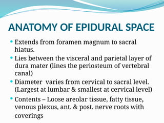
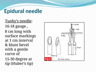
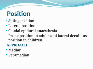
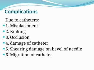
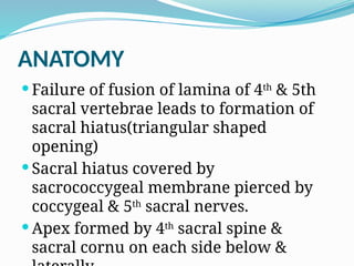
EPIDURAL ANAESTHESIA
Dr. P V. Bhale
Prof. Anesthesia
Dept. Of Anesthesia.
ANATOMY OF EPIDURAL SPACE
 Extends from foramen magnum to sacral
hiatus.
 Lies between the visceral and parietal layer of
dura mater (lines the periosteum of vertebral
canal)
 Diameter varies from cervical to sacral level.
(Largest at lumbar & smallest at cervical level)
 Contents – Loose areolar tissue, fatty tissue,
venous plexus, ant. & post. nerve roots with
coverings
ANATOMY
BOUNDED:
Anteriorly :
posterior longitudinal
ligament
Laterally :
Pedicles &
intervertebral foramina
Posteriorly:
Anterior surface of lamina
Ligamentum flavum.
NEGATIVE PRESSURE IN EXTRADURAL SPACE
 Relative to atmospheric pressure
epidural pressure is negative (lumbar
0.5-1 cmH2O and thoracic 1-3cmH2O)
Causes:
 Dimpling of dura by needle.
 Transfer of negative pressure from
thorax via para vertebral spaces
(especially in thoracic region)
SITE OF ACTION
 Nerve roots in extradural space
 Nerve roots in paravertebral spaces
 Nerve roots in intradural or
subarachnoid space after inward
diffusion of drug across the dura.
 Sensory block is complete & motor
paralysis is less (depending upon the
concentration of local anaesthetic)
SPREAD OF SOLUTION
Depends on :
1) Volume of solution injected
2) Age : Old pt. require less dose due to a
decrease in the size and compliance of the
epidural space, Less lat. spread
3) Height : Taller patients may require higher
dose.
4) Level of injection
5) Length of vertebral column
.
ADVANTAGES
1. Well defined area of anaesthesia –
segmental anaesthesia
2. Duration of anaesthesia can be
prolonged as per the duration of
surgery by giving repeated bolus drug
through the epidural catheter.
3. Minimize infections of spinal cord
4. Less incidence of nausea and
vomiting
5. Less problem of urinary retention
6. Provide post op. analgesia.
7. Less danger of neurological sequel.
8. Suitable in pts. with asthma,
bronchitis or emphysema
DISADVANTAGES
1. Technically difficult
2. Incomplete muscle relaxation
3. Large volumes of LA injected.
4. Chances of accidental dural puncture &
injecting large amount of drug in
subarachnoid space -> resulting in total
spinal block.
5. Risk of entering veins during catheter
insertion -> inadvertent I.V. injection -> L.A
toxicity.
6. Spread of block may be non uniform and
INDICATIONS
 Lower limb surgeries
 Hip and knee surgery :
E.A. reduces the blood loss. Incidence of
DVT is reduced
 Obstetrics.
Epidural labour analgesia is indicated in
pre-eclampsia, valvular heart disease
 In lower abdominal operations
POSTOPERATIVE PERIOD
 Epidural analgesia minimizes the effects of
surgery on cardiopulmonary reserve
 Prevents diaphragmatic splinting and the
inability to cough adequately due to pain
 Helpful in patients with compromised respiratory
function like patients with COPD ,morbid obesity
& elderly.
 Epidural analgesia allows earlier mobilization,
reduces the risk of DVT & allows better
cooperation with chest physiotherapy preventing
chest infections.
CONTRAINDICATIONS
Absolute
 Patient refusal
 Coagulopathy.
 Therapeutic anticoagulation.
 Skin infection at injection site.
 Raised intracranial pressure.
Accidental dural puncture in a patient with raised
ICP may lead to brainstem herniation (coning).
 Severe Hypovolaemia.
 Low fixed cardiac output states
Severe constrictive pericarditis, cardiac
tamponade, severe M.S., Severe A.S.
Relative
 Uncooperative patients
 Pre-existing neurological disorders like
multiple sclerosis
 Spinal deformity : congenital,
traumatic, post laminectomy
 Mild to moderate stenotic valvular
heart diseases.
Epidural needle
Tuohy’s needle:
16-18 gauge ,
8 cm long with
surface markings
at 1 cm interval
& blunt bevel
with a gentle
curve of
15-30 degree at
tip (Huber’s tip)
EPIDURAL CATHETERS
Epidural catheters :
single end-hole or a
number of side holes
at the distal end .
Incidence of
inadequate analgesia
is less with multiport
catheters.
DRUGS
 Lignocaine - 1-2%
 Bupivacaine - 0.25-0.5%
 Ropivacaine - 0.2%- 0.5%
 Adrenaline:
Used as 1 in 200000 solution.
Causes less systemic absorption of local
anaesthetic drug.
Prolongs the duration of action.
May cause more intense blockade of nerve
fibres.
Position
 Sitting position
 Lateral position
 Caudal epidural anaesthesia
Prone position in adults and lateral decubitus
position in children.
APPROACH
 Median
 Paramedian
TECHNIQUE
 Loss of resistance technique:
Insert needle into ligamentum flavum & attach
syringe filled with normal saline at hub of needle.
Advance with repeated attempts at injection.
Needle enters epidural space  sudden loss of
resistance  flow of solution without resistance.
 Hanging drop technique :
After removing the stylet, hub of needle filled with
solution so that a drop hangs from the opening.
Needle enters epidural space negative pressure 
drop immediately sucked in the needle hub .
Test dose
 After introduction of needle in to the space,
aspirate and confirm negative aspiration and
Test dose given to avoid subarachanoid or
intravascular injection.
 3 ml of 1.5% lidocaine with 1:200000 epinephrine
(5mcg/ml). After test dose, monitor PR/BP/RR/CNS
if no significant changes , give the entire dose of
L.A solution.
Complications
Due to catheters:
 1. Misplacement
 2. Kinking
 3. Occlusion
 4. damage of catheter
 5. Shearing damage on bevel of needle
 6. Migration of catheter
COMPLICATIONS
 Hypotension & CVS depression.
 Inadequate or patchy block
 Total spinal block
 Local anaesthetic toxicity ->
disorientation, twitching, convulsions or
apnoea
 Hypoapnea: due to medullary
depression by L.A.
 Epidural haematoma / abscess
Difference between Spinal and Epidural
block.
 The dose of L.A is high in epidural and less in
spinal.
 The volume of drug is more in epidural than
spinal.
 In epidural there are chances of intravascular inj
which are not there in spinal.
 Epidural is technically difficult than spinal.
 As epidural block is slow in onset than spinal the
degree of hypotension seen is less.
 Chances of the rare complication of total spinal
are there in epidural but not in spinal.
Caudal epidural anaesthesia
ANATOMY
 Failure of fusion of lamina of 4th
& 5th
sacral vertebrae leads to formation of
sacral hiatus(triangular shaped
opening)
 Sacral hiatus covered by
sacrococcygeal membrane pierced by
coccygeal & 5th
sacral nerves.
 Apex formed by 4th
sacral spine &
sacral cornu on each side below &
INDICATIONS
 One of the most commonly used regional
anaesthetic procedure in paediatric age group.
 Haemorrhoidectomy & other perianal surgeries
in adults
 Combined with general anesthesia to provide
intraoperative & postoperative analgesia .
 Surgery below umbilicus can be performed
under caudal anaesthesia in children.
Drugs
Local anaesthetic agents
 Lignocaine - 1-2%
 Bupivacaine - 0.25-0.5%
DOSE: 0.5- 1 ml/kg
 Ropivacaine - 0.25%
Dose -1 ml/kg
Adjuvants
 Ketamine: 0.25 mg/kg
 Clonidine – 1 mcg/kg

Epidural Anesthesia.................. .pptx

  • 1.
    EPIDURAL ANAESTHESIA Dr. PV. Bhale Prof. Anesthesia Dept. Of Anesthesia.
  • 2.
    ANATOMY OF EPIDURALSPACE  Extends from foramen magnum to sacral hiatus.  Lies between the visceral and parietal layer of dura mater (lines the periosteum of vertebral canal)  Diameter varies from cervical to sacral level. (Largest at lumbar & smallest at cervical level)  Contents – Loose areolar tissue, fatty tissue, venous plexus, ant. & post. nerve roots with coverings
  • 3.
    ANATOMY BOUNDED: Anteriorly : posterior longitudinal ligament Laterally: Pedicles & intervertebral foramina Posteriorly: Anterior surface of lamina Ligamentum flavum.
  • 4.
    NEGATIVE PRESSURE INEXTRADURAL SPACE  Relative to atmospheric pressure epidural pressure is negative (lumbar 0.5-1 cmH2O and thoracic 1-3cmH2O) Causes:  Dimpling of dura by needle.  Transfer of negative pressure from thorax via para vertebral spaces (especially in thoracic region)
  • 5.
    SITE OF ACTION Nerve roots in extradural space  Nerve roots in paravertebral spaces  Nerve roots in intradural or subarachnoid space after inward diffusion of drug across the dura.  Sensory block is complete & motor paralysis is less (depending upon the concentration of local anaesthetic)
  • 6.
    SPREAD OF SOLUTION Dependson : 1) Volume of solution injected 2) Age : Old pt. require less dose due to a decrease in the size and compliance of the epidural space, Less lat. spread 3) Height : Taller patients may require higher dose. 4) Level of injection 5) Length of vertebral column .
  • 7.
    ADVANTAGES 1. Well definedarea of anaesthesia – segmental anaesthesia 2. Duration of anaesthesia can be prolonged as per the duration of surgery by giving repeated bolus drug through the epidural catheter. 3. Minimize infections of spinal cord 4. Less incidence of nausea and vomiting
  • 8.
    5. Less problemof urinary retention 6. Provide post op. analgesia. 7. Less danger of neurological sequel. 8. Suitable in pts. with asthma, bronchitis or emphysema
  • 9.
    DISADVANTAGES 1. Technically difficult 2.Incomplete muscle relaxation 3. Large volumes of LA injected. 4. Chances of accidental dural puncture & injecting large amount of drug in subarachnoid space -> resulting in total spinal block. 5. Risk of entering veins during catheter insertion -> inadvertent I.V. injection -> L.A toxicity. 6. Spread of block may be non uniform and
  • 10.
    INDICATIONS  Lower limbsurgeries  Hip and knee surgery : E.A. reduces the blood loss. Incidence of DVT is reduced  Obstetrics. Epidural labour analgesia is indicated in pre-eclampsia, valvular heart disease  In lower abdominal operations
  • 11.
    POSTOPERATIVE PERIOD  Epiduralanalgesia minimizes the effects of surgery on cardiopulmonary reserve  Prevents diaphragmatic splinting and the inability to cough adequately due to pain  Helpful in patients with compromised respiratory function like patients with COPD ,morbid obesity & elderly.  Epidural analgesia allows earlier mobilization, reduces the risk of DVT & allows better cooperation with chest physiotherapy preventing chest infections.
  • 12.
    CONTRAINDICATIONS Absolute  Patient refusal Coagulopathy.  Therapeutic anticoagulation.  Skin infection at injection site.  Raised intracranial pressure. Accidental dural puncture in a patient with raised ICP may lead to brainstem herniation (coning).  Severe Hypovolaemia.  Low fixed cardiac output states Severe constrictive pericarditis, cardiac tamponade, severe M.S., Severe A.S.
  • 13.
    Relative  Uncooperative patients Pre-existing neurological disorders like multiple sclerosis  Spinal deformity : congenital, traumatic, post laminectomy  Mild to moderate stenotic valvular heart diseases.
  • 14.
    Epidural needle Tuohy’s needle: 16-18gauge , 8 cm long with surface markings at 1 cm interval & blunt bevel with a gentle curve of 15-30 degree at tip (Huber’s tip)
  • 15.
    EPIDURAL CATHETERS Epidural catheters: single end-hole or a number of side holes at the distal end . Incidence of inadequate analgesia is less with multiport catheters.
  • 16.
    DRUGS  Lignocaine -1-2%  Bupivacaine - 0.25-0.5%  Ropivacaine - 0.2%- 0.5%  Adrenaline: Used as 1 in 200000 solution. Causes less systemic absorption of local anaesthetic drug. Prolongs the duration of action. May cause more intense blockade of nerve fibres.
  • 17.
    Position  Sitting position Lateral position  Caudal epidural anaesthesia Prone position in adults and lateral decubitus position in children. APPROACH  Median  Paramedian
  • 18.
    TECHNIQUE  Loss ofresistance technique: Insert needle into ligamentum flavum & attach syringe filled with normal saline at hub of needle. Advance with repeated attempts at injection. Needle enters epidural space  sudden loss of resistance  flow of solution without resistance.  Hanging drop technique : After removing the stylet, hub of needle filled with solution so that a drop hangs from the opening. Needle enters epidural space negative pressure  drop immediately sucked in the needle hub .
  • 19.
    Test dose  Afterintroduction of needle in to the space, aspirate and confirm negative aspiration and Test dose given to avoid subarachanoid or intravascular injection.  3 ml of 1.5% lidocaine with 1:200000 epinephrine (5mcg/ml). After test dose, monitor PR/BP/RR/CNS if no significant changes , give the entire dose of L.A solution.
  • 20.
    Complications Due to catheters: 1. Misplacement  2. Kinking  3. Occlusion  4. damage of catheter  5. Shearing damage on bevel of needle  6. Migration of catheter
  • 21.
    COMPLICATIONS  Hypotension &CVS depression.  Inadequate or patchy block  Total spinal block  Local anaesthetic toxicity -> disorientation, twitching, convulsions or apnoea  Hypoapnea: due to medullary depression by L.A.  Epidural haematoma / abscess
  • 22.
    Difference between Spinaland Epidural block.  The dose of L.A is high in epidural and less in spinal.  The volume of drug is more in epidural than spinal.  In epidural there are chances of intravascular inj which are not there in spinal.  Epidural is technically difficult than spinal.  As epidural block is slow in onset than spinal the degree of hypotension seen is less.  Chances of the rare complication of total spinal are there in epidural but not in spinal.
  • 23.
  • 24.
    ANATOMY  Failure offusion of lamina of 4th & 5th sacral vertebrae leads to formation of sacral hiatus(triangular shaped opening)  Sacral hiatus covered by sacrococcygeal membrane pierced by coccygeal & 5th sacral nerves.  Apex formed by 4th sacral spine & sacral cornu on each side below &
  • 25.
    INDICATIONS  One ofthe most commonly used regional anaesthetic procedure in paediatric age group.  Haemorrhoidectomy & other perianal surgeries in adults  Combined with general anesthesia to provide intraoperative & postoperative analgesia .  Surgery below umbilicus can be performed under caudal anaesthesia in children.
  • 26.
    Drugs Local anaesthetic agents Lignocaine - 1-2%  Bupivacaine - 0.25-0.5% DOSE: 0.5- 1 ml/kg  Ropivacaine - 0.25% Dose -1 ml/kg Adjuvants  Ketamine: 0.25 mg/kg  Clonidine – 1 mcg/kg