Download as PDF, PPTX

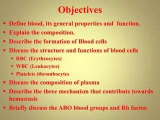
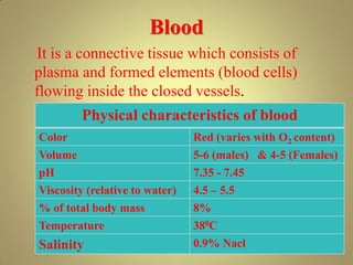
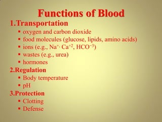
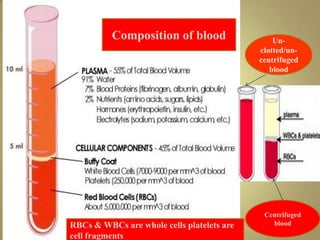
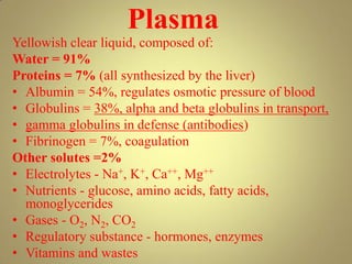
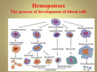
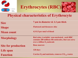
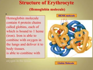

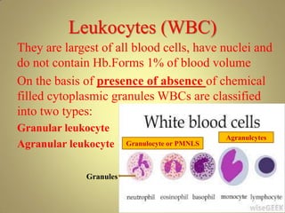
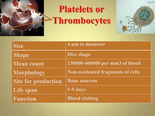
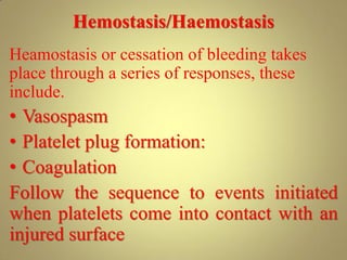
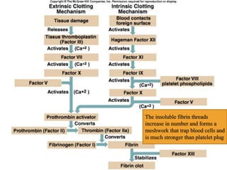
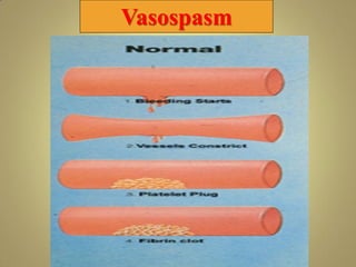
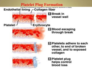
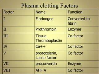
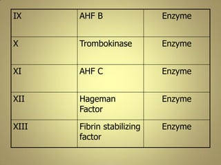
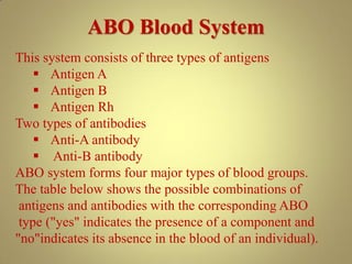
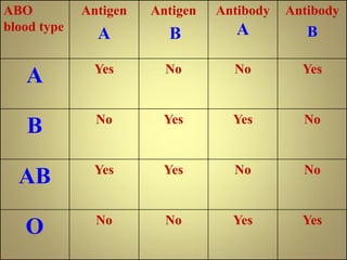
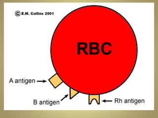

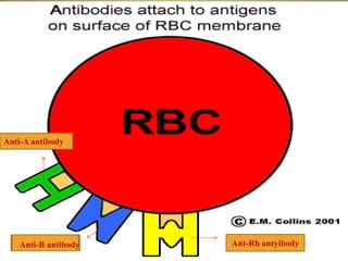
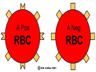
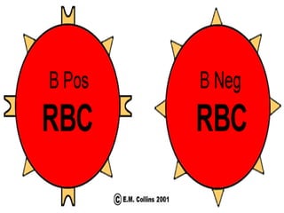
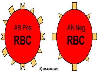
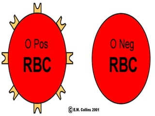
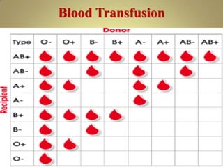


Blood consists of plasma and formed elements including red blood cells, white blood cells, and platelets. It functions to transport oxygen, nutrients, waste, hormones and more throughout the body. Red blood cells contain hemoglobin which carries oxygen. White blood cells help fight infection. Platelets assist in blood clotting. The document discusses blood composition and formation, cell types and functions, hemostasis, and blood groups.


![Polymer [ बहुलक ] Chemistry Notes PDF - Irfanullah Mehar - JJ Sir Chemistry.pdf](https://cdn.slidesharecdn.com/ss_thumbnails/polymerchemistrynotespdf-irfanullahmehar-jjsirchemistry-260210172118-3f9b37f7-thumbnail.jpg?width=640&height=640&fit=bounds)


