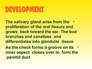
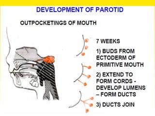
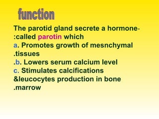
The document details the anatomy and physiology of the parotid gland, the largest salivary gland located near the ear. It describes its structure, boundaries, blood supply, innervation, and clinical notes related to parotid gland conditions such as tumors, parotitis, and Frey's syndrome. The parotid duct's function and potential injuries during surgery are also emphasized.


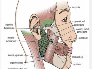
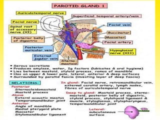
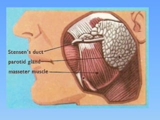
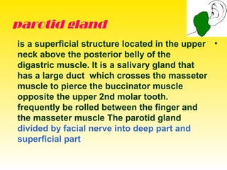
![•]parotis-by the side of ear ]is the
largest of the main salivary glands
,lobulated and yellowish brown in color
. it is predominantly serous ,with only
few scattered mucous acini . The
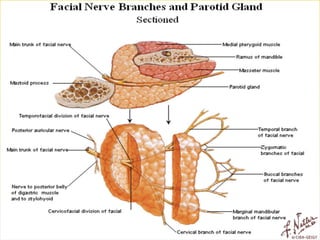
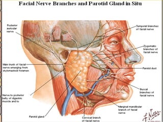
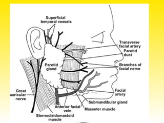
parotid gland divided by facial nerve
into deep part and superficial part ,lies
in space called parotid bed or
reteromandibular fossa
The parotid gland](https://image.slidesharecdn.com/parotidregion-150331074413-conversion-gate01/85/Parotid-region-5-320.jpg)
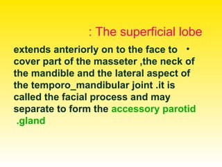
![• **]If the parotid gland is carefully
removed , you can identify the
structures located within it. The first
plane is the venous plane and consists
of the retromandibular vein (rm) and its
tributaries and branches](https://image.slidesharecdn.com/parotidregion-150331074413-conversion-gate01/85/Parotid-region-9-320.jpg)