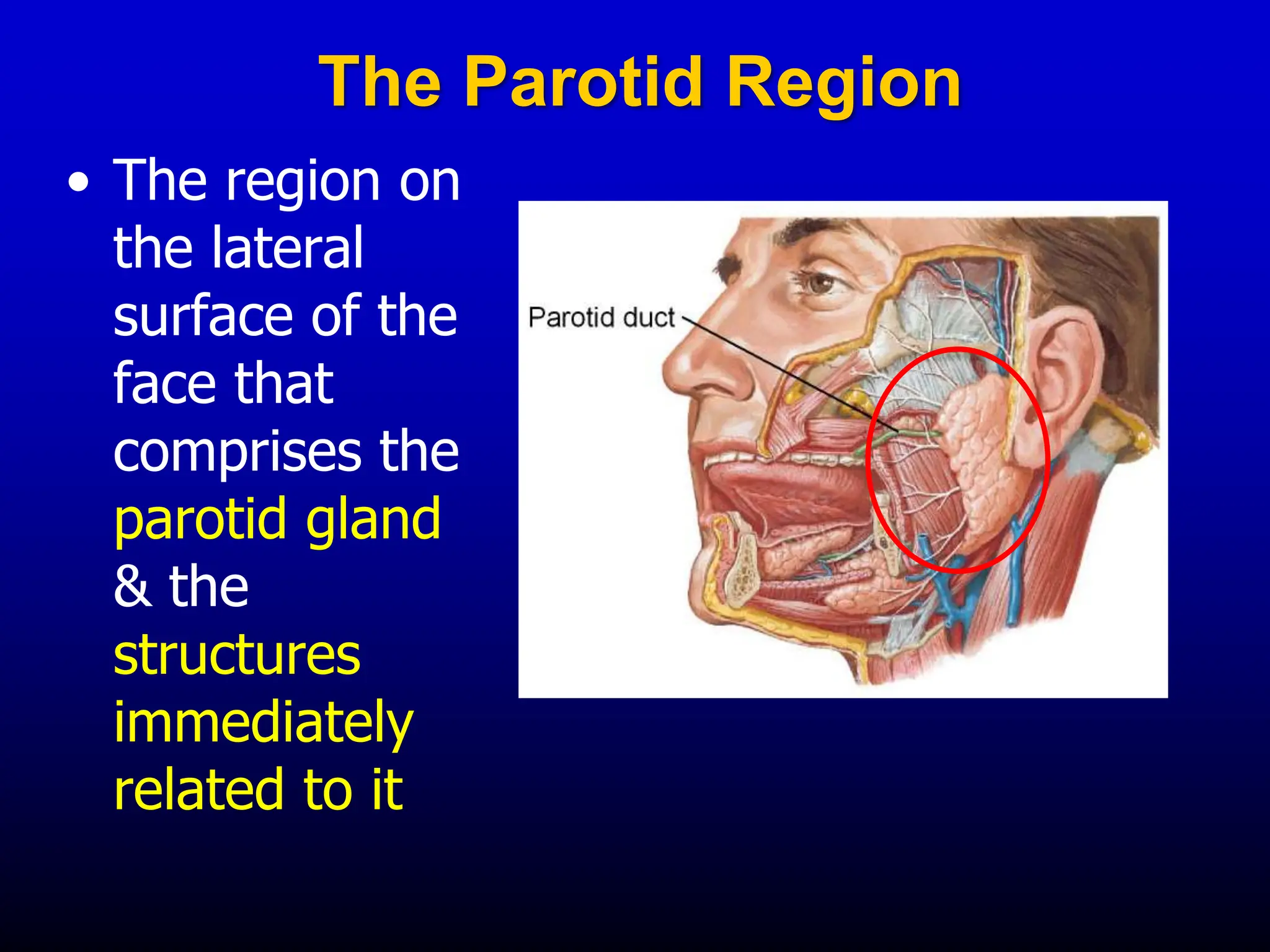
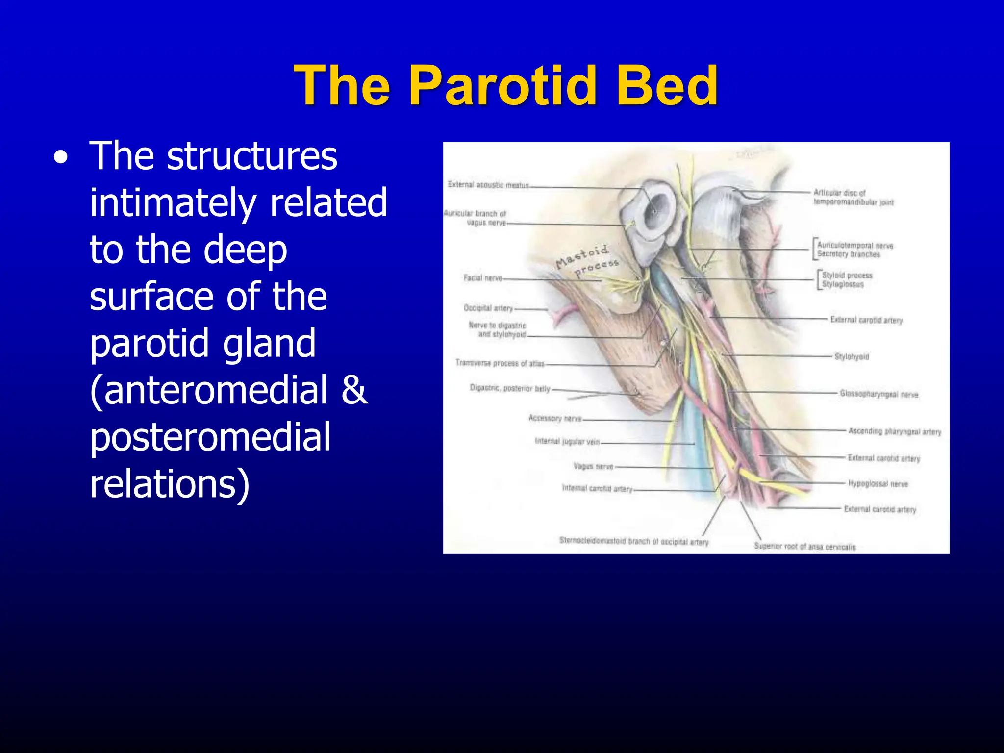
The Parotid Region
Dr. Kaushik
The Parotid Region
• The region on
the lateral
surface of the
face that
comprises the
parotid gland
& the
structures
immediately
related to it
Parotid Gland
• Largest of the salivary glands
• Located subcutaneously,
below and in front of the
external auditory meatus
• Occupies the deep hollow
behind the ramus of the
mandible
• Wedge-shaped when viewed
externally, with the base
above & the apex behind the
angle of the mandible
• Wedge-shaped in
horizontal section with
the base in the lateral
position and apex
against the pharyngeal
wall.
• It exhibits 3 surfaces:
 Lateral
 Anteromedial
 Posteromedial
Lobes
• The facial nerve
courses horizontally
through the gland
and divides it into:
 Superficial lobe
 Deep lobe
Superficial lobe
Deep lobe
Facial nerve
Processes
 Glenoid process, that extends
upward behind the temporo-
mandibular joint, in front of
external auditory meatus
 Facial process, that extends
anteriorly onto the masseter
muscle
 Accessory process (part), small
part of facial process lying along
the parotid duct
 Pterygoid process, that extends
forward from the deeper part, lies
between the medial pterygoid
muscle & the ramus of mandible
 Carotid process, that lies posterior
to the external carotid artery
The gland is an irregular lobulated mass, sends ‘processes’ in
various directions. These include:
Capsules
• The parotid gland is enclosed in two capsules:
 An inner connective tissue capsule
 An outer dense fibrous capsule derived from
the investing layer of the deep cervical fascia
• The deep cervical fascia extends upward,
reaches the inferior border of parotid gland,
splits into the superficial & the deep layer, to
enclose the gland
• Above the gland, the:
 Superficial layer gets attached to the
zygomatic arch
 Deep layer gets attached to the tympanic
plate of temporal bone
A portion of fascia extending from the styloid process to the
angle of mandible is called stylomandibular ligament. It
separates the parotid gland from the submandibular gland
Relations
• Superficial (lateral):
• Skin & superficial fascia
• Great auricular nerve
• Parotid lymph nodes
• Superior:
• External auditory meatus
• Temporomandibular
joint
• Its glenoid process is
related to the auriculo-
temporal nerve
• Anteromedial:
• Stylomandibular
ligament
• Medial pterygoid
• Posterior border
of the ramus of
mandible
• Massater
• Terminal
branches of the
facial nerve
• Temporo-
mandibular joint
• Posteromedial:
• Carotid sheath with
its contents
• Styloid process &
attached muscles
• Facial nerve
• Posterior belly of
digastric muscle
• Mastoid process
• Sternocleidomastoid
The Parotid Bed
• The structures
intimately related
to the deep
surface of the
parotid gland
(anteromedial &
posteromedial
relations)
Structures Coursing Within the Parotid Gland
 Auriculotemporal nerve
 External carotid artery
 Retromandibular vein
 Facial nerve
A few lymph nodes are
scattered in the
substance of the gland
Deep
Superficial
Parotid (Stensen’s) Duct
• About 2 inches long
• Emerges from the facial process of
the gland
• Passes forward over the lateral
surface of the masseter muscle
 about a fingerbreadth below the
zygomatic arch
 accompanied by the:
 transverse facial vessels &
upper zygomatic branches of
facial nerve above
 lower zygomatic branches of
facial nerve below
• Turns around the anterior
border of masseter
muscle
• Pierces the:
• Buccal pad of fat
• Buccopharyngeal
fascia
• Buccinator muscle &
• Buccal mucosa
• Opens into the vestibule
of mouth on a small
papilla, opposite the
second upper molar tooth
Parotid duct
Buccinator
Masseter
• The oblique passage of the duct in the buccinator muscle
acts as a valve-like mechanism & prevents inflation of the
duct during blowing
Parotid Duct
• The duct can be rolled over the
clenched masseter muscle
• The duct is represented by the
middle 1/3 of a line extending
from the tragus of the auricle to a
point midway between the ala of
nose & upper lip
Venous drainage: Into
the retro-mandibular vein
Arterial supply:
External carotid artery
& its terminal branches
Retromandibular v.
External
carotid a.
Maxillary a.
Superficial
temporal a.
Lymph Drainage: Into the
parotid & then into the
deep cervical lymph nodes
Parotid n.
Deep cervical n.
Nerve Supply
• Sensory :
 Auriculotemporal n.
• Autonomic:
• Sympathetic through plexus
around the arteries (T1→SCG
→ plexus around ECA)
• Parasympthetic through otic
ganglion (CN9 → tympanic n.
→ tympanic plexus → lesser
petrosal n. → otic ganglion
→ auriculotemporal n.)
Clinical Anatomy
• Parotid duct being a superficial structure, is prone
to get damaged in injuries, or during surgical
procedures on the face
• Parotid neoplasms (malignant) are very invasive
and quickly involve the facial nerve causing facial
palsy
• Inflammation of parotid gland results in painful
swelling because of a tight capsule enclosing the
gland. The swollen glenoid process exaggerates
this pain on chewing
• Frey’s syndrome: a disorder
characterized by recurrent episodes
of localized facial flushing and/or
sweating in the area over the
parotid gland in response to
gustatory stimuli
• This is due to aberrant nerve
regeneration after injury (a
communication develops between
the auriculo-temporal & greater
auricular nerves such that
parasympathetic fibers migrate into
the cutaneous sympathetic nerves
that supply the sweat glands)
04. Parotid Region.ppt

04. Parotid Region.ppt

  • 1.
  • 2.
    The Parotid Region •The region on the lateral surface of the face that comprises the parotid gland & the structures immediately related to it
  • 3.
    Parotid Gland • Largestof the salivary glands • Located subcutaneously, below and in front of the external auditory meatus • Occupies the deep hollow behind the ramus of the mandible • Wedge-shaped when viewed externally, with the base above & the apex behind the angle of the mandible
  • 4.
    • Wedge-shaped in horizontalsection with the base in the lateral position and apex against the pharyngeal wall. • It exhibits 3 surfaces:  Lateral  Anteromedial  Posteromedial
  • 5.
    Lobes • The facialnerve courses horizontally through the gland and divides it into:  Superficial lobe  Deep lobe Superficial lobe Deep lobe Facial nerve
  • 6.
    Processes  Glenoid process,that extends upward behind the temporo- mandibular joint, in front of external auditory meatus  Facial process, that extends anteriorly onto the masseter muscle  Accessory process (part), small part of facial process lying along the parotid duct  Pterygoid process, that extends forward from the deeper part, lies between the medial pterygoid muscle & the ramus of mandible  Carotid process, that lies posterior to the external carotid artery The gland is an irregular lobulated mass, sends ‘processes’ in various directions. These include:
  • 7.
    Capsules • The parotidgland is enclosed in two capsules:  An inner connective tissue capsule  An outer dense fibrous capsule derived from the investing layer of the deep cervical fascia • The deep cervical fascia extends upward, reaches the inferior border of parotid gland, splits into the superficial & the deep layer, to enclose the gland • Above the gland, the:  Superficial layer gets attached to the zygomatic arch  Deep layer gets attached to the tympanic plate of temporal bone A portion of fascia extending from the styloid process to the angle of mandible is called stylomandibular ligament. It separates the parotid gland from the submandibular gland
  • 8.
    Relations • Superficial (lateral): •Skin & superficial fascia • Great auricular nerve • Parotid lymph nodes • Superior: • External auditory meatus • Temporomandibular joint • Its glenoid process is related to the auriculo- temporal nerve
  • 9.
    • Anteromedial: • Stylomandibular ligament •Medial pterygoid • Posterior border of the ramus of mandible • Massater • Terminal branches of the facial nerve • Temporo- mandibular joint
  • 10.
    • Posteromedial: • Carotidsheath with its contents • Styloid process & attached muscles • Facial nerve • Posterior belly of digastric muscle • Mastoid process • Sternocleidomastoid
  • 11.
    The Parotid Bed •The structures intimately related to the deep surface of the parotid gland (anteromedial & posteromedial relations)
  • 12.
    Structures Coursing Withinthe Parotid Gland  Auriculotemporal nerve  External carotid artery  Retromandibular vein  Facial nerve A few lymph nodes are scattered in the substance of the gland Deep Superficial
  • 13.
    Parotid (Stensen’s) Duct •About 2 inches long • Emerges from the facial process of the gland • Passes forward over the lateral surface of the masseter muscle  about a fingerbreadth below the zygomatic arch  accompanied by the:  transverse facial vessels & upper zygomatic branches of facial nerve above  lower zygomatic branches of facial nerve below
  • 14.
    • Turns aroundthe anterior border of masseter muscle • Pierces the: • Buccal pad of fat • Buccopharyngeal fascia • Buccinator muscle & • Buccal mucosa • Opens into the vestibule of mouth on a small papilla, opposite the second upper molar tooth Parotid duct Buccinator Masseter
  • 15.
    • The obliquepassage of the duct in the buccinator muscle acts as a valve-like mechanism & prevents inflation of the duct during blowing Parotid Duct • The duct can be rolled over the clenched masseter muscle • The duct is represented by the middle 1/3 of a line extending from the tragus of the auricle to a point midway between the ala of nose & upper lip
  • 16.
    Venous drainage: Into theretro-mandibular vein Arterial supply: External carotid artery & its terminal branches Retromandibular v. External carotid a. Maxillary a. Superficial temporal a.
  • 17.
    Lymph Drainage: Intothe parotid & then into the deep cervical lymph nodes Parotid n. Deep cervical n.
  • 18.
    Nerve Supply • Sensory:  Auriculotemporal n. • Autonomic: • Sympathetic through plexus around the arteries (T1→SCG → plexus around ECA) • Parasympthetic through otic ganglion (CN9 → tympanic n. → tympanic plexus → lesser petrosal n. → otic ganglion → auriculotemporal n.)
  • 19.
    Clinical Anatomy • Parotidduct being a superficial structure, is prone to get damaged in injuries, or during surgical procedures on the face • Parotid neoplasms (malignant) are very invasive and quickly involve the facial nerve causing facial palsy • Inflammation of parotid gland results in painful swelling because of a tight capsule enclosing the gland. The swollen glenoid process exaggerates this pain on chewing
  • 20.
    • Frey’s syndrome:a disorder characterized by recurrent episodes of localized facial flushing and/or sweating in the area over the parotid gland in response to gustatory stimuli • This is due to aberrant nerve regeneration after injury (a communication develops between the auriculo-temporal & greater auricular nerves such that parasympathetic fibers migrate into the cutaneous sympathetic nerves that supply the sweat glands)