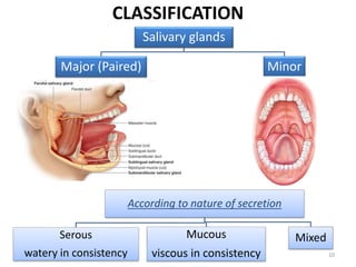
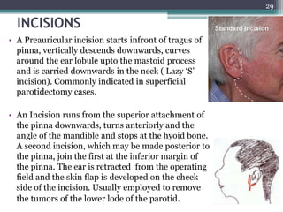
1. The document provides information on the salivary glands, including their embryology, microstructure, classification, and the surgical anatomy of the major salivary glands - the parotid and submandibular glands.
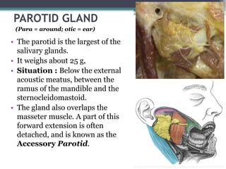
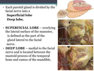
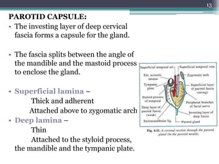
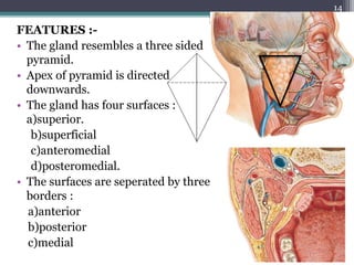
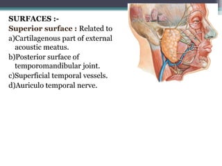
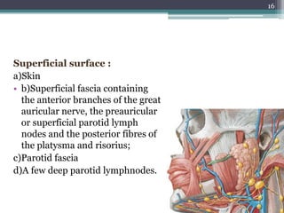
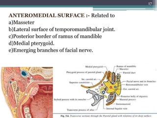
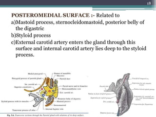
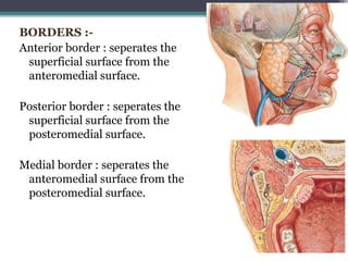
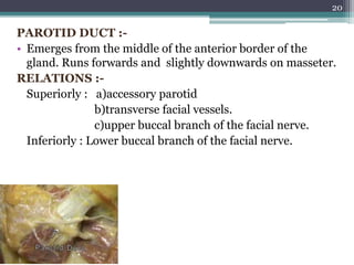
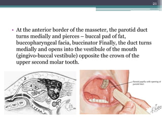
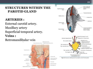
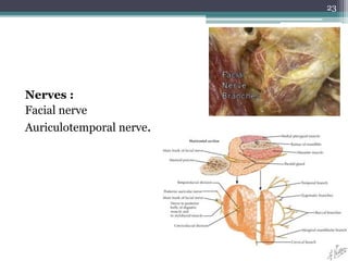
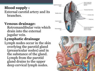
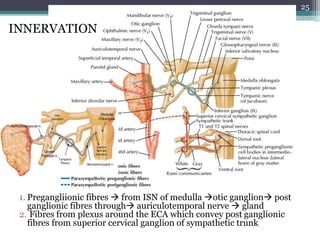
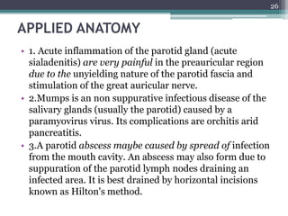
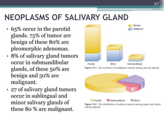
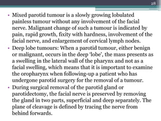
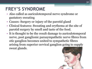
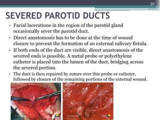
2. It describes the parotid gland as the largest salivary gland, located below the external ear. It discusses the gland's lobes, surfaces, borders, duct, blood supply and innervation.
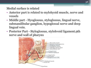
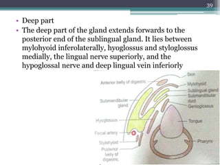
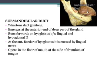
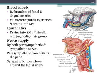
3. It also summarizes the submandibular gland, located in the submandibular triangle, discussing its parts, surfaces, duct, blood supply and innervation.
INTRODUCTIONSalivary glands are compound tubuloacinar, exocrine gland and the ducts opens in the oral cavity.
Salivary glands secretes a fluid called saliva that coats the teeth and the mucosa.
Saliva is a complex fluid, produced by the salivary glands, the most important function of which is to maintain the well- being of mouth.
Individuals with a deficiency of salivary secretion experience difficulty in eating, speaking, and swallowing and become prone to mucosal infections and dental caries.
Salivary glands Disorders and management.Manish Shetty
Short, brief description of the salivary gland disorders.
it explain the basic anatomy, physiology of the salivary glands.
all the 3 salivary gland are individually explained with appropriate management of it disorders.
Detailed discussion on surgical anatomy of salivary glands with special focus on major glands. Relationship of facial nerve and its branhes to parotid gland is also discussed. Complications are also discussed. Surgical approaches are also discussed.
CONTENT
INTRODUCTION
DEVELOPMENT
PAROTID CAPSULE
EXTERNAL FEATURES
RELATIONS
STRUCTURE WITHIN THE PAROTID GLAND
PAROTID DUCT
NERVE SUPPLY
LYMPHATIC DRAINAGE AND LYMPH NODES
FUNCTIONS OF PAROTID GLAND
ROLE OF PUBLIC HEALTH DENTIST
CONCLUSION
REFERENCES
The surgical anatomy of major salivary glands has many significant applications in maxillofacial surgery. Understanding these important anatomic relations- variations enables surgeons to perform the surgical procedures safely. Knowledge of these concepts helps us to recognize the problems and complications as and when they occur and manage them accordingly.
A gland consists of specialized type of cells, wherein they produce products which are used elsewhere in the body. Salivary glands are complex, tubulo acinar, exocrine or merocrine glands secreting mainly saliva.
Saliva is the product of the major and minor salivary gland dispersed throughout the oral cavity
It is a complex mixture of organic, inorganic components and water, carrying out several functions
There are three pairs of major salivary glands namely parotid, sub mandibular and sublingual glands in addition to numerous minor salivary glands in the oral cavity
The prostate is an exocrine gland of the male mammalian reproductive system
It is a walnut-sized gland that forms part of the male reproductive system and is located in front of the rectum and just below the urinary bladder
Function is to store and secrete a clear, slightly alkaline fluid that constitutes 10-30% of the volume of the seminal fluid that along with the spermatozoa, constitutes semen
A healthy human prostate measures (4cm-vertical, by 3cm-horizontal, 2cm ant-post ).
It surrounds the urethra just below the urinary bladder. It has anterior, median, posterior and two lateral lobes
It’s work is regulated by androgens which are responsible for male sex characteristics
Generalised disease of the prostate due to hormonal derangement which leads to non malignant enlargement of the gland (increase in the number of epithelial cells and stromal tissue)to cause compression of the urethra leading to symptoms (LUTS
Couples presenting to the infertility clinic- Do they really have infertility...Sujoy Dasgupta
Dr Sujoy Dasgupta presented the study on "Couples presenting to the infertility clinic- Do they really have infertility? – The unexplored stories of non-consummation" in the 13th Congress of the Asia Pacific Initiative on Reproduction (ASPIRE 2024) at Manila on 24 May, 2024.
Lung Cancer: Artificial Intelligence, Synergetics, Complex System Analysis, S...Oleg Kshivets
RESULTS: Overall life span (LS) was 2252.1±1742.5 days and cumulative 5-year survival (5YS) reached 73.2%, 10 years – 64.8%, 20 years – 42.5%. 513 LCP lived more than 5 years (LS=3124.6±1525.6 days), 148 LCP – more than 10 years (LS=5054.4±1504.1 days).199 LCP died because of LC (LS=562.7±374.5 days). 5YS of LCP after bi/lobectomies was significantly superior in comparison with LCP after pneumonectomies (78.1% vs.63.7%, P=0.00001 by log-rank test). AT significantly improved 5YS (66.3% vs. 34.8%) (P=0.00000 by log-rank test) only for LCP with N1-2. Cox modeling displayed that 5YS of LCP significantly depended on: phase transition (PT) early-invasive LC in terms of synergetics, PT N0—N12, cell ratio factors (ratio between cancer cells- CC and blood cells subpopulations), G1-3, histology, glucose, AT, blood cell circuit, prothrombin index, heparin tolerance, recalcification time (P=0.000-0.038). Neural networks, genetic algorithm selection and bootstrap simulation revealed relationships between 5YS and PT early-invasive LC (rank=1), PT N0—N12 (rank=2), thrombocytes/CC (3), erythrocytes/CC (4), eosinophils/CC (5), healthy cells/CC (6), lymphocytes/CC (7), segmented neutrophils/CC (8), stick neutrophils/CC (9), monocytes/CC (10); leucocytes/CC (11). Correct prediction of 5YS was 100% by neural networks computing (area under ROC curve=1.0; error=0.0).
CONCLUSIONS: 5YS of LCP after radical procedures significantly depended on: 1) PT early-invasive cancer; 2) PT N0--N12; 3) cell ratio factors; 4) blood cell circuit; 5) biochemical factors; 6) hemostasis system; 7) AT; 8) LC characteristics; 9) LC cell dynamics; 10) surgery type: lobectomy/pneumonectomy; 11) anthropometric data. Optimal diagnosis and treatment strategies for LC are: 1) screening and early detection of LC; 2) availability of experienced thoracic surgeons because of complexity of radical procedures; 3) aggressive en block surgery and adequate lymph node dissection for completeness; 4) precise prediction; 5) adjuvant chemoimmunoradiotherapy for LCP with unfavorable prognosis.
micro teaching on communication m.sc nursing.pdfAnurag Sharma
Microteaching is a unique model of practice teaching. It is a viable instrument for the. desired change in the teaching behavior or the behavior potential which, in specified types of real. classroom situations, tends to facilitate the achievement of specified types of objectives.
TEST BANK for Operations Management, 14th Edition by William J. Stevenson, Ve...kevinkariuki227
TEST BANK for Operations Management, 14th Edition by William J. Stevenson, Verified Chapters 1 - 19, Complete Newest Version.pdf
Tom Selleck Health: A Comprehensive Look at the Iconic Actor’s Wellness Journeygreendigital
Tom Selleck, an enduring figure in Hollywood. has captivated audiences for decades with his rugged charm, iconic moustache. and memorable roles in television and film. From his breakout role as Thomas Magnum in Magnum P.I. to his current portrayal of Frank Reagan in Blue Bloods. Selleck's career has spanned over 50 years. But beyond his professional achievements. fans have often been curious about Tom Selleck Health. especially as he has aged in the public eye.
Follow us on: Pinterest
Introduction
Many have been interested in Tom Selleck health. not only because of his enduring presence on screen but also because of the challenges. and lifestyle choices he has faced and made over the years. This article delves into the various aspects of Tom Selleck health. exploring his fitness regimen, diet, mental health. and the challenges he has encountered as he ages. We'll look at how he maintains his well-being. the health issues he has faced, and his approach to ageing .
Early Life and Career
Childhood and Athletic Beginnings
Tom Selleck was born on January 29, 1945, in Detroit, Michigan, and grew up in Sherman Oaks, California. From an early age, he was involved in sports, particularly basketball. which played a significant role in his physical development. His athletic pursuits continued into college. where he attended the University of Southern California (USC) on a basketball scholarship. This early involvement in sports laid a strong foundation for his physical health and disciplined lifestyle.
Transition to Acting
Selleck's transition from an athlete to an actor came with its physical demands. His first significant role in "Magnum P.I." required him to perform various stunts and maintain a fit appearance. This role, which he played from 1980 to 1988. necessitated a rigorous fitness routine to meet the show's demands. setting the stage for his long-term commitment to health and wellness.
Fitness Regimen
Workout Routine
Tom Selleck health and fitness regimen has evolved. adapting to his changing roles and age. During his "Magnum, P.I." days. Selleck's workouts were intense and focused on building and maintaining muscle mass. His routine included weightlifting, cardiovascular exercises. and specific training for the stunts he performed on the show.
Selleck adjusted his fitness routine as he aged to suit his body's needs. Today, his workouts focus on maintaining flexibility, strength, and cardiovascular health. He incorporates low-impact exercises such as swimming, walking, and light weightlifting. This balanced approach helps him stay fit without putting undue strain on his joints and muscles.
Importance of Flexibility and Mobility
In recent years, Selleck has emphasized the importance of flexibility and mobility in his fitness regimen. Understanding the natural decline in muscle mass and joint flexibility with age. he includes stretching and yoga in his routine. These practices help prevent injuries, improve posture, and maintain mobilit
HOT NEW PRODUCT! BIG SALES FAST SHIPPING NOW FROM CHINA!! EU KU DB BK substit...GL Anaacs
Contact us if you are interested:
Email / Skype : kefaya1771@gmail.com
Threema: PXHY5PDH
New BATCH Ku !!! MUCH IN DEMAND FAST SALE EVERY BATCH HAPPY GOOD EFFECT BIG BATCH !
Contact me on Threema or skype to start big business!!
Hot-sale products:
NEW HOT EUTYLONE WHITE CRYSTAL!!
5cl-adba precursor (semi finished )
5cl-adba raw materials
ADBB precursor (semi finished )
ADBB raw materials
APVP powder
5fadb/4f-adb
Jwh018 / Jwh210
Eutylone crystal
Protonitazene (hydrochloride) CAS: 119276-01-6
Flubrotizolam CAS: 57801-95-3
Metonitazene CAS: 14680-51-4
Payment terms: Western Union,MoneyGram,Bitcoin or USDT.
Deliver Time: Usually 7-15days
Shipping method: FedEx, TNT, DHL,UPS etc.Our deliveries are 100% safe, fast, reliable and discreet.
Samples will be sent for your evaluation!If you are interested in, please contact me, let's talk details.
We specializes in exporting high quality Research chemical, medical intermediate, Pharmaceutical chemicals and so on. Products are exported to USA, Canada, France, Korea, Japan,Russia, Southeast Asia and other countries.
Pulmonary Thromboembolism - etilogy, types, medical- Surgical and nursing man...VarunMahajani
Disruption of blood supply to lung alveoli due to blockage of one or more pulmonary blood vessels is called as Pulmonary thromboembolism. In this presentation we will discuss its causes, types and its management in depth.
Ethanol (CH3CH2OH), or beverage alcohol, is a two-carbon alcohol
that is rapidly distributed in the body and brain. Ethanol alters many
neurochemical systems and has rewarding and addictive properties. It
is the oldest recreational drug and likely contributes to more morbidity,
mortality, and public health costs than all illicit drugs combined. The
5th edition of the Diagnostic and Statistical Manual of Mental Disorders
(DSM-5) integrates alcohol abuse and alcohol dependence into a single
disorder called alcohol use disorder (AUD), with mild, moderate,
and severe subclassifications (American Psychiatric Association, 2013).
In the DSM-5, all types of substance abuse and dependence have been
combined into a single substance use disorder (SUD) on a continuum
from mild to severe. A diagnosis of AUD requires that at least two of
the 11 DSM-5 behaviors be present within a 12-month period (mild
AUD: 2–3 criteria; moderate AUD: 4–5 criteria; severe AUD: 6–11 criteria).
The four main behavioral effects of AUD are impaired control over
drinking, negative social consequences, risky use, and altered physiological
effects (tolerance, withdrawal). This chapter presents an overview
of the prevalence and harmful consequences of AUD in the U.S.,
the systemic nature of the disease, neurocircuitry and stages of AUD,
comorbidities, fetal alcohol spectrum disorders, genetic risk factors, and
pharmacotherapies for AUD.
Title: Sense of Taste
Presenter: Dr. Faiza, Assistant Professor of Physiology
Qualifications:
MBBS (Best Graduate, AIMC Lahore)
FCPS Physiology
ICMT, CHPE, DHPE (STMU)
MPH (GC University, Faisalabad)
MBA (Virtual University of Pakistan)
Learning Objectives:
Describe the structure and function of taste buds.
Describe the relationship between the taste threshold and taste index of common substances.
Explain the chemical basis and signal transduction of taste perception for each type of primary taste sensation.
Recognize different abnormalities of taste perception and their causes.
Key Topics:
Significance of Taste Sensation:
Differentiation between pleasant and harmful food
Influence on behavior
Selection of food based on metabolic needs
Receptors of Taste:
Taste buds on the tongue
Influence of sense of smell, texture of food, and pain stimulation (e.g., by pepper)
Primary and Secondary Taste Sensations:
Primary taste sensations: Sweet, Sour, Salty, Bitter, Umami
Chemical basis and signal transduction mechanisms for each taste
Taste Threshold and Index:
Taste threshold values for Sweet (sucrose), Salty (NaCl), Sour (HCl), and Bitter (Quinine)
Taste index relationship: Inversely proportional to taste threshold
Taste Blindness:
Inability to taste certain substances, particularly thiourea compounds
Example: Phenylthiocarbamide
Structure and Function of Taste Buds:
Composition: Epithelial cells, Sustentacular/Supporting cells, Taste cells, Basal cells
Features: Taste pores, Taste hairs/microvilli, and Taste nerve fibers
Location of Taste Buds:
Found in papillae of the tongue (Fungiform, Circumvallate, Foliate)
Also present on the palate, tonsillar pillars, epiglottis, and proximal esophagus
Mechanism of Taste Stimulation:
Interaction of taste substances with receptors on microvilli
Signal transduction pathways for Umami, Sweet, Bitter, Sour, and Salty tastes
Taste Sensitivity and Adaptation:
Decrease in sensitivity with age
Rapid adaptation of taste sensation
Role of Saliva in Taste:
Dissolution of tastants to reach receptors
Washing away the stimulus
Taste Preferences and Aversions:
Mechanisms behind taste preference and aversion
Influence of receptors and neural pathways
Impact of Sensory Nerve Damage:
Degeneration of taste buds if the sensory nerve fiber is cut
Abnormalities of Taste Detection:
Conditions: Ageusia, Hypogeusia, Dysgeusia (parageusia)
Causes: Nerve damage, neurological disorders, infections, poor oral hygiene, adverse drug effects, deficiencies, aging, tobacco use, altered neurotransmitter levels
Neurotransmitters and Taste Threshold:
Effects of serotonin (5-HT) and norepinephrine (NE) on taste sensitivity
Supertasters:
25% of the population with heightened sensitivity to taste, especially bitterness
Increased number of fungiform papillae
These lecture slides, by Dr Sidra Arshad, offer a quick overview of physiological basis of a normal electrocardiogram.
Learning objectives:
1. Define an electrocardiogram (ECG) and electrocardiography
2. Describe how dipoles generated by the heart produce the waveforms of the ECG
3. Describe the components of a normal electrocardiogram of a typical bipolar leads (limb II)
4. Differentiate between intervals and segments
5. Enlist some common indications for obtaining an ECG
Study Resources:
1. Chapter 11, Guyton and Hall Textbook of Medical Physiology, 14th edition
2. Chapter 9, Human Physiology - From Cells to Systems, Lauralee Sherwood, 9th edition
3. Chapter 29, Ganong’s Review of Medical Physiology, 26th edition
4. Electrocardiogram, StatPearls - https://www.ncbi.nlm.nih.gov/books/NBK549803/
5. ECG in Medical Practice by ABM Abdullah, 4th edition
6. ECG Basics, http://www.nataliescasebook.com/tag/e-c-g-basics
3. INTRODUCTION
• Salivary glands are exocrine glands that secrete saliva into
oral cavity.
• Saliva normally ranges between 800 and 1500 ml/day.
• Proper understanding of surgical anatomy, various
diseases & their nature is must.
• Accurate diagnosis & treatment of these conditions
should be addressed.
3
5. EMBRYOLOGY
• The salivary glands originate by oral ectodermal epithelial buds
invading the underlying mesenchyme.
5
Initial bud formation
Epithelial cell cord elongation
Branching morphogenesis
Development of a lumen
Terminal buds differentiation
6. • Autonomic innervation of the parenchymal cells is a key event
in development.
– parasympathetic nerve stimulation – overall gland growth
– Sympathetic nerve stimulation - Acinar differentiation
6
GLAND ORIGIN I.U. LIFE
Parotid Corners of the
stomodeum
6th week
Submandibular Floor of the
mouth
End of 6th
week
Sublingual Lateral to the
submandibula
r primordium
8th week
Minor salivary
glands
buccal
epithelium
12th week
7. MICROSTRUCTURE
• Salivary glands have numerous lobes
composed of smaller lobules separated
by dense connective tissue which
contains excretory duct, blood vessels,
lymph vessels and nerve fibre
• Each lobule has single duct whose
branches terminates at secretory end
piece
• The secretory end piece are of 2 types
– serous (spherical) and mucous
(Tubular)
• Myoepithelial cells are contractile cells
associated with SEP & intercalated
duct
7
8. • Secretion produced by SEP passes first
through intercalated duct
• Several intercalated ducts form striated
duct
• Striated duct is the main ductal component
located within the lobules of gland
• From striated duct saliva drain into
excretory duct which is located in
connective tissue.
• Then excretory duct transport saliva to the
main duct which opens into mucosa
surface of the oral cavity
8
In secretory end pieces, canaliculi are present which are
extensions of lumen of the end piece that serve to increase the
luminal surface for secretion.
11. PAROTID GLAND
• The parotid is the largest of the
salivary glands.
• It weighs about 25 g,
• Situation : Below the external
acoustic meatus, between the
ramus of the mandible and the
sternocleidomastoid.
• The gland also overlaps the
masseter muscle. A part of this
forward extension is often
detached, and is known as the
Accessory Parotid.
11
(Para = around; otic = ear)
12. • Each parotid gland is divided by the
facial nerve into a
Superficial lobe
Deep lobe.
• SUPERFICIAL LOBE – overlying
the lateral surface of the masseter,
is defined as the part of the
gland lateral to the facial
nerve.
• DEEP LOBE – medial to the facial
nerve and is located between the
mastoid process of the temporal
bone and ramus of the mandible.
12
13. PAROTID CAPSULE:
• The investing layer of deep cervical
fascia forms a capsule for the gland.
• The fascia splits between the angle of
the mandible and the mastoid process
to enclose the gland.
• Superficial lamina –
Thick and adherent
Attached above to zygomatic arch
• Deep lamina –
Thin
Attached to the styloid process,
the mandible and the tympanic plate.
13
14. FEATURES :-
• The gland resembles a three sided
pyramid.
• Apex of pyramid is directed
downwards.
• The gland has four surfaces :
a)superior.
b)superficial
c)anteromedial
d)posteromedial.
• The surfaces are seperated by three
borders :
a)anterior
b)posterior
c)medial
14
15. SURFACES :-
Superior surface : Related to
a)Cartilagenous part of external
acoustic meatus.
b)Posterior surface of
temporomandibular joint.
c)Superficial temporal vessels.
d)Auriculo temporal nerve.
15
16. Superficial surface :
a)Skin
• b)Superficial fascia containing
the anterior branches of the great
auricular nerve, the preauricular
or superficial parotid lymph
nodes and the posterior fibres of
the platysma and risorius;
c)Parotid fascia
d)A few deep parotid lymphnodes.
16
17. ANTEROMEDIAL SURFACE :- Related to
a)Masseter
b)Lateral surface of temporomandibular joint.
c)Posterior border of ramus of mandible
d)Medial pterygoid.
e)Emerging branches of facial nerve.
17
18. POSTEROMEDIAL SURFACE :- Related to
a)Mastoid process, sternocleidomastoid, posterior belly of
the digastric
b)Styloid process
c)External carotid artery enters the gland through this
surface and internal carotid artery lies deep to the styloid
process.
18
19. BORDERS :-
Anterior border : seperates the
superficial surface from the
anteromedial surface.
Posterior border : seperates the
superficial surface from the
posteromedial surface.
Medial border : seperates the
anteromedial surface from the
posteromedial surface.
19
20. PAROTID DUCT :-
• Emerges from the middle of the anterior border of the
gland. Runs forwards and slightly downwards on masseter.
RELATIONS :-
Superiorly : a)accessory parotid
b)transverse facial vessels.
c)upper buccal branch of the facial nerve.
Inferiorly : Lower buccal branch of the facial nerve.
20
21. • At the anterior border of the masseter, the parotid duct
turns medially and pierces – buccal pad of fat,
buccopharyngeal facia, buccinator Finally, the duct turns
medially and opens into the vestibule of the mouth
(gingivo-buccal vestibule) opposite the crown of the
upper second molar tooth.
21
24. Blood supply :
External carotid artery and its
branches.
Venous drainage:
Retromandibular vein which
drain into the external
jugular vein.
Lymphatic drainage
Lymph nodes occur in the skin
overlying the parotid gland
(preauricular nodes) and in
the substance of the gland.
Lymph from the parotid
gland drains to the upper
deep cervical lymph nodes.
24
25. 1. Pregangliionic fibres from ISN of medulla otic ganglion post
ganglionic fibres through auriculotemporal nerve gland
2. Fibres from plexus around the ECA which convey post ganglionic
fibres from superior cervical ganglion of sympathetic trunk
25
INNERVATION
26. APPLIED ANATOMY
• 1. Acute inflammation of the parotid gland (acute
sialadenitis) are very painful in the preauricular region
due to the unyielding nature of the parotid fascia and
stimulation of the great auricular nerve.
• 2.Mumps is an non suppurative infectious disease of the
salivary glands (usually the parotid) caused by a
paramyovirus virus. Its complications are orchitis arid
pancreatitis.
• 3.A parotid abscess maybe caused by spread of infection
from the mouth cavity. An abscess may also form due to
suppuration of the parotid lymph nodes draining an
infected area. It is best drained by horizontal incisions
known as Hilton's method.
26
27. NEOPLASMS OF SALIVARY GLAND
• 65% occur in the parotid
glands. 75% of tumor are
benign of these 80% are
pleomorphic adenomas.
• 8% of salivary gland tumors
occur in submandibular
glands, of these 50% are
benign and 50% are
malignant.
• 27 of salivary gland tumors
occur in sublingual and
minor salivary glands of
these 80 % are malignant.
27
28. • Mixed parotid tumour is a slowly growing lobulated
painless tumour without any involvement of the facial
nerve. Malignant change of such a tumour is indicated by
pain, rapid growth, fixity with hardness, involvement of the
facial nerve, and enlargement of cervical lymph nodes.
• Deep lobe tumours: When a parotid tumour, either benign
or malignant, occurs in the deep ‘lobe', the mass presents as
a swelling in the lateral wall of the pharynx and not as a
facial swelling, which means that it is important to examine
the oropharynx when following-up a patient who has
undergone parotid surgery for the removal of a tumour.
• During surgical removal of the parotid gland or
parotidectomy, the facial nerve is preserved by removing
the gland in two parts, superficial and deep separately. The
plane of cleavage is defined by tracing the nerve from
behind forwards.
28
29. INCISIONS
• A Preauricular incision starts infront of tragus of
pinna, vertically descends downwards, curves
around the ear lobule upto the mastoid process
and is carried downwards in the neck ( Lazy ‘S’
incision). Commonly indicated in superficial
parotidectomy cases.
• An Incision runs from the superior attachment of
the pinna downwards, turns anteriorly and the
angle of the mandible and stops at the hyoid bone.
A second incision, which may be made posterior to
the pinna, join the first at the inferior margin of
the pinna. The ear is retracted from the operating
field and the skin flap is developed on the cheek
side of the incision. Usually employed to remove
the tumors of the lower lode of the parotid.
29
30. FREY’S SYNDROME
• Also called as auriculotemporal nerve syndrome or
gustatory sweating
• Causes: Surgery or injury of the parotid gland
• Clinical features: Sweating and erythema at the site of
parotid surgery by smell and taste of the food.
• It is thought to be the result damage to auriculotemporal
nerve, post ganglionic parasympathetic nerve fibres from
otic ganglion becomes united to sympathetic fibres
arising from superior cervical ganglion going to supply
sweat glands.
30
31. • Investigations: Strach iodine test – During
mastication the area turns blue in colour coated with
starch and iodine.
• Prevention: Sternomastoid muscle flap, temporalis
facial flap and Artifical membrane – Forms barrier
between skin and parotid bed to minimise inappropriate
regeneration of autonomic nerve fibres
• Treatment: Mostly recover in 6 months, use
Antiperspirants, Denervation of tympanic neurectomy,
Injection of botulinum toxin into affected skin.
31
32. SEVERED PAROTID DUCTS
• Facial lacerations in the region of the parotid gland
occasionally sever the parotid duct.
• Direct anastomosis has to be done at the time of wound
closure to prevent the formation of an external salivary fistula.
• If both ends of the duct are visible, direct anastomosis of the
severed ends is possible. A metal probe or polyethylene
catheter is placed into the lumen of the duct, bridging across
the severed portion.
• The duct is then repaired by suture over this probe or catheter,
followed by closure of the remaining portions of the external wound.
32
33. • The probe or catheter is removed in approximately 3
days and the flow of saliva stimulated.
• Once salivary flow has started through the repaired duct,
danger of stricture or stoppage of salivary flow is
minimal.
33
34. SUBMANDIBULAR GLAND
34
The submandibular gland is irregular in shape and about
the size of a walnut. It consists of a larger superficial and
a smaller deep part, continuous with each other around
the posterior border of mylohyoid. It is a seromucous (but
predominantly serous) gland.
The superficial part of the gland is situated in the
digastric triangle where it reaches forward to the anterior
belly of digastric and back to the stylomandibular
ligament, by which it is separated from the parotid gland.
Above, it extends medial to the body of the mandible.
Below, it usually overlaps the intermediate tendon of
digastric and the insertion of stylohyoid.
35. • It has (a) inferior; (b) lateral; and (c) medial surfaces and
is partially enclosed between two layers of deep cervical
fascia that extend from the greater cornu of the hyoid
bone.
• The superficial layer is attached to the lower border of the
mandible and covers the inferior surface of the gland.
• The deep layer is attached to the mylohyoid line on the
medial surface of the mandible and covers the medial
surface of the gland.
35
36. • Inferior- covered by
• Skin, Superficial fascia containing platysma and cervical
branches of facial N
• Deep Fascia
• Facial Vein, Submandibular Nodes
36
37. Lateral surface is related to the
• Submandibular fossa on the medial surface of the body
of the mandible and
• The mandibular attachment of medial pterygoid.
• The facial artery grooves its posterosuperior part
37
38. Medial surface is related
• Anterior part is related to mylohyoid muscle, nerve and
vessels
• Middle part - Hyoglossus, styloglossus, lingual nerve,
submandibular ganglion, hypoglossal nerve and deep
lingual vein.
• Posterior Part - Styloglossus, stylohvoid ligament,9th
nerve and wall of pharynx
38
39. • Deep part
• The deep part of the gland extends forwards to the
posterior end of the sublingual gland. It lies between
mylohyoid inferolaterally, hyoglossus and styloglossus
medially, the lingual nerve superiorly, and the
hypoglossal nerve and deep lingual vein inferiorly
39
40. SUBMANDIBULAR DUCT
• Whartons duct 5cmlong
• Emerges at the anterior end of deep part of the gland
• Runs forwards on hyoglossus b/w lingual and
hypoglossal N
• At the ant. Border of hyoglossus it is crossed by lingual
nerve
• Opens in the floor of mouth at the side of frenulum of
tongue
40
41. Blood supply
• By branches of facial &
lingual arteries
• Veins corresponds to arteries
& drains into IJV
Lymphatics
• Drains into SML & finally
into jugulodigastric group
Nerve supply
By both parasympathetic &
sympathetic nerves
Parasympathetic from SSN in
the pons
Sympathetic from plexus
around the facial artery
41
42. APPLIED ANATOMY
• Excision of the
submandibular gland for
calculus or tumour is done by
an incision below the angle of
the jaw.
• Since the marginal
mandibular branch of the
facial nerve passes
posteroinferior to the angle of
the jaw before crossing it, the
incision must be placed more
than 2.5 cm below the angle to
preserve the nerve.
42
43. • The chorda tympani supplying secretomotor fibres to
submandibular and sublingual salivary glands lies
medial to the spine of sphenoid. The auriculotemporal
nerve supplying secretomotor fibres to the parotid gland
is related to lateral aspect of spine of sphenoid. Injury to
spine may involve both these nerves with loss of
secretion from all three salivary glands.
43
44. SUBLINGUAL SALIVARY GLAND
• The sublingual gland is the smallest salivary glands: each
gland is narrow, flat, shaped like an almond, and weighs
approximately 4 g.
• The sublingual gland lies on mylohyoid, and is covered
by the mucosa of the floor of the mouth, which is raised
as a sublingual fold .
• The sublingual glands are seromucous, but
predominantly mucous.
44
45. Relations
Above
• Mucosa of oral floor, raised as
sublingual fold
Below
• Mvelohvoid Infront
• Anterior end of its fellow
Behind
• Deep part of Submandibular gland
Lateral
• Mandible above the anterior part of mylohyoid line
Medial
• Genioglossus and separated from it by lingual nerve and
submandibular duct
45
46. Sublingual ducts
• The sublingual gland has 8–20 excretory ducts open,
usually separately, from the posterior part of the gland
onto the summit of the sublingual fold
• Small rami from the anterior part of the gland
sometimes form a major sublingual duct (Bartholin's
duct), which opens with, or near to, the orifice of the
submandibular duct.
46
47. Blood supply
By sublingual & submental arteries
Lymphatics
Drains into submental & submandibular nodes
Nerve supply
By both parasympathetic & sympathetic nerves
Parasympathetic from SSN in the pons
Sympathetic from plexus around the facial artery
47
48. MINOR SALIVARY GLANDS
• The minor salivary glands of the
mouth include the labial, buccal,
palatoglossal, palatal and lingual
glands.
• The labial and buccal glands
contain both mucous and serous
elements.
• The palatoglossal glands are
mucous glands and are located
around the pharyngeal isthmus.
• The palatal glands are mucous
glands and occur in both the soft
and hard palates.
48
49. • The anterior and posterior lingual glands
are mainly mucous. The anterior glands
are embedded within muscle near the
ventral surface of the tongue and open
by means of four or five ducts near the
lingual frenulum and the posterior
glands are located in the root of the
tongue. The deep posterior lingual
glands are predominantly serous.
• Serous glands (of von Ebner) occur
around the circumvallate papillae, their
secretion is watery, and they probably
assist in gustation by spreading taste
stimuli over the taste buds and then
washing them away.
49
50. APPLIED ANATOMY
Sublingual and Minor salivary gland
• The structures at risk during dissection of the gland are
submandibular duct and lingual nerve.
• The duct lies superficially in the floor of the mouth
medial to the sublingual fold and crossed inferiorly by
the nerve then enters the tongue.
• The sublingual artery and vein also lies on the medial
aspect of the gland closed to the submandibular duct and
lingual nerve.
• So vertical incision should be placed on lateral aspect of
the submandibular duct.
50
51. MUCOCELE
• Mucocele is the most common cystic lesion of the minor
salivary gland. A vertical incision over the lesion with
underlying minor salivary gland removal is the
treatment of the choice for mucocele.
51
52. RANULA
• Presents as a blue dome shaped swelling in the floor
of mouth (FOM).
• They tend to be larger than mucoceles & can fill the
FOM & elevate tongue.
• Marsupialization has fallen into disfavor due to the
excessive recurrence rate of 60-90%
• Sublingual gland removal via intraoral approach
Baurmash HD. Mucoceles and Ranulas. J
Oral Maxillo Surg. 2003, vol 61, 369
53. REFERENCES
• Gray’s Anatomy – 40th Edition
• Human Anatomy – Head, Neck and Brain Vol-3 – BD Chaurasia
• Ten Cate’s Oral Histology – Development, Structure and Function,
6th Edition, 2004.
• Orban’s Oral Histology and Embryology - 11th Edition.
• Salivary Glands – Diseases, Disorders and Surgery - 1st Edition -
John E DeB Norman
• Peterson’s principles of oral and Maxillofacial surgery – 2nd
Edition
• Operative Maxillofacial Surgery. 1st Edition - J D Langdon
• Netter’s Head and Neck Anatomy – 2nd Edition
• Fonseca – Oral and Maxillofacial Surgery – vol –II
• Text book of oral and maxillofacial surgery - Kruger – 5th edition
53
0.5 milliliter of saliva, almost entirely of the mucous type, is secreted each minute, The pH of saliva from resting glands is slightly less than 7.0
The fluid nature of saliva provides a washing action that flushes away nonadherent bacteria and other debris. mucins and other glycoproteins provide lubrication and barrier against noxious stimuli, microbial toxins, and minor trauma.
The bicarbonate and phosphate, ions in saliva provide a buffering action that helps to protect the teeth from demineralization caused by bacterial acids produced during sugar metabolism.
Calcium, phosphate ions, acidic proline-rich proteins and statherin helps in Enamel maturation & repair
Lysozyme, lactoferrin, peroxidase, and secretory leukocyte protease inhibitor have antimicrobial activity along with Ig A
Growth factors and proteins promote tissue growth & differentiation and wound healing
Enzymes such as amylase and lipase begin the digestive process allow the formation and swallowing of a food bolus. Amylase Converts starch into maltose, Lingual lipase Converts triglycerides of milk fat into fatty acids and diacylglycerol
Saliva produced by minor glands in the vicinity of the circumvallate papillae contains proteins that are believed to bind taste substances and present them to the taste receptors.
Vasoactive intestinal peptide
Control of Salivary Secretion
Salivary secretion is under neural control. The salivary glands are controlled mainly by parasympathetic nervous signals all the way from the superior and inferior salivatory nuclei in the brain stem. Stimulation of the parasympathetic nerve supply causes profuse secretion of watery saliva with a relatively low content of organic material. Associated with this secretion is a pronounced vasodilation in the gland, which appears to be due to the local release of VIP. This polypeptide is a cotransmitter with acetylcholine in some of the postganglionic parasympathetic neurons. Atropine and other cholinergic blocking agents reduce salivary secretion. The sympathetic nerves originate from the superior cervical ganglia and travel along the surfaces of the blood vessel walls to the salivary glands. Sympathetic stimulation can also increase salivation a slight amount, much less so than does parasympathetic stimulation saliva rich in organic constituents
Parenchymal tissue (secretory) of the glands arises from the proliferation of oral epithelium which is either ectodermal (for the major glands) or endodermal (for the lingual glands) in origin.
The stroma (capsule and septae) of the glands originates from mesenchyme that may be mesodermal or neural crest in origin.
The epithelial buds of each gland enlarge, elongate to form the main duct primordium and initiation of branching in terminal part of Epithelial cell cord forming solid structures.
Continuation of this process called Branching morphogenesis
Each branch terminates in one or two solid end bulbs.
Elongation of the end bulb follows and lumina appears in their centers, transforming the end bulbs into terminal tubules.
These tubules join the canalizing ducts to the peripheral acini.
Canalization results from mitotic activity of the outer layers of the cord outpacing that of the inner cell layers
Canalization is complete by 6th month post conception.
At around the 7-8th month in utero, secretory cells (acini) begin to develop around the ductal system.
Development of the lumen in the terminal buds, the epithelium consists of two layers of cells. outer layer form the contractile myoepithelial cells & inner layer differentiate into the secretory cells
The parotid gland buds appear, at the 6th week after conception. On the inner cheek near the angles of the mouth and then grow back towards the ear. The maxillary and mandibular prominences merge, displacing the opening of the duct on the inside of the cheek to some distance from the corner of the mouth. In the “par-otid” or ear region, the epithelial cord of cells branches between the divisions of the facial nerve and canalizes to provide the acini and ducts of the gland. The ductal and acinar system is embedded in a mesenchymal stroma that is organized into lobules, and the whole gland becomes encapsulated by fibrous connective tissue. The parotid gland duct, repositioned upward, traces the path of the embryonic epithelial cord in the adult. The parotid ducts are canalized at 10 weeks post conception, the terminal buds are canalized at 16 weeks, and secretions commence at 18 weeks.
The submandibular salivary gland buds appear late in the 6th week as a grouped series, forming epithelial outgrowths on either side of the midline in the linguogingival groove of the floor of the mouth at the sites of the future papillae. An epithelial cord proliferates dorsally into the mesenchyme beneath the developing mylohyoid muscle, turning ventrally while branching and canalizing, to form the acini and duct of the submandibular gland. Differentiation of the acini starts at 12 weeks; serous secretory activity starts at 16 weeks, increases until the 28th week, and then diminishes. The serous secretions during the 16th to 28th weeks contribute to the amniotic fluid and contain amylase and possibly nerve and epidermal growth factors. Growth of the submandibular gland continues postnatally with the formation of mucous acini. The mesenchymal stroma separates off the parenchymal lobules and provides the capsule of the gland.
The sublingual glands arise in the 8th week post conception as a series of about 10 epithelial buds just lateral to the submandibular gland anlagen. These branch and canalize to provide a number of ducts that open independently beneath the tongue.
A great number of smaller salivary glands arise from the oral ectodermal and endodermal epithelium and remain as discrete acini and ducts scattered throughout the mouth. Labial salivary glands (on the inner aspect of the lips) arise during the 9th week post conception and are morphologically mature by the 25th week.
Salivary gland secretory unit Composed of terminal acini, Intercalated, striated and excretory ducts, Myoepithelial cells
Deep Lobe
The remaining 20% extends medially through the stylomandibular tunnel, which is formed
ventrally by the posterior edge of the ramus
dorsally by the anterior border of the SCM & posterior digastric muscle
deeply and dorsally by the stylomandibular ligament. (Silvers AR, Som PM. Head and Neck Imaging: the Salivary Glands. 1998, 36:941)
The stylomandibular ligament (portion of the DCF) separates the parotid and submandibular gland.
It extends from the anterior part of the superior surface to the apex. The following structures emerge at this border: (a) The parotid duct; (b) most of the terminal branches of the facial nerve; and (c) the transverse facial vessels. In addition, the accessory parotid gland lies on the parotid duct close to this border
It overlaps the sternocleidomastoid
It is related to the lateral wall of the pharynx
The external carotid artery, retromandibular vein and facial nerve, either in part or in whole, traverse the gland and branch within it. The external carotid artery enters the posteromedial surface, and divides into the maxillary artery, which emerges from the anteromedial surface, and the superficial temporal artery, which gives off its transverse facial branch in the gland and ascends to leave its upper limit. The posterior auricular artery may also branch from the external carotid artery within the gland, leaving by its posteromedial surface.
The retromandibular vein, formed by the union of the maxillary and superficial temporal veins (which enter near the points of exit of the corresponding arteries), is superficial to the external carotid artery. It descends in the parotid gland and emerges behind the apex of the gland, where it usually divides into an anterior branch, which passes forwards to join the facial vein, and a posterior branch, which joins the posterior auricular vein to form the external jugular vein. Occasionally it is not connected to the external jugular vein, which is then small, and the anterior jugular vein is enlarged.
There are usually 10 lymph nodes present in the gland, the majority lie in the superficial part of the gland above the plane related to the facial nerve.
Acute inflammation of the parotid gland (acute sialadenitis) may cause exquisite pain in the preauricular region as a result of stretching of the capsule and stimulation of the great auricular nerve. The pain is usually exacerbated at mealtimes when the gustatory stimulus to the gland results in further turgor within the capsule. Causes of acute sialadenitis include parotid duct obstruction (calculus, mucus plug and duct stricture) and mumps.
The lateral surface is related to the submandibular fossa on the medial surface of the body of the mandible and the mandibular attachment of medial pterygoid. The facial artery grooves its posterosuperior part, lies at first deep to the gland and then emerges between its lateral surface and the mandibular attachment of the medial pterygoid to reach the lower border of the mandible.
The medial surface is related anteriorly to mylohyoid, from which it is separated by the mylohyoid nerve and vessels and branches of the submental vessels.
More posteriorly, it is related to styloglossus, the stylohyoid ligament and the glossopharyngeal nerve, which separate it from the pharynx. In its intermediate part the medial surface is related to hyoglossus, from which it is separated by styloglossus, the lingual nerve, submandibular ganglion, hypoglossal nerve and deep lingual vein (sequentially from above down). Below, the medial surface is related to the stylohyoid muscle and the posterior belly of digastric.
Submandibular duct
The submandibular duct is usually 5 cm long and has a thinner wall than the parotid duct. It begins from numerous tributaries in the superficial part of the gland and emerges from the medial surface of this part of the gland behind the posterior border of mylohyoid. It traverses the deep part of the gland, and then passes at first up and slightly back for approximately 5 mm, this sharp bend over the free edge of mylohyoid being known as the genu of the duct. It then runs forwards between mylohyoid and hyoglossus passing between the sublingual gland and genioglossus to open in the floor of the mouth on the summit of the sublingual papilla at the side of the frenulum of the tongue (Figs 30.4, 30.10). It lies between the lingual and hypoglossal nerves on hyoglossus, but, at the anterior border of the muscle, it is crossed laterally by the lingual nerve, terminal branches of which ascend on its medial side. As the duct traverses the deep part of the gland it receives small tributaries draining this part of the gland. It has been suggested previously that the genu of the duct predisposes to the stasis of saliva and thereby encourages salivary stone (sialolith) formation, but this is somewhat controversial and largely unproven.
This duct may be visualized occasionally in a submandibular sialogram