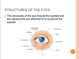
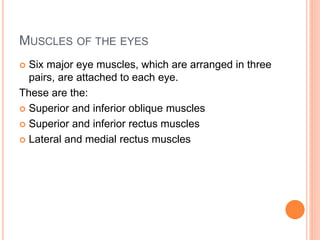
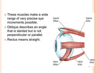
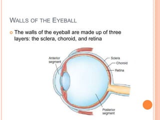
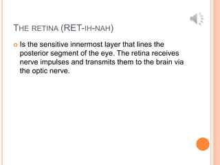
 The eyes are the receptor organs of sight, and their
functions are to receive images and transmit them
to the brain.
 Oculus means eye, and the plural is oculi
STRUCTURES OF THE EYES
 The structures of the eye include the eyeball and
the adnexa that are attached to or surround the
eyeball
THE ADNEXA OF THE EYES
 The adnexa of the eyes, also known as adnexa
oculi, are the structures outside the eyeball. These
include the orbit, eye muscles, eyelids, eyelashes,
conjunctiva, and lacrimal apparatus.
(ad-NECK-sah)
THE ORBIT
 The orbit, also known as the eye socket, is the bony
cavity of the skull that contains and protects the
eyeball and its associated muscles, blood vessels,
and nerves.
MUSCLES OF THE EYES
 Six major eye muscles, which are arranged in three
pairs, are attached to each eye.
These are the:
 Superior and inferior oblique muscles
 Superior and inferior rectus muscles
 Lateral and medial rectus muscles
 These muscles make a wide
range of very precise eye
movements possible.
 Oblique describes an angle
that is slanted but is not
perpendicular or parallel.
 Rectus means straight.
 The upper and lower eyelids, together with the
eyebrows and eyelashes, help protect the eyeball
from foreign matter, excessive light, and injuries
due to other causes.
 The canthus (KAN-thus) is the angle where the
upper and lower eyelids meet (canth means corner
of the eye, and -us is a singular noun ending)
(plural,canthi).
 The cilia (SIL-ee-ah), which are small hairs, make
upthe eyebrows and eyelashes. Cilia are also
present inthe nose to prevent foreign matter from
being inhaled
 The tarsus (TAHR-suhs), also known as the tarsal
plate, is the framework within the upper and lower
eyelids that provides the necessary stiffness and
shape (tars means edge of the eyelid, and -us is a
singular noun ending) (plural, tarsi)
THE CONJUNCTIVA (KON-JUNK-TYE-VAH
 Is the transparent mucous membrane that lines the
underside of each eyelid and continues to form a
protective covering over the exposed surface of the
eyeball
THE LACRIMAL APPARATUS (LACK-RIH-MAL)
 Also known as the tear apparatus, consists of the
structures that produce, store, and remove tears.
 Lacrimation is the secretion of tears.
 The lacrimal glands, which secrete lacrimal fluid
(tears), are located on the underside of the upper
eyelid just above the outer corner of each eye
 The function of lacrimal fluid, commonly known as
tears, is to maintain moisture on the anterior
surface of the eyeball. Blinking distributes the
lacrimal fluid across the eye.
 The lacrimal canal consists of a duct at the inner
corner of each eye. These ducts collect tears and
empty them into the lacrimal sacs. Crying is the
overflowing of tears from the lacrimal canals.
 The lacrimal sac, also known the tear sac, is an
enlargement of the upper portion of the lacrimal
duct.
 The lacrimal duct, also known as the nasolacrimal
duct, is the passageway that drains excess tears
into the nose.
THE EYEBALL
 The eyeball, also known as the globe, is a 1-inch
sphere with only about one-sixth of its surface
visible
 The term optic (OP-tik) means pertaining to the eye
or sight (opt means sight, and -ic means pertaining
to).
 Ocular (OCK-you-lar) means pertaining to the eye
(ocul means eye, and -ar means pertaining to).
 Extraocular (eck-strah-OCK-you-lar) means outside
the eyeball (extra- means on the outside, ocul
means eye, and -ar means pertaining to).
 Intraocular (in-trah-OCK-you-lar) means within the
eyeball (intra- means within, ocul means eye, and -
ar means pertaining to).
WALLS OF THE EYEBALL
 The walls of the eyeball are made up of three
layers: the sclera, choroid, and retina
THE SCLERA (SKLEHR-AH)
 Also known as the white of the eye, maintains the
shape of the eye and protects the delicate inner
layers of tissue.
 This tough, fibrous tissue forms the outer layer of
the eye, except for the part covered by the cornea.
THE CHOROID (KOH-ROID
 Also known as the choroid coat, is the opaque
middle layer of the eyeball that contains many
blood vessels and provides the blood supply for the
entire eye.
THE RETINA (RET-IH-NAH)
 Is the sensitive innermost layer that lines the
posterior segment of the eye. The retina receives
nerve impulses and transmits them to the brain via
the optic nerve.
Structures of the Eye

Structures of the Eye

  • 3.
     The eyesare the receptor organs of sight, and their functions are to receive images and transmit them to the brain.  Oculus means eye, and the plural is oculi
  • 4.
    STRUCTURES OF THEEYES  The structures of the eye include the eyeball and the adnexa that are attached to or surround the eyeball
  • 5.
    THE ADNEXA OFTHE EYES  The adnexa of the eyes, also known as adnexa oculi, are the structures outside the eyeball. These include the orbit, eye muscles, eyelids, eyelashes, conjunctiva, and lacrimal apparatus. (ad-NECK-sah)
  • 6.
    THE ORBIT  Theorbit, also known as the eye socket, is the bony cavity of the skull that contains and protects the eyeball and its associated muscles, blood vessels, and nerves.
  • 7.
    MUSCLES OF THEEYES  Six major eye muscles, which are arranged in three pairs, are attached to each eye. These are the:  Superior and inferior oblique muscles  Superior and inferior rectus muscles  Lateral and medial rectus muscles
  • 8.
     These musclesmake a wide range of very precise eye movements possible.  Oblique describes an angle that is slanted but is not perpendicular or parallel.  Rectus means straight.
  • 9.
     The upperand lower eyelids, together with the eyebrows and eyelashes, help protect the eyeball from foreign matter, excessive light, and injuries due to other causes.
  • 10.
     The canthus(KAN-thus) is the angle where the upper and lower eyelids meet (canth means corner of the eye, and -us is a singular noun ending) (plural,canthi).
  • 11.
     The cilia(SIL-ee-ah), which are small hairs, make upthe eyebrows and eyelashes. Cilia are also present inthe nose to prevent foreign matter from being inhaled
  • 12.
     The tarsus(TAHR-suhs), also known as the tarsal plate, is the framework within the upper and lower eyelids that provides the necessary stiffness and shape (tars means edge of the eyelid, and -us is a singular noun ending) (plural, tarsi)
  • 13.
    THE CONJUNCTIVA (KON-JUNK-TYE-VAH Is the transparent mucous membrane that lines the underside of each eyelid and continues to form a protective covering over the exposed surface of the eyeball
  • 14.
    THE LACRIMAL APPARATUS(LACK-RIH-MAL)  Also known as the tear apparatus, consists of the structures that produce, store, and remove tears.  Lacrimation is the secretion of tears.
  • 15.
     The lacrimalglands, which secrete lacrimal fluid (tears), are located on the underside of the upper eyelid just above the outer corner of each eye
  • 16.
     The functionof lacrimal fluid, commonly known as tears, is to maintain moisture on the anterior surface of the eyeball. Blinking distributes the lacrimal fluid across the eye.
  • 17.
     The lacrimalcanal consists of a duct at the inner corner of each eye. These ducts collect tears and empty them into the lacrimal sacs. Crying is the overflowing of tears from the lacrimal canals.
  • 18.
     The lacrimalsac, also known the tear sac, is an enlargement of the upper portion of the lacrimal duct.  The lacrimal duct, also known as the nasolacrimal duct, is the passageway that drains excess tears into the nose.
  • 19.
    THE EYEBALL  Theeyeball, also known as the globe, is a 1-inch sphere with only about one-sixth of its surface visible  The term optic (OP-tik) means pertaining to the eye or sight (opt means sight, and -ic means pertaining to).
  • 20.
     Ocular (OCK-you-lar)means pertaining to the eye (ocul means eye, and -ar means pertaining to).  Extraocular (eck-strah-OCK-you-lar) means outside the eyeball (extra- means on the outside, ocul means eye, and -ar means pertaining to).
  • 21.
     Intraocular (in-trah-OCK-you-lar)means within the eyeball (intra- means within, ocul means eye, and - ar means pertaining to).
  • 22.
    WALLS OF THEEYEBALL  The walls of the eyeball are made up of three layers: the sclera, choroid, and retina
  • 23.
    THE SCLERA (SKLEHR-AH) Also known as the white of the eye, maintains the shape of the eye and protects the delicate inner layers of tissue.  This tough, fibrous tissue forms the outer layer of the eye, except for the part covered by the cornea.
  • 24.
    THE CHOROID (KOH-ROID Also known as the choroid coat, is the opaque middle layer of the eyeball that contains many blood vessels and provides the blood supply for the entire eye.
  • 25.
    THE RETINA (RET-IH-NAH) Is the sensitive innermost layer that lines the posterior segment of the eye. The retina receives nerve impulses and transmits them to the brain via the optic nerve.