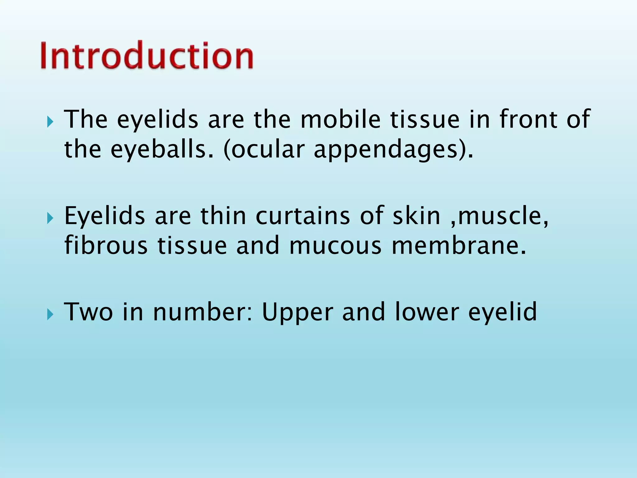
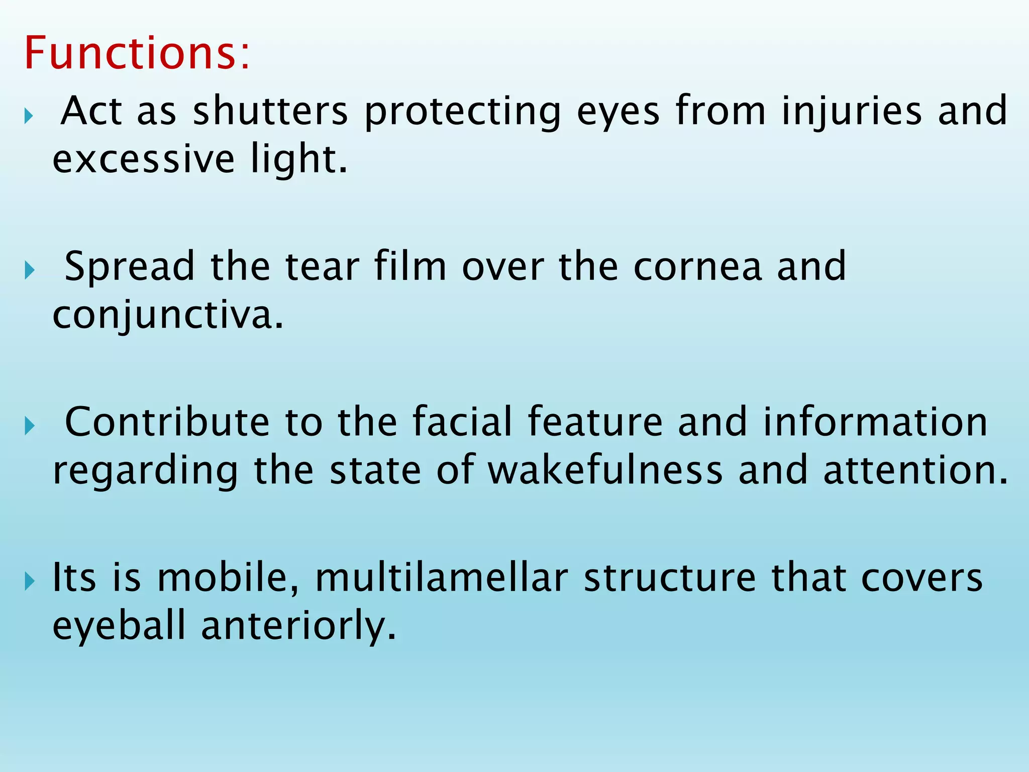
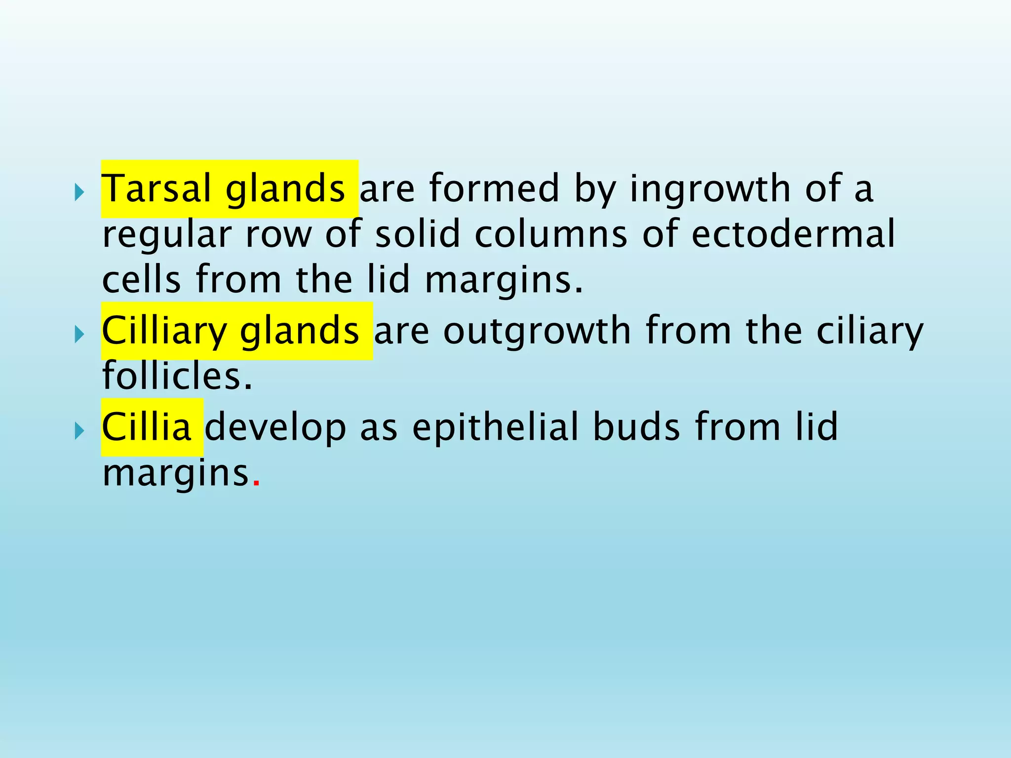
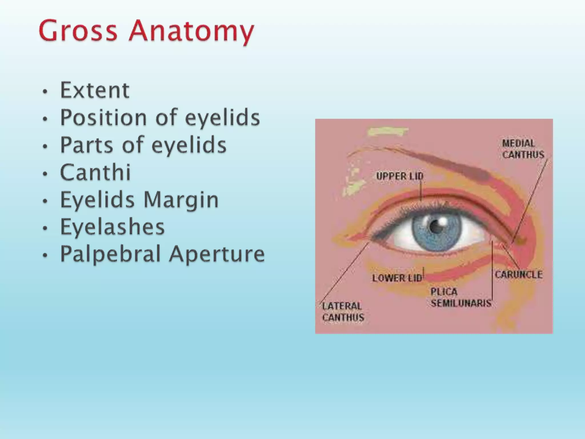
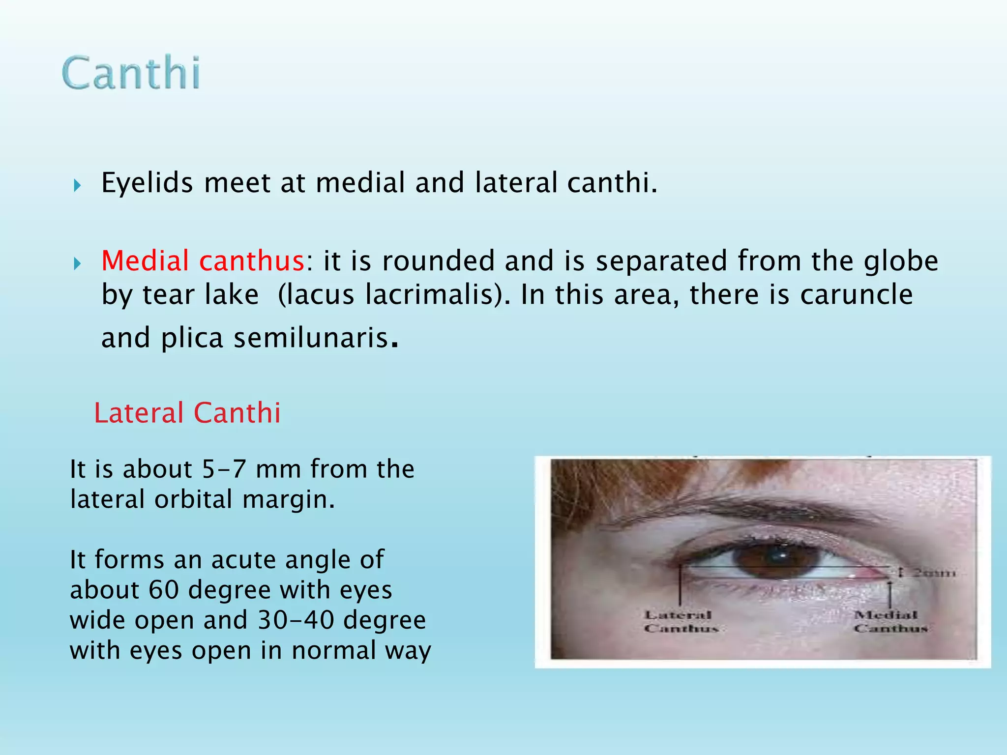
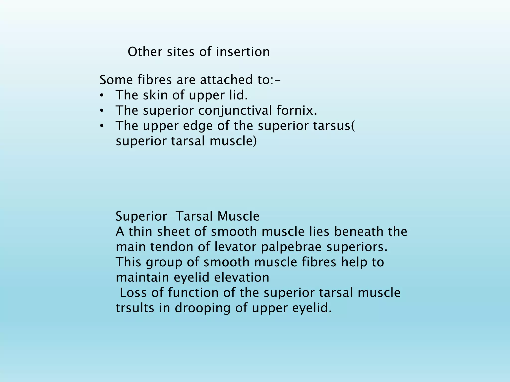
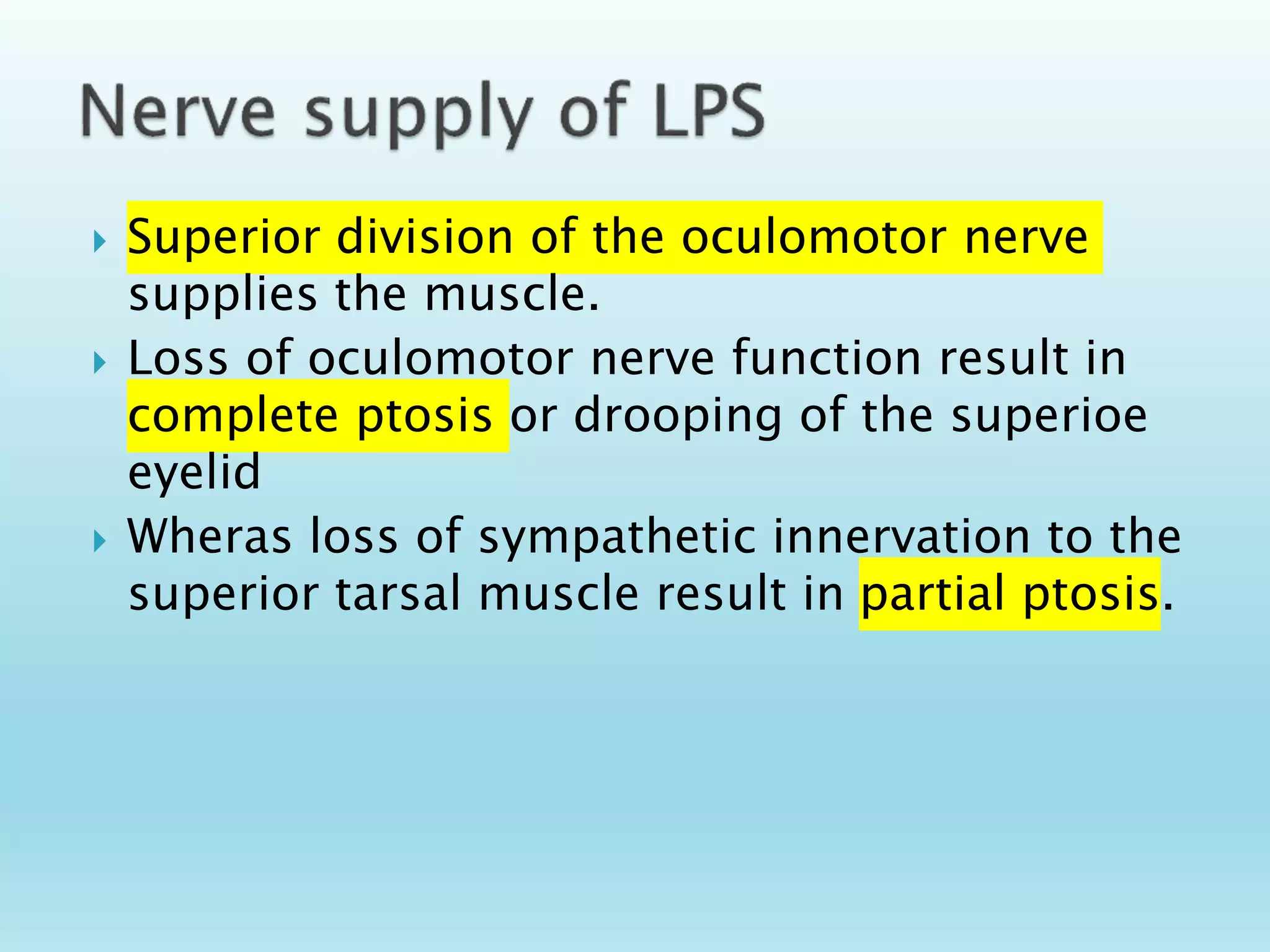
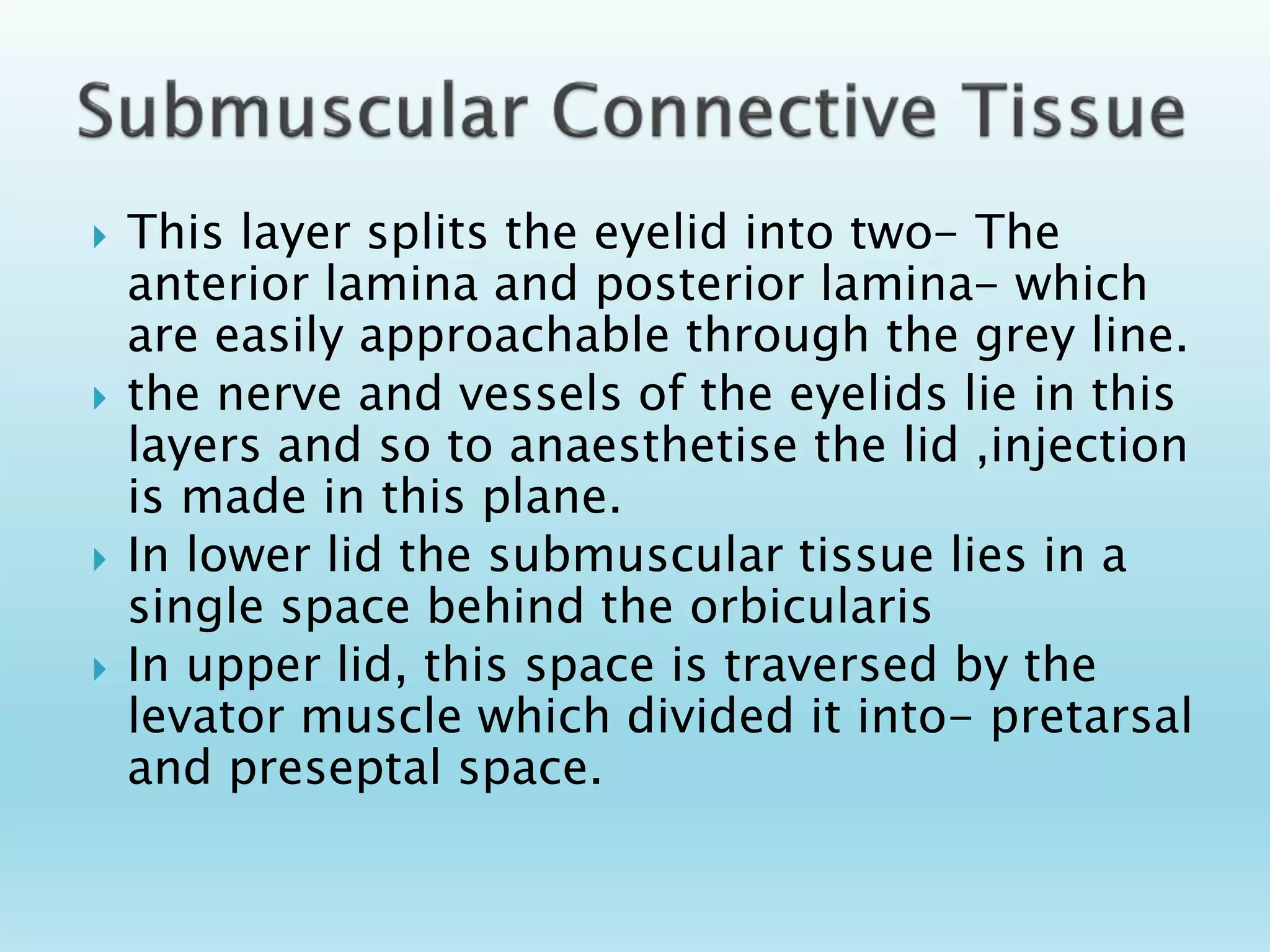
Presented By
Dr. Suruchi Baluni
P.G. Scholar 1st year
Dept -Shalakyatantra
Contents
 Inroduction
 Embryology
 Anatomy
 Layers of Eyelid
 Muscle of Eyelid
 Gland of Eyelid
 Function of Eyelid
 Nerve supply
 Vascular supply
 Venous drainage
 Lymphatic drainage
 The eyelids are the mobile tissue in front of
the eyeballs. (ocular appendages).
 Eyelids are thin curtains of skin ,muscle,
fibrous tissue and mucous membrane.
 Two in number: Upper and lower eyelid
Functions:
 Act as shutters protecting eyes from injuries and
excessive light.
 Spread the tear film over the cornea and
conjunctiva.
 Contribute to the facial feature and information
regarding the state of wakefulness and attention.
 Its is mobile, multilamellar structure that covers
eyeball anteriorly.
 It is formed by reduplication of surface ectoderm
above and below the cornea during 2nd month of
gestation.
 The folds enlarge and their margin meet and fuse
with each other by 3rd month.
 A closed space, conjunctival sac is formed.
 The folds thus formed contain some mesoderm
which would form the muscles of the lid and the
tarsal plate.
 Lid separate after seventh month of intra uterine
life
8 Weeks
5-7 month
7 weeks
 Tarsal glands are formed by ingrowth of a
regular row of solid columns of ectodermal
cells from the lid margins.
 Cilliary glands are outgrowth from the ciliary
follicles.
 Cillia develop as epithelial buds from lid
margins.
Extent :
 Upper eyelid extends from
the eyebrow downward to end
in a free margin which forms
the superior boundary of
palpebral fissure.
 Lower eyelid merge into the
skin of the cheek.
Parts
 Each eyelid is divided by an
horizontal furrow into an
orbital and tarsal plate.
 Additional folds in lower lid are
Nasojugal fold medially and the
malar fold laterally.
 These folds limit the spread of
blood downward from eyelids
to cheek.
 Upper eyelid covers 1/6th part of the cornea.
 Lower lid just touchs the limbus.
Palpebral aperture
It is the elliptical space between the
upper and the lower lid. When the
eyes are open,it measures about
vertically-10-11mm.
horizontally 28-30 mm.
At Birth
horizontal 18-21mm.
vertical 8 mm.
 Eyelids meet at medial and lateral canthi.
 Medial canthus: it is rounded and is separated from the globe
by tear lake (lacus lacrimalis). In this area, there is caruncle
and plica semilunaris.
Lateral Canthi
It is about 5-7 mm from the
lateral orbital margin.
It forms an acute angle of
about 60 degree with eyes
wide open and 30-40 degree
with eyes open in normal way
 caruncula lacrimalis:
its is a small
pink,globular nodule at
the inner corner (medial
canthus) of the eye
.consist of skin,hair
follicle,sweat gland and
sebaceous gland.
 A semilunar fold called plica
semilunaris lies on lateral
side of caruncle represents
the third eye lid of other
vertebrae.
 Each lid margin is divided into two parts by the
lacrimal papilla(a small elevation present on the
medial side, which contains a hole- the lacrimal
punctum in its centre).
 The medial portion of the eyelid margin,
extending from the punctum medially to the
medial canthal angle termed as lacrimal portion
is rounded and devoid of lashes or glands.
 The lateral, ciliary portion of the eyelid margin
consists of a rounded anterior border, a sharp
posterior border (placed against the globe) and
an intermarginal strip between the two borders.
Grey line which marks the junction of skin
and conjunctiva,divides the intermarginal
strip into an anterior strip bearing lashes
and a posterior strip which contains
opening of meibomian glands arranged in a
row and a lipid strip.
 Arranged in 2-3 row.
 Upper lid 100-150 directed forward,upward and
backward
 Lower lid 50-75 directed forward downward and
backward.
Cilia
 Cillia vary in size ranging from 20-120mm in diameter
and from 6-12mm in length
 Taper throughout their length to end in fine sharp point.
 Each cilium has a life span of some 3- 4 months. At the
termination of this period the old cilium drops away the
follicle test for several week and then a new cilium grows
out.
 Each follicle is surrounded by a dense plexus of vessels
and nerves, the latter provide the tactile sensibility to
each cilium.
 Trichiasis:Acquired misdirection of
eyelashes.
 Madarosis: Decrease in number of
eyelashes.
 Lash Poliosis: Premature graying of the
lashes.
 Trichomegaly: Excessive eyelash
growth.
CONGENITAL /DEVELOPMENTAL ANOMALIES
Coloboma of lid: Notch in the edge of eyelid
Cryptophthalmos: partial or complete loss of
brows, palpebral fissure, lashes,conjuctiva
and absence of eyelid.
Ectropion: Eversion of eyelid margin
Entropion: Eyelid margin inversion
Euryblepharon: vertical shortening
and horizontal lengthening of
eyelids.
Epiblepharon: lower lid Pretarsal
muscle and skin ride above the
lower lid margin to form a
horizontal fold of tissue.
Ankyloblepharon: partial or
complete fusion of eyelids by
webs of skin.
Symblepharon: adhesion of lid
to the gobe.
Distichiasis : extra
rows of
eyelashes.
Blepherophimosis:
condition in
which palpebral
fissure appear to
be smaller
Skin
The skin covering the eyelid is elastic, having a
fine texture, is thinnest in the body and folds
easily thereby contributing to the ease and
speed of mobility of the upper eyelid.
Nasal part of the skin is smooth,shining and
greasy in comparison to temporal part.
Fine hair are seen on the temporal part of skin.
 Beneath the skin is a layer of loose aerolar connective
tissue, containing no fat. It is thus readily distended by
oedema or blood.
 This layer is non- existent near the ciliary margin, at the
lid folds and at medial and lateral angles where the skin
is attached to the underlying ligaments.
Layer of striated muscle
• This layer consists of orbicularis muscle which forms a thin oval sheet
across the eyelids. It comprises three portions: the orbital, palpebral and
lacrimal.
• The Orbital part forms the most peripheral fibres of the
orbicularis which arise from the anterior part of the medial
palpebral ligament and the adjacent bones( upper orbital
margin,the maxillary process of frontal bone, frontal process of
maxilla and the lower orbital margin medial to the infra-orbital
foramen.
• The Palpebral part of orbicularis are preseptal and pretarsal
portions. The fibres of pretarsal portions helps in drainage of tear
by lacrimal sac and are called as pars lacrimalis.(horners muscle)
• It closes the eyelid and is supplied by zygomatic branch of facial
nerve. Therefore in paralysis of facial nerve there occurs
lagophthalmos.
Levator palpebrae
Superioris
Origin- Arises from the
undersurface of the lesser wing of
the sphenoid from the roof of the
orbit, just anterior to the optic
canal.
It is a flat muscle that broadens as it
passes forwards.
Primary insertion- Primary point of
insertion is into superior surface of
the tarsus.
At the anterior end the muscle form
an aponeurotic tendon that is
widened on each side up to a
crescentric margin.
This broad tendon penetrates the
orbital septum and is inserted into
the front of the superior tarsal plate
Other sites of insertion
Some fibres are attached to:-
• The skin of upper lid.
• The superior conjunctival fornix.
• The upper edge of the superior tarsus(
superior tarsal muscle)
Superior Tarsal Muscle
A thin sheet of smooth muscle lies beneath the
main tendon of levator palpebrae superiors.
This group of smooth muscle fibres help to
maintain eyelid elevation
Loss of function of the superior tarsal muscle
trsults in drooping of upper eyelid.
 Superior division of the oculomotor nerve
supplies the muscle.
 Loss of oculomotor nerve function result in
complete ptosis or drooping of the superioe
eyelid
 Wheras loss of sympathetic innervation to the
superior tarsal muscle result in partial ptosis.
Whitnall ligament- it is located at transition
zone- act asa fulcrum for levator transferring
its vector from ant-post to sup-inf direction.
Its analogue in lower lid is Lockwood Ligament
 This layer splits the eyelid into two- The
anterior lamina and posterior lamina- which
are easily approachable through the grey line.
 the nerve and vessels of the eyelids lie in this
layers and so to anaesthetise the lid ,injection
is made in this plane.
 In lower lid the submuscular tissue lies in a
single space behind the orbicularis
 In upper lid, this space is traversed by the
levator muscle which divided it into- pretarsal
and preseptal space.
THANK YOU

Eyelid Anatomy.pptx

  • 1.
    Presented By Dr. SuruchiBaluni P.G. Scholar 1st year Dept -Shalakyatantra
  • 2.
    Contents  Inroduction  Embryology Anatomy  Layers of Eyelid  Muscle of Eyelid  Gland of Eyelid  Function of Eyelid  Nerve supply  Vascular supply  Venous drainage  Lymphatic drainage
  • 3.
     The eyelidsare the mobile tissue in front of the eyeballs. (ocular appendages).  Eyelids are thin curtains of skin ,muscle, fibrous tissue and mucous membrane.  Two in number: Upper and lower eyelid
  • 4.
    Functions:  Act asshutters protecting eyes from injuries and excessive light.  Spread the tear film over the cornea and conjunctiva.  Contribute to the facial feature and information regarding the state of wakefulness and attention.  Its is mobile, multilamellar structure that covers eyeball anteriorly.
  • 5.
     It isformed by reduplication of surface ectoderm above and below the cornea during 2nd month of gestation.  The folds enlarge and their margin meet and fuse with each other by 3rd month.  A closed space, conjunctival sac is formed.  The folds thus formed contain some mesoderm which would form the muscles of the lid and the tarsal plate.  Lid separate after seventh month of intra uterine life
  • 6.
  • 7.
     Tarsal glandsare formed by ingrowth of a regular row of solid columns of ectodermal cells from the lid margins.  Cilliary glands are outgrowth from the ciliary follicles.  Cillia develop as epithelial buds from lid margins.
  • 9.
    Extent :  Uppereyelid extends from the eyebrow downward to end in a free margin which forms the superior boundary of palpebral fissure.  Lower eyelid merge into the skin of the cheek. Parts  Each eyelid is divided by an horizontal furrow into an orbital and tarsal plate.  Additional folds in lower lid are Nasojugal fold medially and the malar fold laterally.  These folds limit the spread of blood downward from eyelids to cheek.
  • 10.
     Upper eyelidcovers 1/6th part of the cornea.  Lower lid just touchs the limbus. Palpebral aperture It is the elliptical space between the upper and the lower lid. When the eyes are open,it measures about vertically-10-11mm. horizontally 28-30 mm. At Birth horizontal 18-21mm. vertical 8 mm.
  • 11.
     Eyelids meetat medial and lateral canthi.  Medial canthus: it is rounded and is separated from the globe by tear lake (lacus lacrimalis). In this area, there is caruncle and plica semilunaris. Lateral Canthi It is about 5-7 mm from the lateral orbital margin. It forms an acute angle of about 60 degree with eyes wide open and 30-40 degree with eyes open in normal way
  • 12.
     caruncula lacrimalis: itsis a small pink,globular nodule at the inner corner (medial canthus) of the eye .consist of skin,hair follicle,sweat gland and sebaceous gland.  A semilunar fold called plica semilunaris lies on lateral side of caruncle represents the third eye lid of other vertebrae.
  • 13.
     Each lidmargin is divided into two parts by the lacrimal papilla(a small elevation present on the medial side, which contains a hole- the lacrimal punctum in its centre).  The medial portion of the eyelid margin, extending from the punctum medially to the medial canthal angle termed as lacrimal portion is rounded and devoid of lashes or glands.  The lateral, ciliary portion of the eyelid margin consists of a rounded anterior border, a sharp posterior border (placed against the globe) and an intermarginal strip between the two borders.
  • 14.
    Grey line whichmarks the junction of skin and conjunctiva,divides the intermarginal strip into an anterior strip bearing lashes and a posterior strip which contains opening of meibomian glands arranged in a row and a lipid strip.
  • 15.
     Arranged in2-3 row.  Upper lid 100-150 directed forward,upward and backward  Lower lid 50-75 directed forward downward and backward. Cilia  Cillia vary in size ranging from 20-120mm in diameter and from 6-12mm in length  Taper throughout their length to end in fine sharp point.  Each cilium has a life span of some 3- 4 months. At the termination of this period the old cilium drops away the follicle test for several week and then a new cilium grows out.  Each follicle is surrounded by a dense plexus of vessels and nerves, the latter provide the tactile sensibility to each cilium.
  • 16.
     Trichiasis:Acquired misdirectionof eyelashes.  Madarosis: Decrease in number of eyelashes.  Lash Poliosis: Premature graying of the lashes.  Trichomegaly: Excessive eyelash growth. CONGENITAL /DEVELOPMENTAL ANOMALIES Coloboma of lid: Notch in the edge of eyelid Cryptophthalmos: partial or complete loss of brows, palpebral fissure, lashes,conjuctiva and absence of eyelid.
  • 17.
    Ectropion: Eversion ofeyelid margin Entropion: Eyelid margin inversion
  • 18.
    Euryblepharon: vertical shortening andhorizontal lengthening of eyelids. Epiblepharon: lower lid Pretarsal muscle and skin ride above the lower lid margin to form a horizontal fold of tissue. Ankyloblepharon: partial or complete fusion of eyelids by webs of skin. Symblepharon: adhesion of lid to the gobe.
  • 19.
    Distichiasis : extra rowsof eyelashes. Blepherophimosis: condition in which palpebral fissure appear to be smaller
  • 20.
    Skin The skin coveringthe eyelid is elastic, having a fine texture, is thinnest in the body and folds easily thereby contributing to the ease and speed of mobility of the upper eyelid. Nasal part of the skin is smooth,shining and greasy in comparison to temporal part. Fine hair are seen on the temporal part of skin.
  • 21.
     Beneath theskin is a layer of loose aerolar connective tissue, containing no fat. It is thus readily distended by oedema or blood.  This layer is non- existent near the ciliary margin, at the lid folds and at medial and lateral angles where the skin is attached to the underlying ligaments.
  • 22.
    Layer of striatedmuscle • This layer consists of orbicularis muscle which forms a thin oval sheet across the eyelids. It comprises three portions: the orbital, palpebral and lacrimal. • The Orbital part forms the most peripheral fibres of the orbicularis which arise from the anterior part of the medial palpebral ligament and the adjacent bones( upper orbital margin,the maxillary process of frontal bone, frontal process of maxilla and the lower orbital margin medial to the infra-orbital foramen.
  • 23.
    • The Palpebralpart of orbicularis are preseptal and pretarsal portions. The fibres of pretarsal portions helps in drainage of tear by lacrimal sac and are called as pars lacrimalis.(horners muscle) • It closes the eyelid and is supplied by zygomatic branch of facial nerve. Therefore in paralysis of facial nerve there occurs lagophthalmos.
  • 24.
    Levator palpebrae Superioris Origin- Arisesfrom the undersurface of the lesser wing of the sphenoid from the roof of the orbit, just anterior to the optic canal. It is a flat muscle that broadens as it passes forwards. Primary insertion- Primary point of insertion is into superior surface of the tarsus. At the anterior end the muscle form an aponeurotic tendon that is widened on each side up to a crescentric margin. This broad tendon penetrates the orbital septum and is inserted into the front of the superior tarsal plate
  • 25.
    Other sites ofinsertion Some fibres are attached to:- • The skin of upper lid. • The superior conjunctival fornix. • The upper edge of the superior tarsus( superior tarsal muscle) Superior Tarsal Muscle A thin sheet of smooth muscle lies beneath the main tendon of levator palpebrae superiors. This group of smooth muscle fibres help to maintain eyelid elevation Loss of function of the superior tarsal muscle trsults in drooping of upper eyelid.
  • 26.
     Superior divisionof the oculomotor nerve supplies the muscle.  Loss of oculomotor nerve function result in complete ptosis or drooping of the superioe eyelid  Wheras loss of sympathetic innervation to the superior tarsal muscle result in partial ptosis.
  • 27.
    Whitnall ligament- itis located at transition zone- act asa fulcrum for levator transferring its vector from ant-post to sup-inf direction. Its analogue in lower lid is Lockwood Ligament
  • 28.
     This layersplits the eyelid into two- The anterior lamina and posterior lamina- which are easily approachable through the grey line.  the nerve and vessels of the eyelids lie in this layers and so to anaesthetise the lid ,injection is made in this plane.  In lower lid the submuscular tissue lies in a single space behind the orbicularis  In upper lid, this space is traversed by the levator muscle which divided it into- pretarsal and preseptal space.
  • 29.