Downloaded 1,075 times
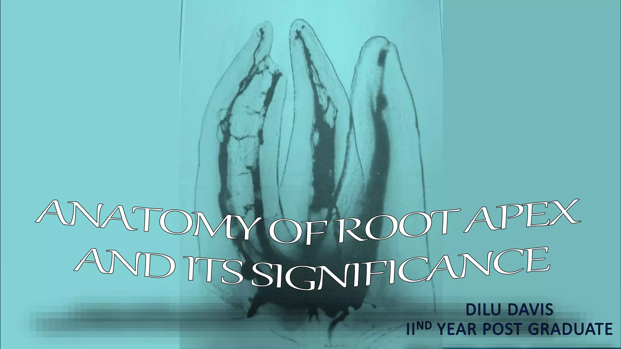
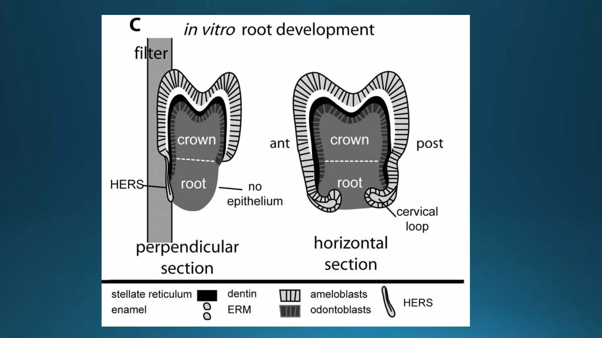
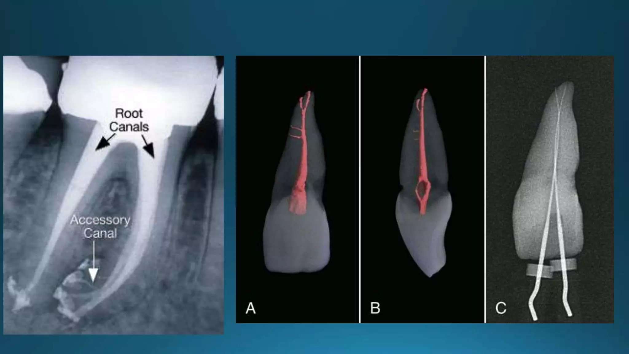
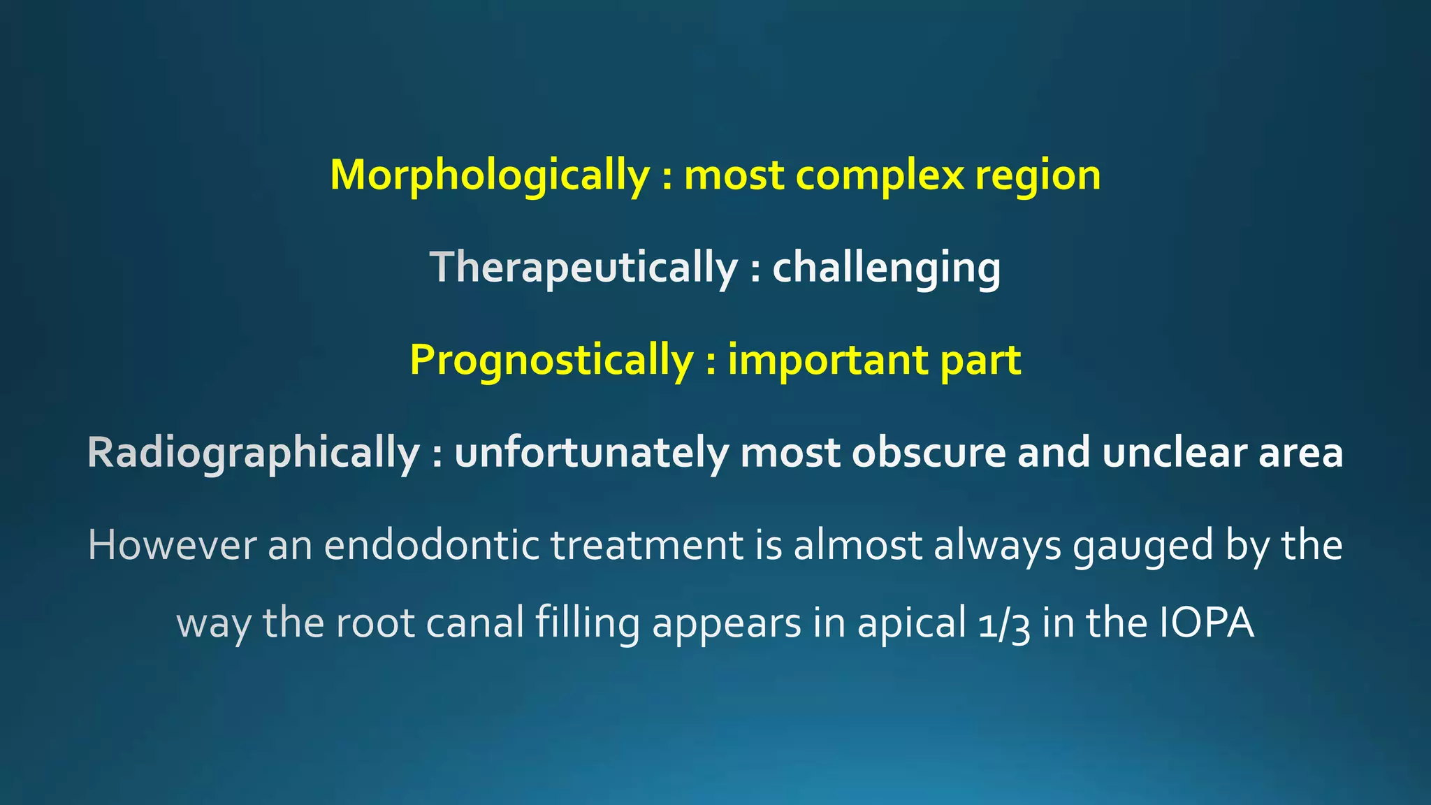
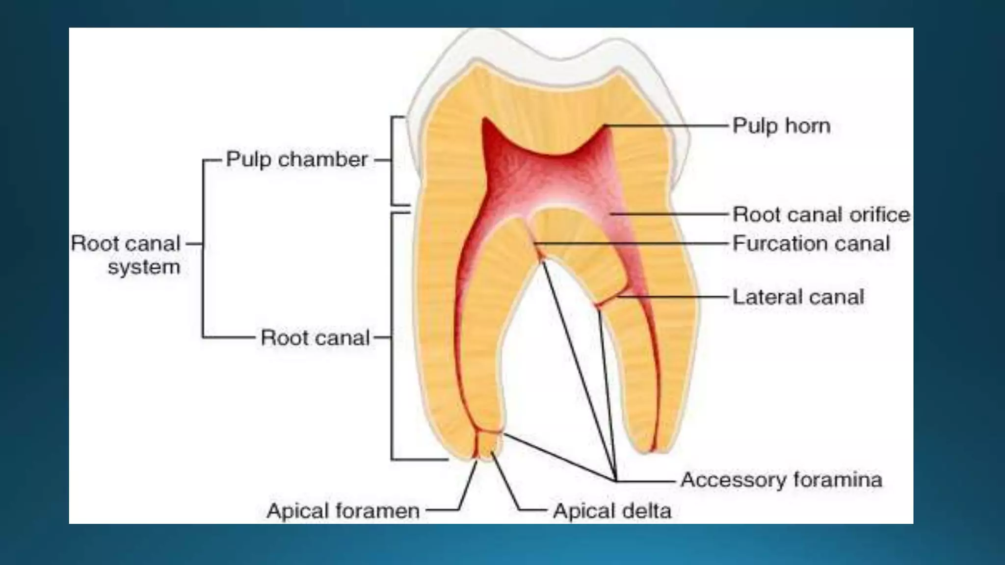
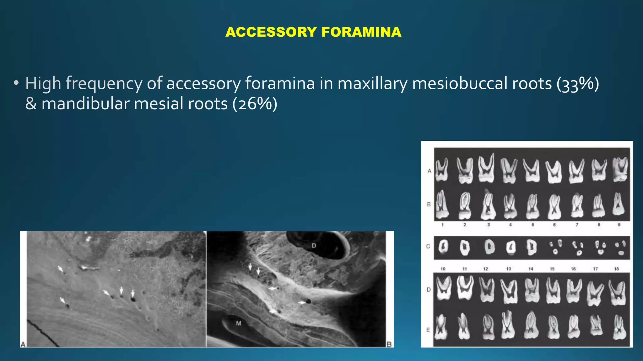
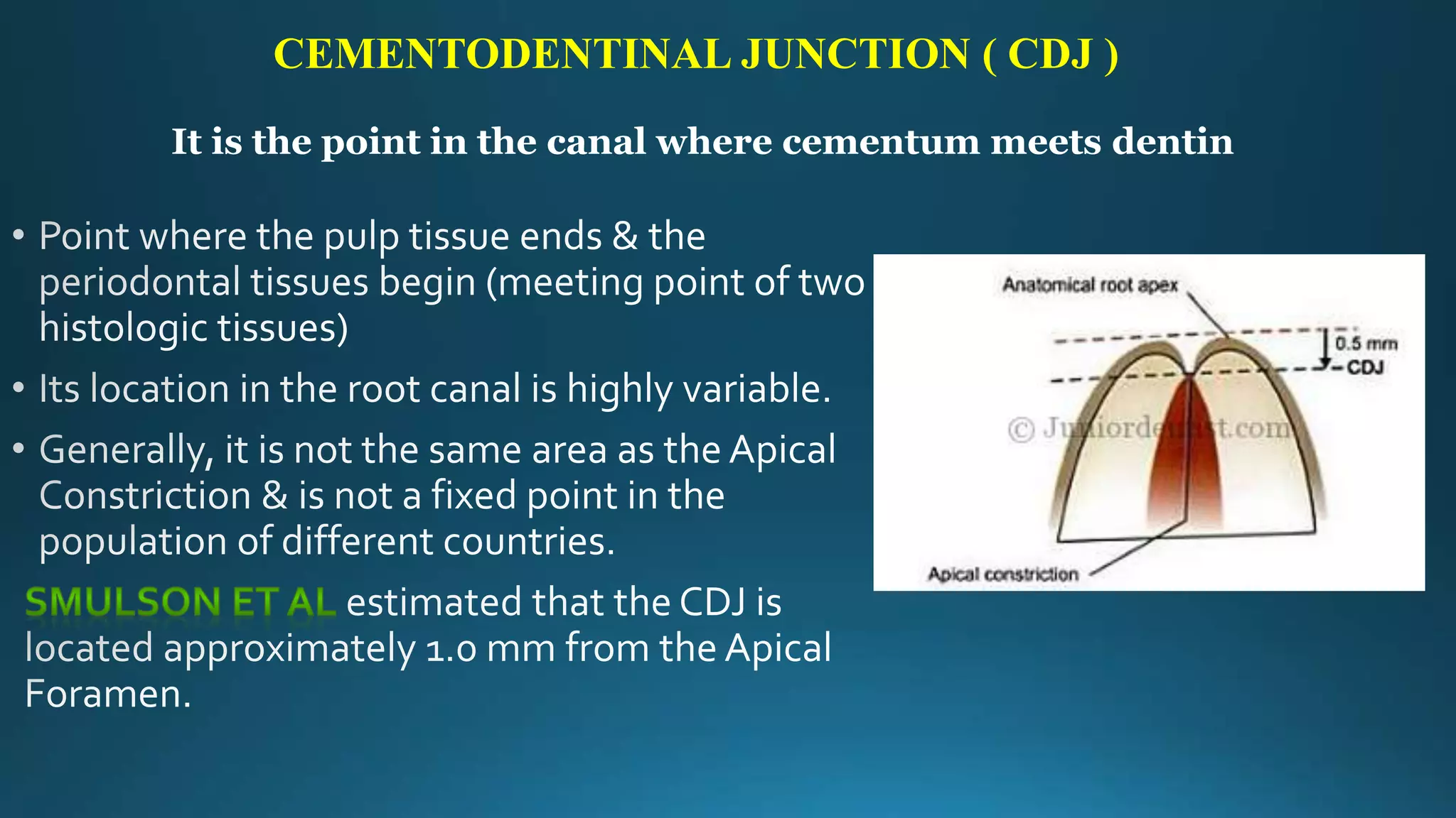

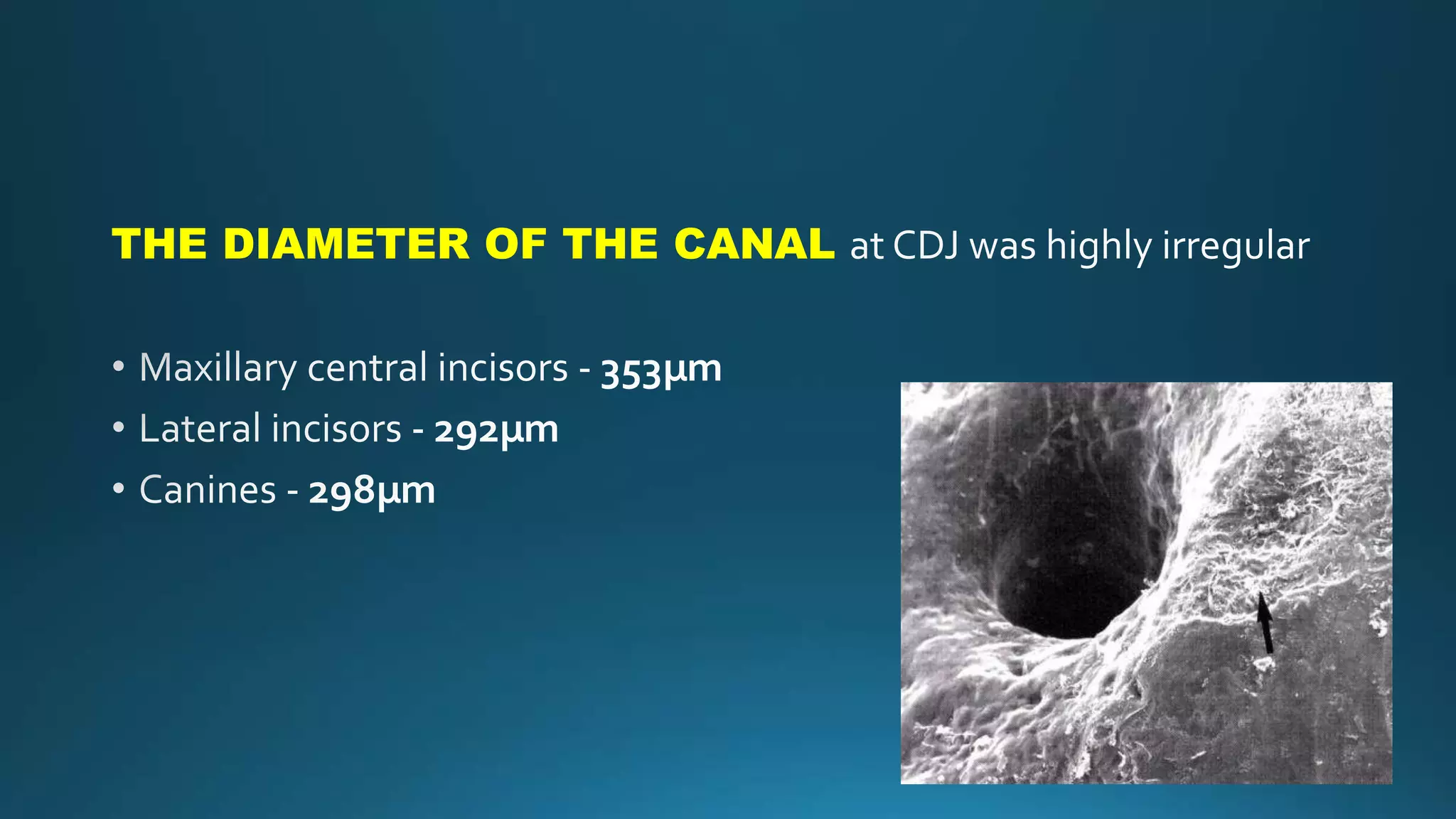
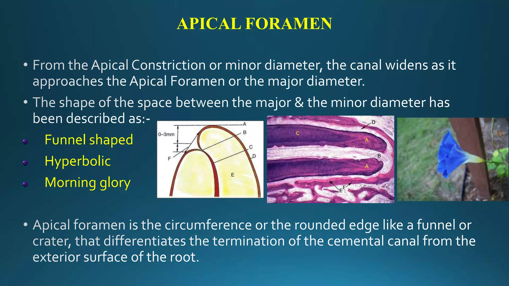
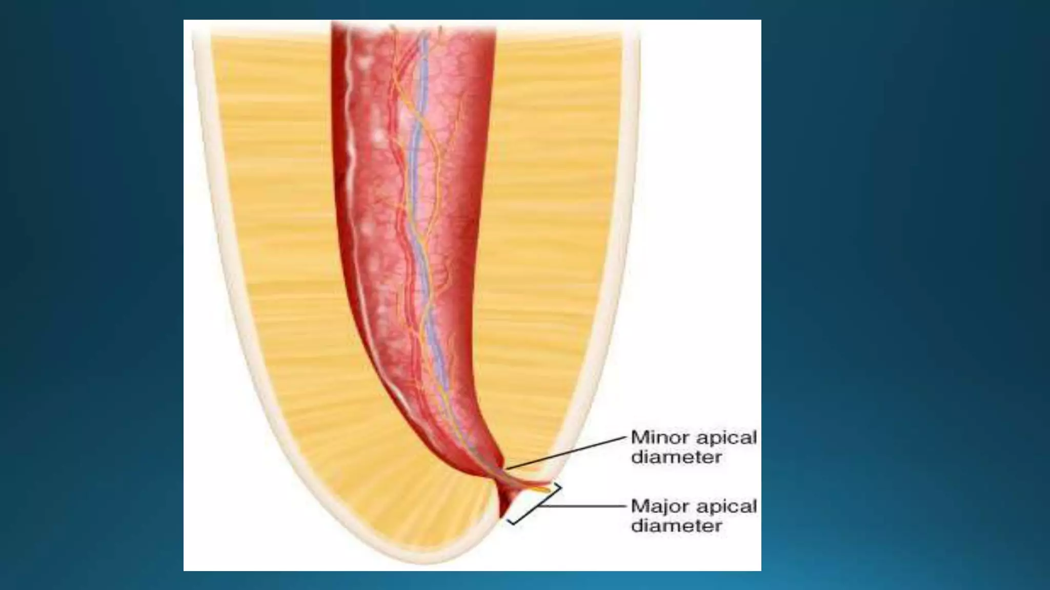
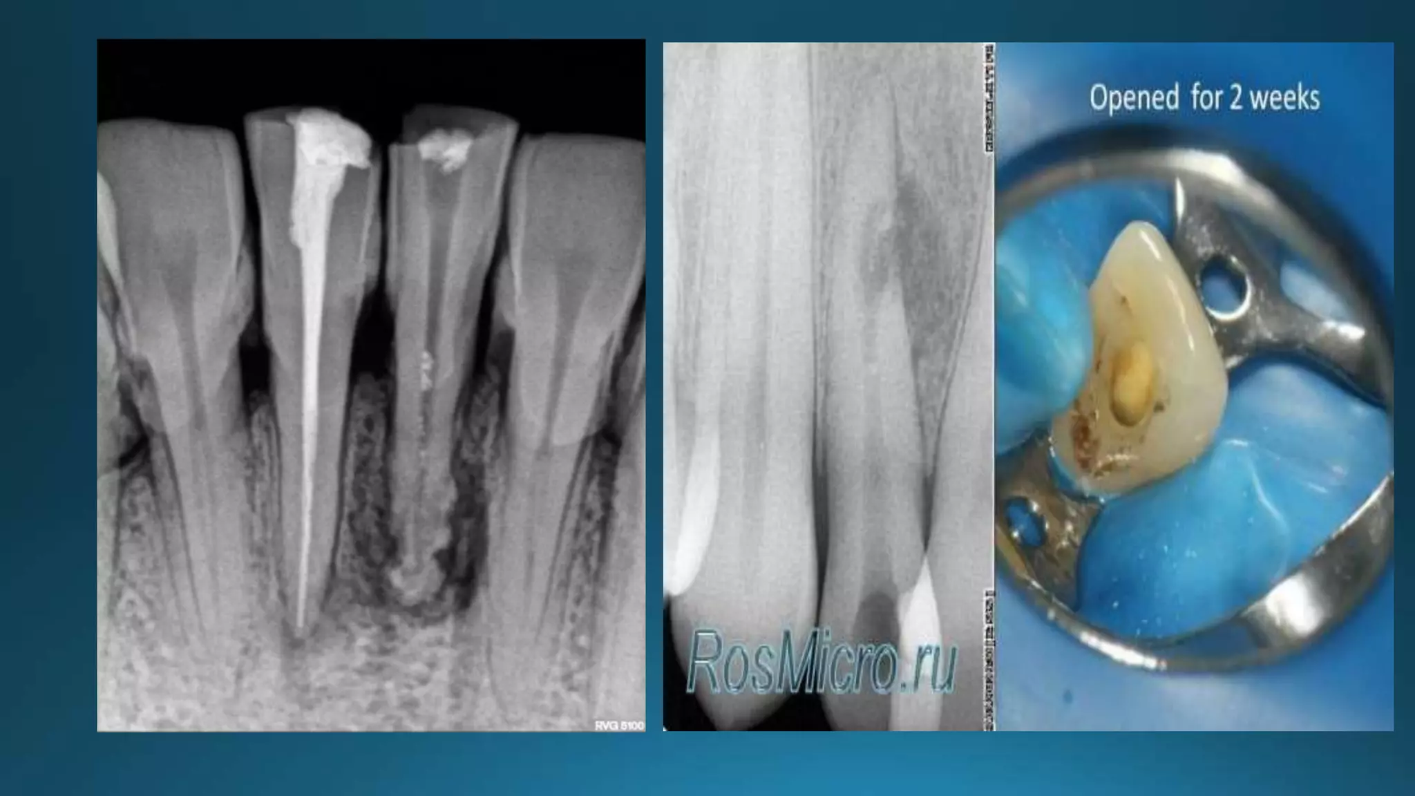
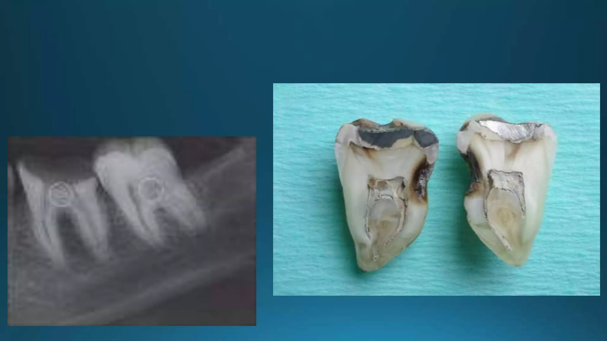
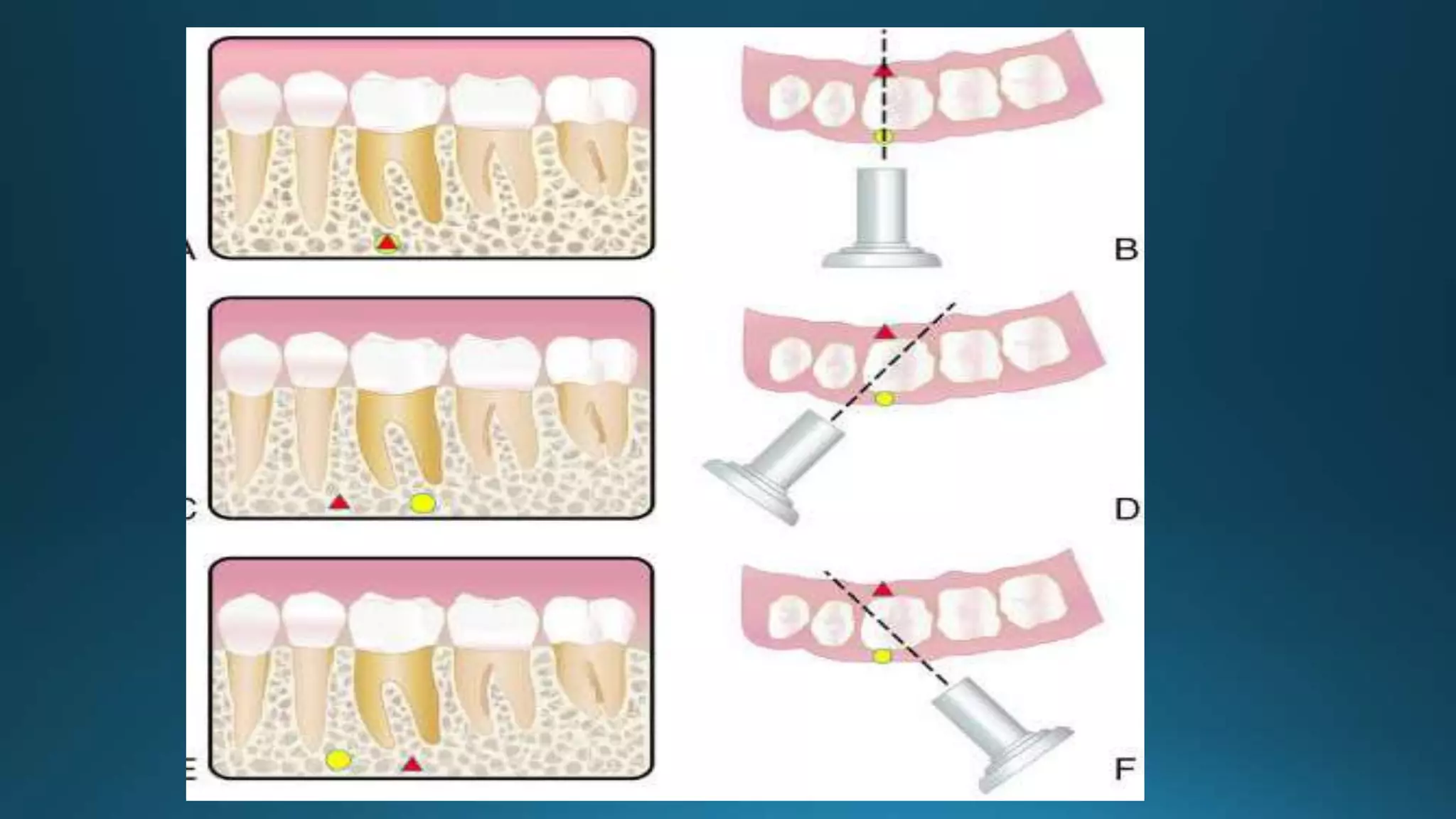
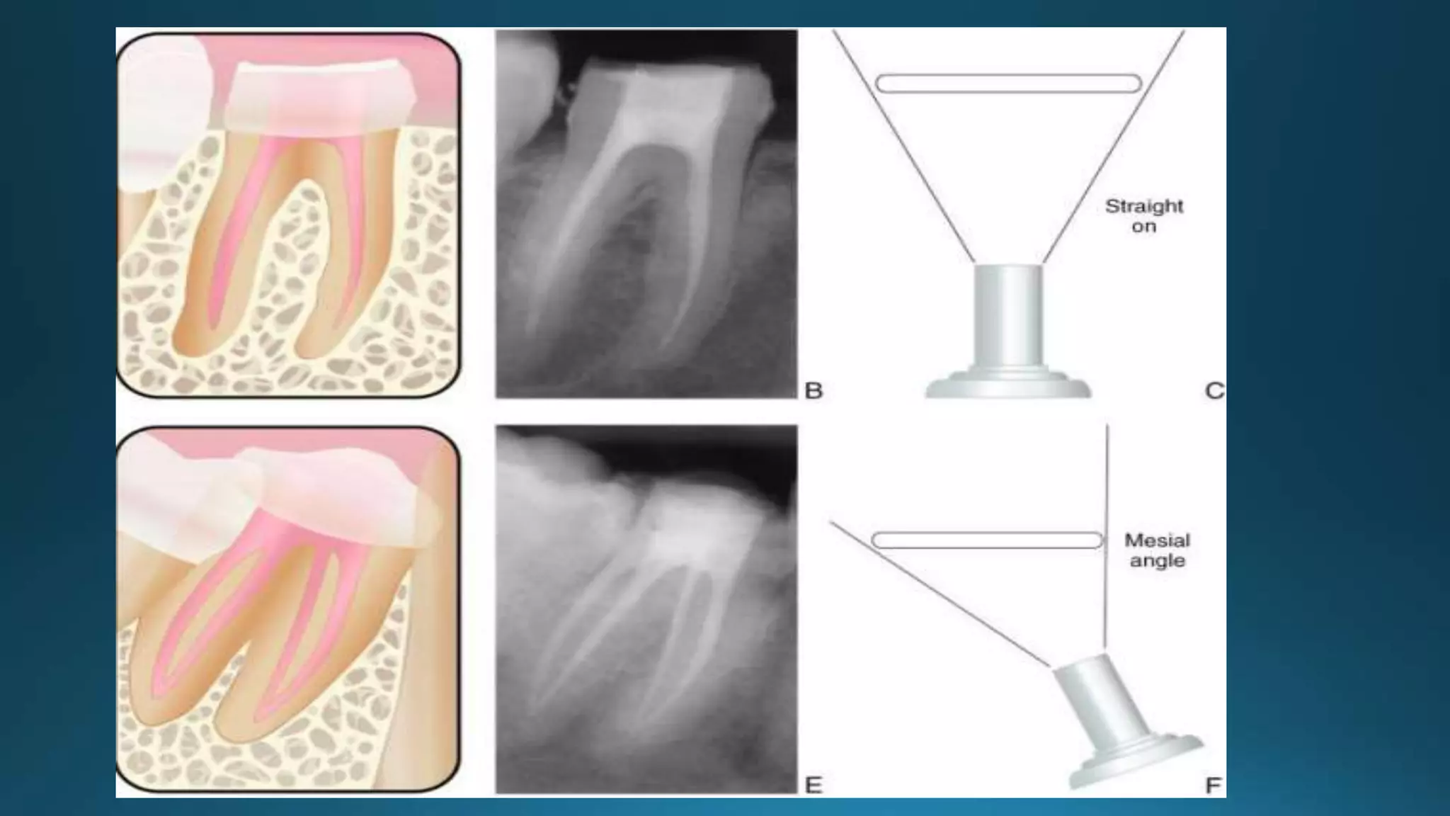
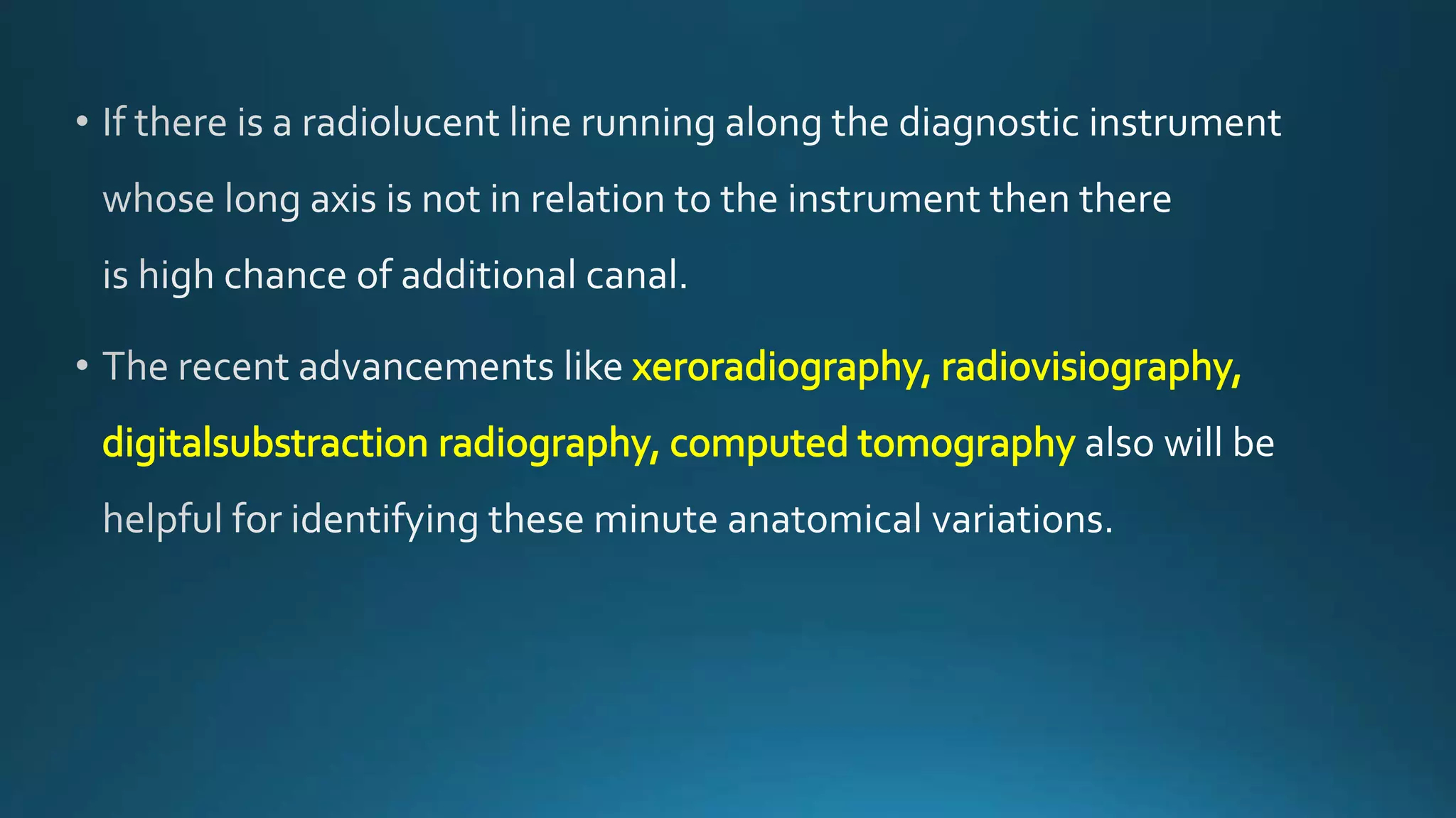
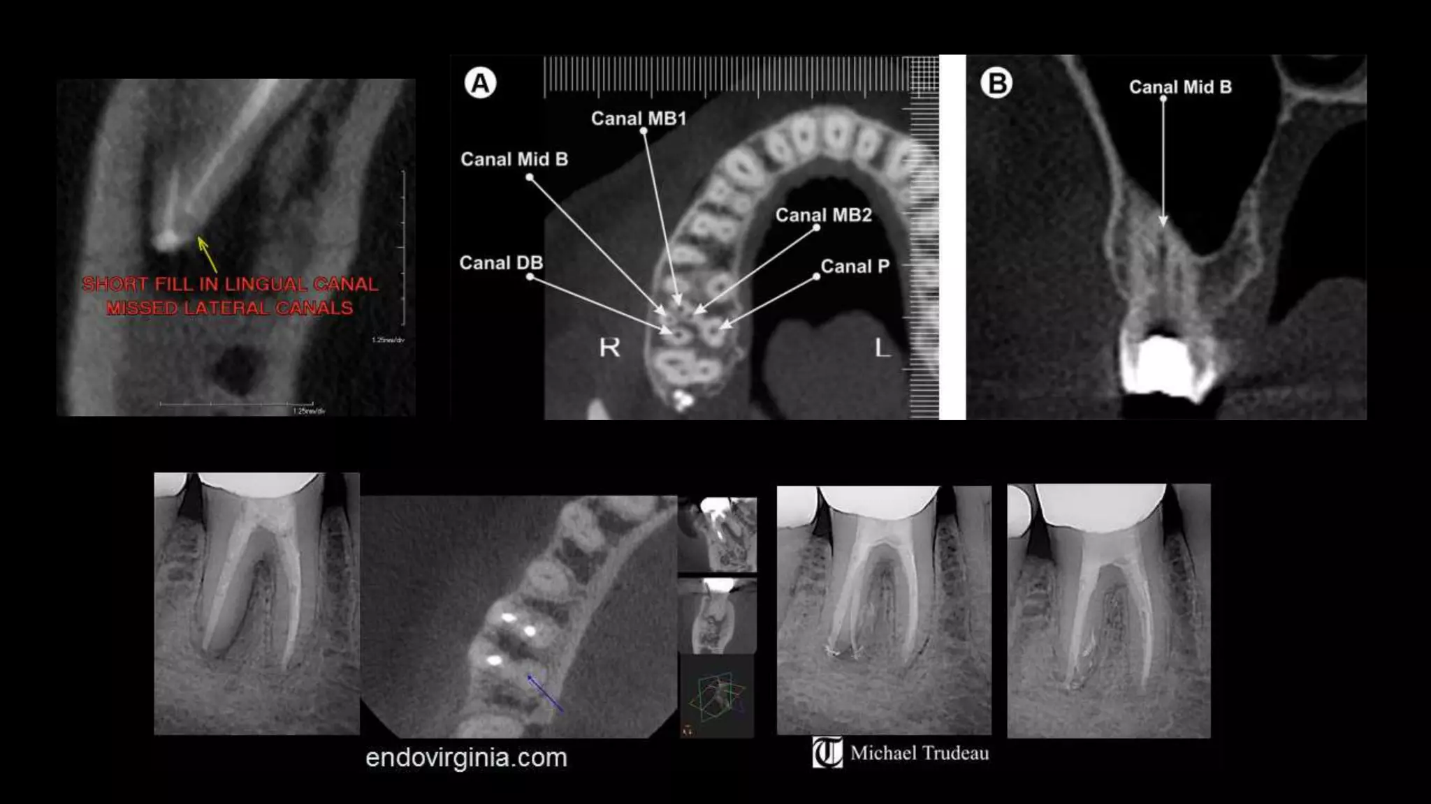
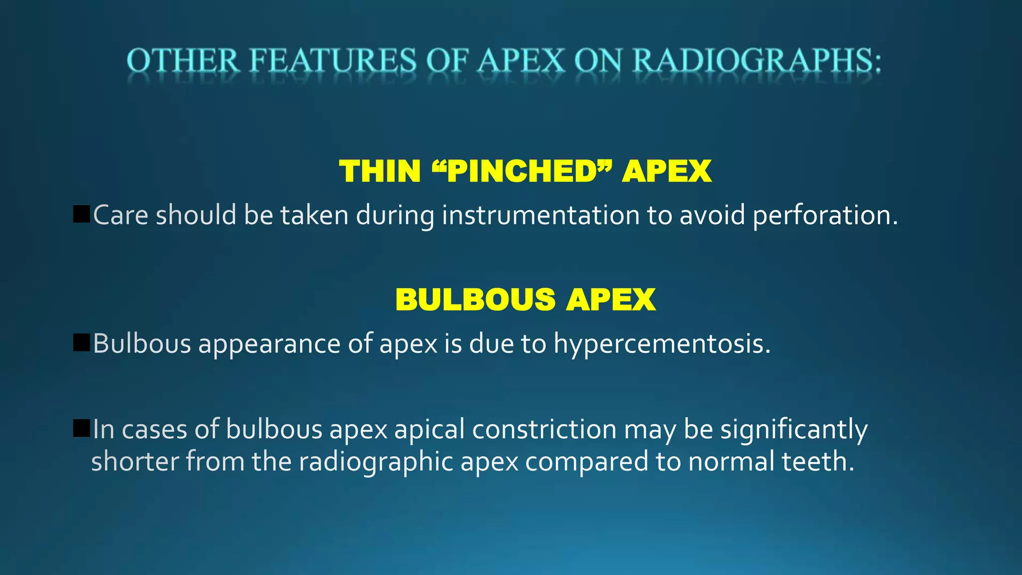
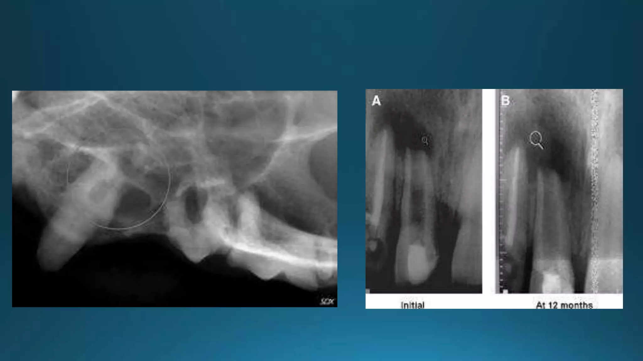

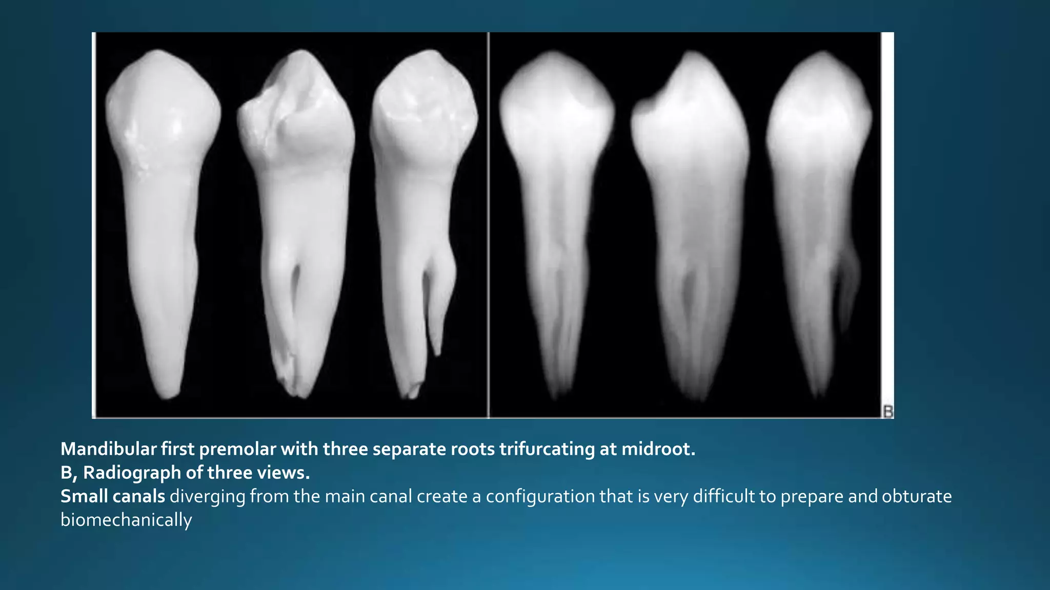
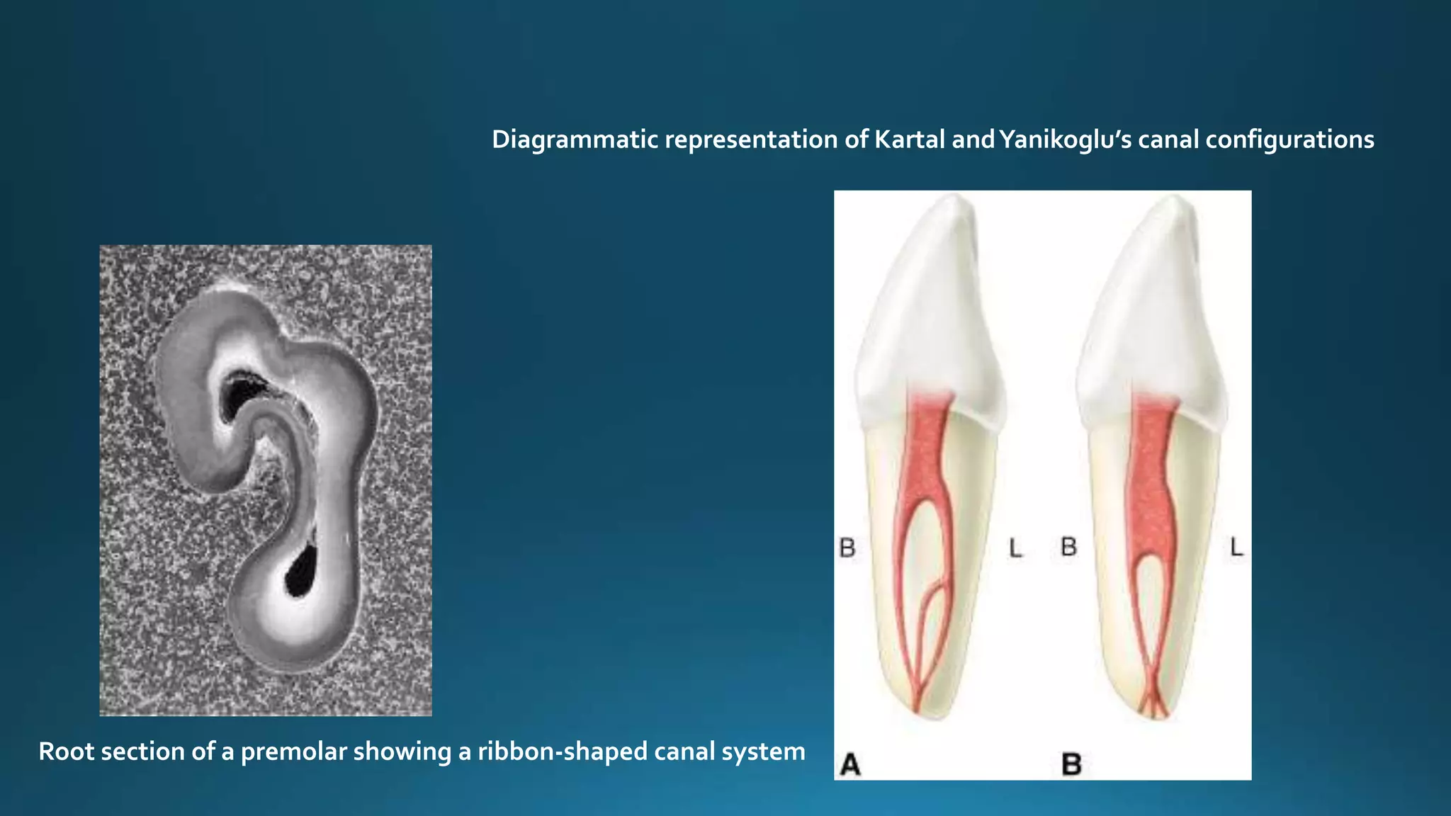
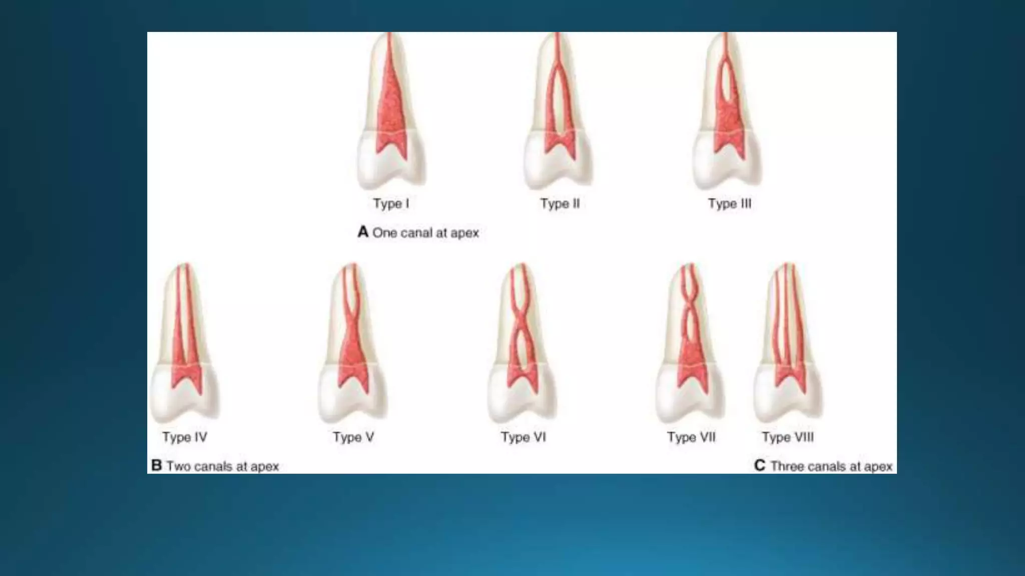
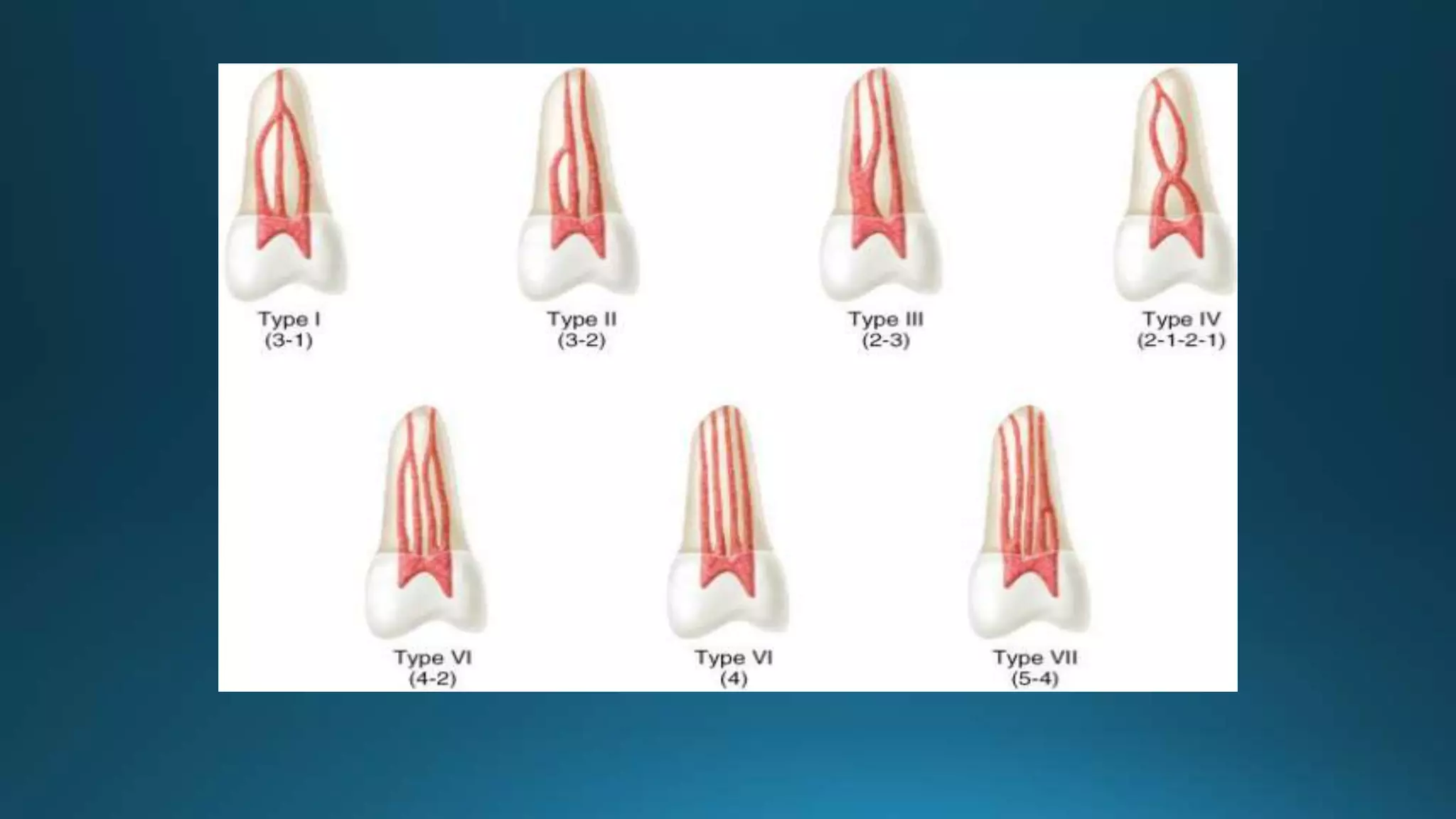
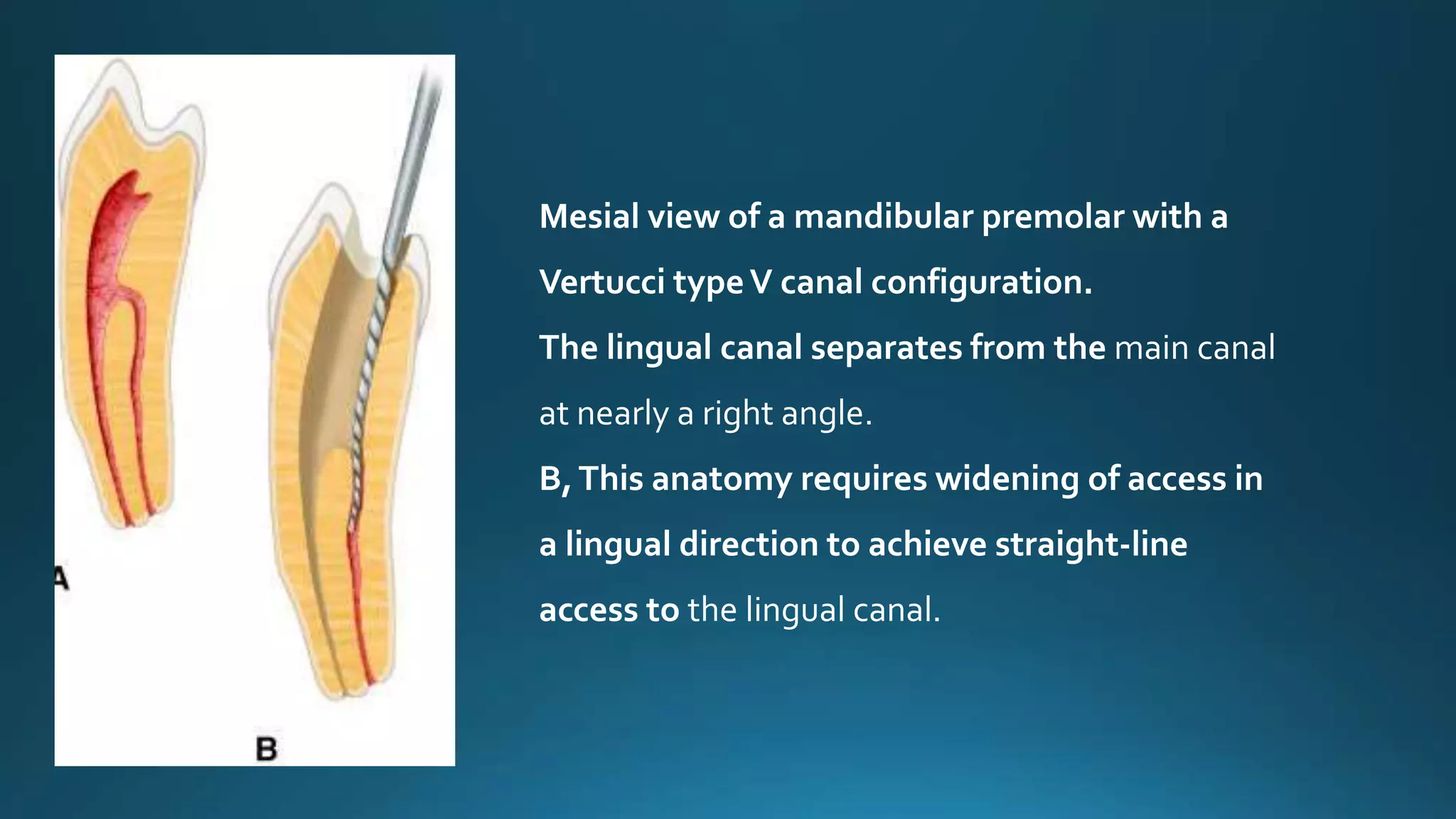
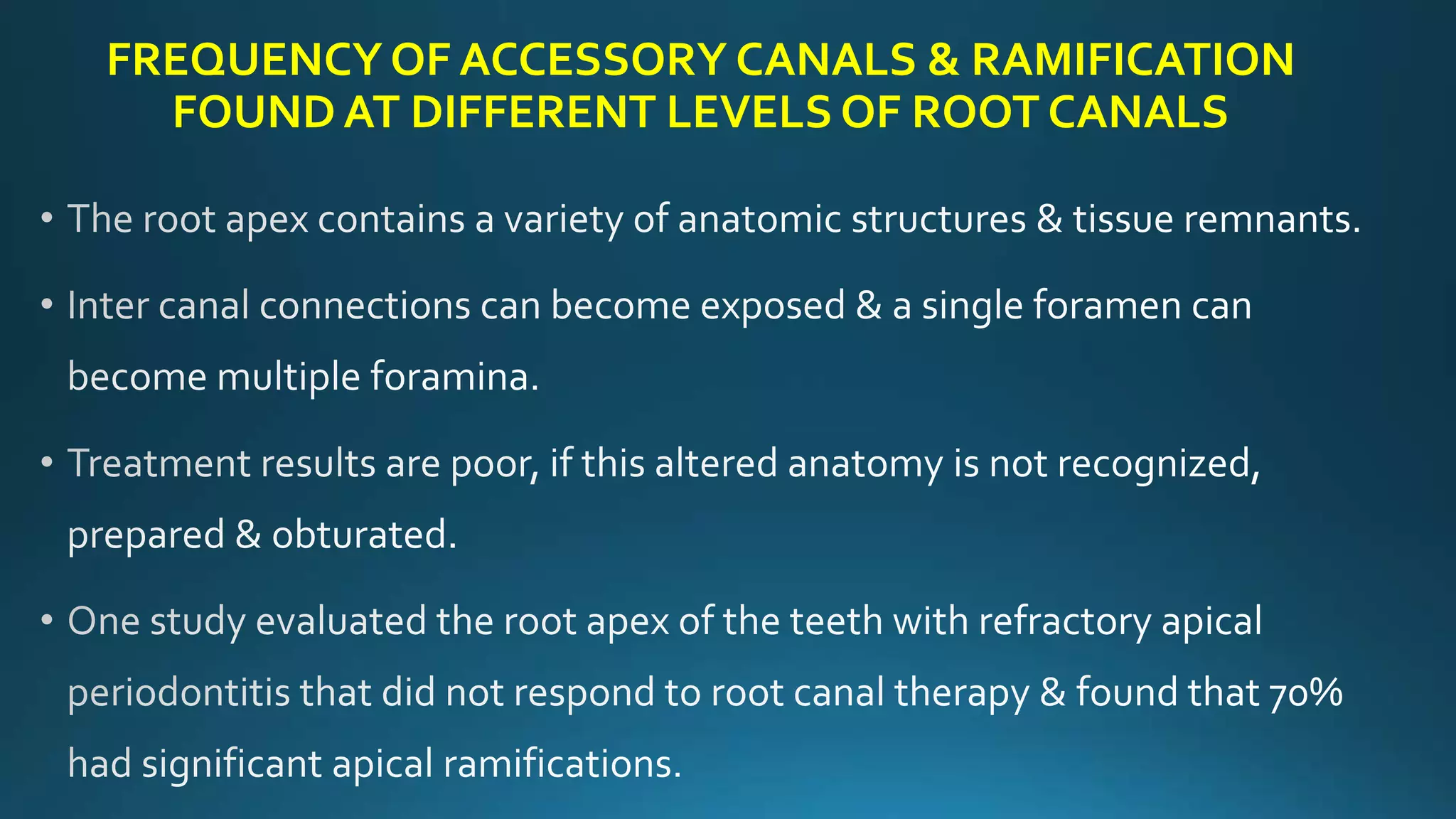


The document discusses the anatomy and morphology of the apical third of teeth roots. It notes that this region is the most complex part of teeth and is important prognostically for endodontic procedures. The apical third can display variations like accessory canals, isthmuses, additional root canals and foramen, and curved or ribbon-shaped canal systems. These anatomical variations make cleaning, shaping, filling and surgery in this region challenging for endodontists. Proper instrumentation techniques and materials are required to navigate the complex apical third anatomy.


