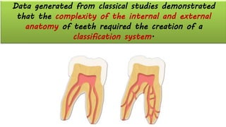
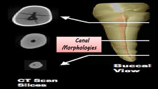
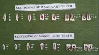
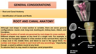
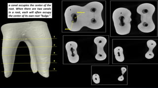
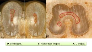
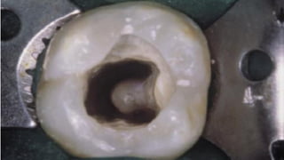
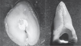
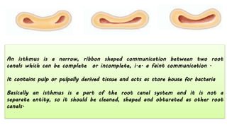
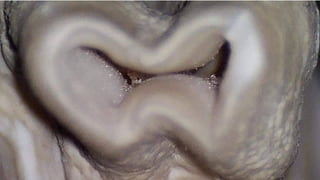
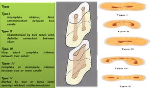
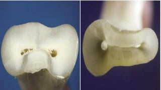
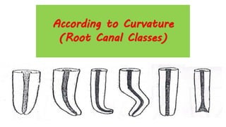
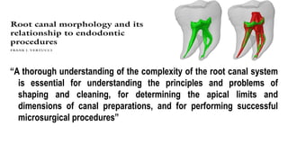
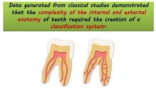
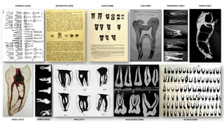
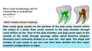
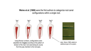
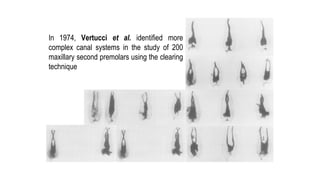
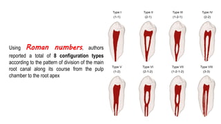
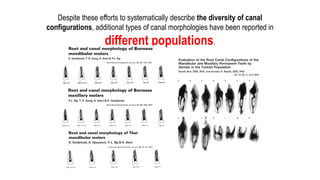
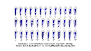
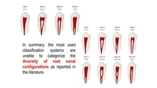
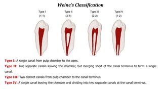
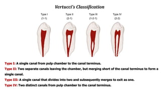
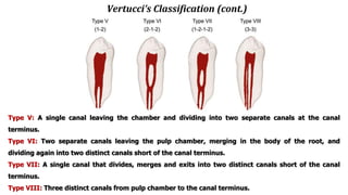
SlideShare a Scribd company logo
Dr. Hadil Abdallah Altilbani
BDS Santiago de Compostela University Spain.
MSc. University of Valencia Spain.
Department of Endodontics University of Palestine .
INTERNAL
ANATOMY
INTRODUCTION
In Terms Of Success Of Treatment,
✓ knowledge of pulp anatomy cannot be overstated.
✓ For the success of endodontic therapy, the knowledge of Pulp Anatomy Cannot Be Ruled Out.
✓ As a cause of treatment failures, Lack Of A Working Knowledge of pulp anatomy ranks second only
to errors in diagnosis and treatment planning.
✓ It is critical to know the Normal Or Usual Configuration of the pulp and to be aware of Variations.
✓ Special techniques are required to determine the internal anatomy of the tooth under treatment.
✓ Knowledge of the pulp anatomy must be Threedimensional.
✓ The Pulp Cavity Must Be Mentally Visualized Three Dimensionally.
✓ The pulp cavity must be mentally visualized both longitudinally (from coronal aspect to apical foramen)
and in cross section.
✓ In addition to general morphologic features, Irregularities And "Hidden" regions of pulp are present
within each canal.
✓ To clean and shape the pulp system maximally, intracanal instruments must reach as many of these
regions as possible to plane the walls to loosen tissue and tissue remmants.
✓ Lack of attention to this important principle may lead to treatment failure.
Data generated from classical studies demonstrated
that the complexity of the internal and external
anatomy of teeth required the creation of a
classification system.
Appreciable knowledge of the morphology of this ‘small zone’ and its variance, ability to interpret it
correctly in radiograph, and to ‘feel’ it through tactile sensation during instrumentation are essential for an
effective rendering of the treatment of root canals.
THE ROOT
APEX
Anatomy of the apical third
INTRODUCTION
✓ Morphologically-most complex region
✓ Therapeutically-most challenging zone
✓ Prognostically- most important part
✓ Radiographically-most obscure and
unclear area
Thorough comprehension of apical region
of tooth
is essential to determine the working length
and working width to the most accurate
position biologically .
Scrupulous understanding and knowledge
of the root apex is also a requisite to
perform a successful endodontic surgical
procedure.
A detailed knowledge of the apical part of
the root canal system is vital as it is a
common area for procedural errors during
nstrumentation
Significance of apical third
• The main problems associated with apical part of root
are its variability and unpredictability. Because of great
variation in size and shape, problems may occur during
the endodontic treatment.
• The root canal treatment of apical part of root is
difficult sometimes because of presence of accessory and
lateral canals, pulp stones, varying amounts of irregular
secondary dentin and areas of resorption.
• Most of curvatures occur in apical third, so one has
to be very careful while canal preparation.
• Obturation should end at apical constriction so as to
have optimal results of treatment.
• Apical 3 mm of root is generally resected during
endodontic surgery in order to eliminate canal
aberrations.
ANATOMY OF ROOT APEX
(Kuttler’s studies)
A.Anatomic apex
B.Apical constriction (minor diameter)
F. Apical foramen
The mean distance between the major and minor diameters
0.5 mm in a young person and 0.67 mm in an older individual.
The increased length in older individuals is due to the increased
buildup of cementum
GREEN(1955 1956 1960)-
Major apical foramen are situated directly at
the apex more frequently in maxillary
centrals, laterals, cuspids, first premolars and
mandibular second Premolars
In the maxillary molars and all the
mandibular teeth with the exception of the
2nd PM, the main apical foramina coincide
with the apexes less frequently.
Anatomy of Apical Canal
According to Kuttler, the narrowest diameter of
the canal is definitely not at the site of exiting of the
canal from the tooth but usually occurs within the dentin,
just prior to the initial layers of cementum.
He referred to this position as the minor diameter
of the canal, although others call it the apical constriction.
The diameter of the canal at the site of exiting from the
tooth (major diameter) was found to be approximately
twice as wide as minor diameter. This means that the
longitudinal view of the canal as a tapering funnel to the
tip of the root is incorrect.
Topography of the apical constriction
(DUMMER CLASSIFICATION)
1. Typical Single Constriction
2. Tapering Constriction With The Narrowest Portion Near The Actual Apex
3. Several Constrictions
4. Constriction Followed By A Narrow, Parallel Canal
5. Complete Blockage Of The Apical Canal By Secondary Dentin
1. TYPICAL SINGLE
CONSTRICTION
SEVERAL
CONSTRICTIONS
TYPE OF
ROOT
APEX
THIN PINCHED
APEX
proper care required during
instrumentation
Over enlargement may lead to perforation
BULBOUS APEX
usually due to hypercementosis
proper care required during length determination
Apical constriction is significantly shorter from radiographic apex
▪When heavy stress is placed on tooth, thickened amount of cementum is
elaborated, increasing the area of periodontal attachment and strengthening the
supporting mechanism - this increased deposition of cementum is in response to
function and is known as hypercementosis (hypertrophy).
RESORBED APEX
caused due to advanced
inflammation at the
periapex resorption of
cementum and dentin and
widening of apical
foramen
WL determination
,preparation and
condensation of gutta-
percha is difficult-
Preparation should stop
1-2mm short of
radiographic apex
BLUNDERBUSS APEX
newly erupted tooth showing an
incompletely formed root having a
wide canal and the pulp may get
necrosed due to caries or trauma
and may require root canal
standard instrumentation and
obturation techniques are not
favorable
Open Apex
Endodontic management of the pulpless,
permanent teeth with wide open blunder buss
apex offers dentists a most difficult condition
to treat.
Problem of open apex - the open apex occurs
when trauma or caries cause pulpal exposure
prior to the completion of root development.
An open apex refer to absence of sufficient root
development to provide a conical taper to the
canal - “Blunderbuss” canal.
Since it is necessary to seal the apex to gain
endodontic success, it is physically impossible to
achieve this objective through ordinary procedure
in open apex cases. www.indiandentalacademy.com
Canal
Morphologies
GENERAL CONSIDERATIONS
• Root and Canal Anatomy
• Identification of Canals and Roots
ROOT AND CANAL ANATOMY
Although root shape in cross section is variable, there are seven general
configurations: round, oval, long oval, bowling pin, kidney bean, ribbon, and
hourglass.
Shape and location of canals are governed by root shape (in cross section).
Different shapes may appear at any level in a single root. For example, a
root may be hourglass shaped in cross section at the cervical third, taper to
a deep oval in the middle third, and blend to oval in the apical third; the
number and shape of canals in each level will vary accordingly." Importantly
though, a canal is seldom round at any level.
To assume that it is may result in improper canal preparation
a canal occupies the center of the
root. When there are two canals
in a root, each will often occupy
the center of its own root “bulge.”
IDENTIFICATION OF CANALS AND
ROOTS
Obviously, to clean, shape, and obturate a canal, it must be
located."
In roots that may contain two canals, a basic rule is to
assume that the root contains two canals until proved
otherwise.
Rather than memorize roots that often contain two canals,
it is easier to remember those few that are unlikely to have
two canals.
Shape of the Canals -Torabineajad
6 different shapes have been noted
1. •Round
2. •Oval
3. •Deep oval
4. •Bowling pin
5. •Kidney bean
6. •Hour glass
Canal
Morphologies
They include round, ribbon or figure eight, ovoid, bowling pin, kidney bean, and C shape.
With the exception of the round morphologic shape, each presents unique problems f
or adequate cleaning and shaping.
A, Round. B, Ribbon-shaped (hourglass). C, Ovoid.
D, Bowling pin. E, Kidney bean-shaped. F, C-shaped.
It contains pulp or pulpally derived tissue and acts as store house for bacteria
Types
Type•I
Incomplete isthmus; faint
communication between two
canals.
Type• II
Characterized by two canals with
definite connection between
them.
Type• III
Very short complete isthmus
between two canals.
Types. IV
Complete or incomplete isthmus
between two or more canals.
Type. V
Marked by two or three canal
openings without visibleconnections
According to Curvature
(Root Canal Classes)
When tooth erupts
into oral cavity Its apex Is not
Completely formed
this slow Bodily movement of the incompletely
formed tooth is the cause of Curvatures in the
apical third of the root
as the tooth becomes functional it is
subjected to Biting stresses which may
move the tooth mesially
Curvature formation
Root Canal Curvatures
SCHNEIDER‘ sclassification on the basis of degree
of curvature
➢ Straight: 5 ̊or less
➢Moderate: 10°-20°
➢Severe: 25°-70
Introduction
“A thorough understanding of the complexity of the root canal system
is essential for understanding the principles and problems of
shaping and cleaning, for determining the apical limits and
dimensions of canal preparations, and for performing successful
microsurgical procedures”
Data generated from classical studies demonstrated
that the complexity of the internal and external
anatomy of teeth required the creation of a
classification system.
Introduction
CARABELLI (1842) MUHLREITER (1870) BLACK (1890) GYSI (1892) PREISWERCK (1901) FISHER (1907)
DEWEY (1916)
FASOLI (1913) HESS (1917) PUCCI & REIG (1944) DE DEUS (1960)
ROOT CANALS (PULP CANALS)
Root canals (pulp canals) are the portions of the pulp cavity located within
the root(s) of a tooth. Root canals connect to the pulp chamber through
canal orifices on the floor of the pulp chamber, and pulp canals open to the
outside of the tooth through openings called apical foramina (singular
foramen) most commonly located at or near the root apex. The shape and
number of root canals in any one root have been divided into four major
anatomic configurations or types.
Introduction
Weine et al. (1969) were the first authors to categorize root canal
configurations within a single root.
Type I
(1-1)
Type II
(2-1)
Type III
(2-2)
Type IV
(1-2)
Later, Weine (1982) added an
additional type to his system.
Using Roman numbers, configurations were
classified into 3 types according to the pattern of
division of the main root canal along its course
from the pulp chamber to the root apex.
Introduction
In 1974, Vertucci et al. identified more
complex canal systems in the study of 200
maxillary second premolars using the clearing
technique
Introduction
Using Roman numbers, authors
reported a total of 8 configuration types
according to the pattern of division of the main
root canal along its course from the pulp
chamber to the root apex
Introduction
Despite these efforts to systematically describe the diversity of canal
configurations, additional types of canal morphologies have been reported in
different populations.
Introduction
AhmedHMA, VersianiMA, De-Deus G, Dummer PMH. A new system for classifying root and root canal morphology. International Endodontic Journal 2016
Recently, based on previous reports and anatomical studies using micro-CT technology,
Versiani & Ordinola-Zapata (2015) were able to identify 37 types of root canal configuration.
In summary, the most used
classification systems are
unable to categorize the
diversity of root canal
configurations as reported in
the literature.
Introduction
• Root canal curvature (Schneider 1971, Pruett et al. 1997)
• Canal bifurcation and root fusion (Vertucci 2005)
• Accessory canals and apical ramifications (De Deus 1975)
• Dens invaginatus (Oehlers 1957)
• C-shaped canals (Melton et al. 1991, Fan et al. 2004, Kato et al. 2014)
• Taurodontism (Shaw 1928, Jafarzadeh et al. 2008)
• Supernumerary roots (Christie et al. 1991, Carlsen & Alexandersen 2000, Song et al. 2010)
• And others
Exclusion Criteria
Literature already has many comprehensive classifications categorizing several morphological
aspects and developmental anomalies of the teeth. Therefore, including the following
anatomical variations in the root and root canal system:
Despite such information could be useful, the benefits of any new system must be the
simplicity, so that it can be adopted universally!
Weine’s Classification
Type I: A single canal from pulp chamber to the apex.
Type II: Two separate canals leaving the chamber, but merging short of the canal terminus to form a single
canal.
Type III: Two distinct canals from pulp chamber to the canal terminus.
Type IV: A single canal leaving the chamber and dividing into two separate canals at the canal terminus.
Vertucci’s Classification
Type I: A single canal from pulp chamber to the canal terminus.
Type II: Two separate canals leaving the chamber, but merging short of the canal terminus to form a
single canal.
Type III: A single canal that divides into two and subsequently merges to exit as one.
Type IV: Two distinct canals from pulp chamber to the canal terminus.
Vertucci’s Classification (cont.)
Type V: A single canal leaving the chamber and dividing into two separate canals at the canal
terminus.
Type VI: Two separate canals leaving the pulp chamber, merging in the body of the root, and
dividing again into two distinct canals short of the canal terminus.
Type VII: A single canal that divides, merges and exits into two distinct canals short of the canal
terminus.
Type VIII: Three distinct canals from pulp chamber to the canal terminus.
CLASSIFICATION (1).pdf

More Related Content

Similar to CLASSIFICATION (1).pdf

Access preparation in special situations
Access preparation in special situationsAccess preparation in special situations
Access preparation in special situations
consendosbpdch
 
Anatomy of canines and premolars (ENDO)
Anatomy of canines and premolars  (ENDO)Anatomy of canines and premolars  (ENDO)
Anatomy of canines and premolars (ENDO)
Weam Faroun
 
Periodontal Flap Surgery
Periodontal Flap SurgeryPeriodontal Flap Surgery
Periodontal Flap SurgeryWendy Jeng
 
Access cavity preparation for maxillary canines
Access cavity preparation for maxillary caninesAccess cavity preparation for maxillary canines
Access cavity preparation for maxillary caninesKritika Sarkar
 
Access cavity ppt
Access cavity pptAccess cavity ppt
Access cavity ppt
nehasingh1671
 
Pulp cavities
Pulp cavitiesPulp cavities
Trouble shooting in endodontics
Trouble shooting in endodonticsTrouble shooting in endodontics
Trouble shooting in endodontics
Dr. Meenal Atharkar
 
Internal Anatomy of Pulp cavity
Internal Anatomy of Pulp cavityInternal Anatomy of Pulp cavity
Internal Anatomy of Pulp cavity
Arshad Shamsudeen
 
Internal anatomy of tooth
Internal  anatomy of  toothInternal  anatomy of  tooth
Internal anatomy of tooth
SNEHA RATNANI
 
Internal anatomy of permanent/ orthodontic course by indian dental academy
Internal anatomy of permanent/ orthodontic course by indian dental academyInternal anatomy of permanent/ orthodontic course by indian dental academy
Internal anatomy of permanent/ orthodontic course by indian dental academy
Indian dental academy
 
PULP SPACE (Dr. SONA)
PULP SPACE (Dr. SONA)PULP SPACE (Dr. SONA)
PULP SPACE (Dr. SONA)
MINDS MAHE
 
17
1717
management of non vital open apex roots/ orthodontic course by indian dental...
 management of non vital open apex roots/ orthodontic course by indian dental... management of non vital open apex roots/ orthodontic course by indian dental...
management of non vital open apex roots/ orthodontic course by indian dental...
Indian dental academy
 
Apex and its significance in endodontics
Apex and its significance in endodonticsApex and its significance in endodontics
Apex and its significance in endodontics
RockyJohn6
 
Root canal anatomy
Root canal anatomyRoot canal anatomy
Root canal anatomy
Lama K Banna
 
Anatomy of apical third /certified fixed orthodontic courses by Indian dental...
Anatomy of apical third /certified fixed orthodontic courses by Indian dental...Anatomy of apical third /certified fixed orthodontic courses by Indian dental...
Anatomy of apical third /certified fixed orthodontic courses by Indian dental...
Indian dental academy
 
Anatomy of apical third /certified fixed orthodontic courses by Indian dental...
Anatomy of apical third /certified fixed orthodontic courses by Indian dental...Anatomy of apical third /certified fixed orthodontic courses by Indian dental...
Anatomy of apical third /certified fixed orthodontic courses by Indian dental...
Indian dental academy
 
Access cavity preparation
Access cavity preparationAccess cavity preparation
Access cavity preparationIAU Dent
 
Determination of root canal working length /certified fixed orthodontic cours...
Determination of root canal working length /certified fixed orthodontic cours...Determination of root canal working length /certified fixed orthodontic cours...
Determination of root canal working length /certified fixed orthodontic cours...
Indian dental academy
 

Similar to CLASSIFICATION (1).pdf (20)

Access preparation in special situations
Access preparation in special situationsAccess preparation in special situations
Access preparation in special situations
 
Anatomy of canines and premolars (ENDO)
Anatomy of canines and premolars  (ENDO)Anatomy of canines and premolars  (ENDO)
Anatomy of canines and premolars (ENDO)
 
Periodontal Flap Surgery
Periodontal Flap SurgeryPeriodontal Flap Surgery
Periodontal Flap Surgery
 
Access cavity preparation for maxillary canines
Access cavity preparation for maxillary caninesAccess cavity preparation for maxillary canines
Access cavity preparation for maxillary canines
 
Access cavity ppt
Access cavity pptAccess cavity ppt
Access cavity ppt
 
Pulp cavities
Pulp cavitiesPulp cavities
Pulp cavities
 
Trouble shooting in endodontics
Trouble shooting in endodonticsTrouble shooting in endodontics
Trouble shooting in endodontics
 
Internal Anatomy of Pulp cavity
Internal Anatomy of Pulp cavityInternal Anatomy of Pulp cavity
Internal Anatomy of Pulp cavity
 
Internal anatomy of tooth
Internal  anatomy of  toothInternal  anatomy of  tooth
Internal anatomy of tooth
 
Internal anatomy of permanent/ orthodontic course by indian dental academy
Internal anatomy of permanent/ orthodontic course by indian dental academyInternal anatomy of permanent/ orthodontic course by indian dental academy
Internal anatomy of permanent/ orthodontic course by indian dental academy
 
PULP SPACE (Dr. SONA)
PULP SPACE (Dr. SONA)PULP SPACE (Dr. SONA)
PULP SPACE (Dr. SONA)
 
17
1717
17
 
17
1717
17
 
management of non vital open apex roots/ orthodontic course by indian dental...
 management of non vital open apex roots/ orthodontic course by indian dental... management of non vital open apex roots/ orthodontic course by indian dental...
management of non vital open apex roots/ orthodontic course by indian dental...
 
Apex and its significance in endodontics
Apex and its significance in endodonticsApex and its significance in endodontics
Apex and its significance in endodontics
 
Root canal anatomy
Root canal anatomyRoot canal anatomy
Root canal anatomy
 
Anatomy of apical third /certified fixed orthodontic courses by Indian dental...
Anatomy of apical third /certified fixed orthodontic courses by Indian dental...Anatomy of apical third /certified fixed orthodontic courses by Indian dental...
Anatomy of apical third /certified fixed orthodontic courses by Indian dental...
 
Anatomy of apical third /certified fixed orthodontic courses by Indian dental...
Anatomy of apical third /certified fixed orthodontic courses by Indian dental...Anatomy of apical third /certified fixed orthodontic courses by Indian dental...
Anatomy of apical third /certified fixed orthodontic courses by Indian dental...
 
Access cavity preparation
Access cavity preparationAccess cavity preparation
Access cavity preparation
 
Determination of root canal working length /certified fixed orthodontic cours...
Determination of root canal working length /certified fixed orthodontic cours...Determination of root canal working length /certified fixed orthodontic cours...
Determination of root canal working length /certified fixed orthodontic cours...
 

More from AltilbaniHadil

Disorders of the Dental Pulp.pptx
Disorders of the Dental Pulp.pptxDisorders of the Dental Pulp.pptx
Disorders of the Dental Pulp.pptx
AltilbaniHadil
 
PULP PERIAPICAL PATHOSIS. 111pptx.pdf
PULP  PERIAPICAL PATHOSIS. 111pptx.pdfPULP  PERIAPICAL PATHOSIS. 111pptx.pdf
PULP PERIAPICAL PATHOSIS. 111pptx.pdf
AltilbaniHadil
 
payalseminar-150519204542-lva1-app6892.pdf
payalseminar-150519204542-lva1-app6892.pdfpayalseminar-150519204542-lva1-app6892.pdf
payalseminar-150519204542-lva1-app6892.pdf
AltilbaniHadil
 
NiTi Briefly - Matt Brock(1).ppt
NiTi Briefly - Matt Brock(1).pptNiTi Briefly - Matt Brock(1).ppt
NiTi Briefly - Matt Brock(1).ppt
AltilbaniHadil
 
advancementsincleaningshaping-200815071852 (1).pptx
advancementsincleaningshaping-200815071852 (1).pptxadvancementsincleaningshaping-200815071852 (1).pptx
advancementsincleaningshaping-200815071852 (1).pptx
AltilbaniHadil
 
endodonticmotorsandrotaryfiles-220204115618.pptx
endodonticmotorsandrotaryfiles-220204115618.pptxendodonticmotorsandrotaryfiles-220204115618.pptx
endodonticmotorsandrotaryfiles-220204115618.pptx
AltilbaniHadil
 
differential_diagnosis_of_orofacial_pain.ppt
differential_diagnosis_of_orofacial_pain.pptdifferential_diagnosis_of_orofacial_pain.ppt
differential_diagnosis_of_orofacial_pain.ppt
AltilbaniHadil
 
teethdiscoloration-160429235959.pdf
teethdiscoloration-160429235959.pdfteethdiscoloration-160429235959.pdf
teethdiscoloration-160429235959.pdf
AltilbaniHadil
 
19387634-Discoloration.ppt
19387634-Discoloration.ppt19387634-Discoloration.ppt
19387634-Discoloration.ppt
AltilbaniHadil
 
revascularization.pdf
revascularization.pdfrevascularization.pdf
revascularization.pdf
AltilbaniHadil
 
Cracked Tooth Syndrome.pdf
Cracked Tooth Syndrome.pdfCracked Tooth Syndrome.pdf
Cracked Tooth Syndrome.pdf
AltilbaniHadil
 
The traumatic dental injuries.pdf
The traumatic  dental injuries.pdfThe traumatic  dental injuries.pdf
The traumatic dental injuries.pdf
AltilbaniHadil
 
Irrigation in Endodontics.pdf
Irrigation in Endodontics.pdfIrrigation in Endodontics.pdf
Irrigation in Endodontics.pdf
AltilbaniHadil
 
pulp protection.pdf
pulp protection.pdfpulp protection.pdf
pulp protection.pdf
AltilbaniHadil
 
horizontal fracture .pdf
horizontal fracture .pdfhorizontal fracture .pdf
horizontal fracture .pdf
AltilbaniHadil
 
tooth avulsion.pdf
tooth avulsion.pdftooth avulsion.pdf
tooth avulsion.pdf
AltilbaniHadil
 
Diagnostic Terminology.pdf
Diagnostic Terminology.pdfDiagnostic Terminology.pdf
Diagnostic Terminology.pdf
AltilbaniHadil
 
Cracks.pdf
Cracks.pdfCracks.pdf
Cracks.pdf
AltilbaniHadil
 
ACPrep.pdf
ACPrep.pdfACPrep.pdf
ACPrep.pdf
AltilbaniHadil
 
nitiendo-2-140902012325-phpapp02.pdf
nitiendo-2-140902012325-phpapp02.pdfnitiendo-2-140902012325-phpapp02.pdf
nitiendo-2-140902012325-phpapp02.pdf
AltilbaniHadil
 

More from AltilbaniHadil (20)

Disorders of the Dental Pulp.pptx
Disorders of the Dental Pulp.pptxDisorders of the Dental Pulp.pptx
Disorders of the Dental Pulp.pptx
 
PULP PERIAPICAL PATHOSIS. 111pptx.pdf
PULP  PERIAPICAL PATHOSIS. 111pptx.pdfPULP  PERIAPICAL PATHOSIS. 111pptx.pdf
PULP PERIAPICAL PATHOSIS. 111pptx.pdf
 
payalseminar-150519204542-lva1-app6892.pdf
payalseminar-150519204542-lva1-app6892.pdfpayalseminar-150519204542-lva1-app6892.pdf
payalseminar-150519204542-lva1-app6892.pdf
 
NiTi Briefly - Matt Brock(1).ppt
NiTi Briefly - Matt Brock(1).pptNiTi Briefly - Matt Brock(1).ppt
NiTi Briefly - Matt Brock(1).ppt
 
advancementsincleaningshaping-200815071852 (1).pptx
advancementsincleaningshaping-200815071852 (1).pptxadvancementsincleaningshaping-200815071852 (1).pptx
advancementsincleaningshaping-200815071852 (1).pptx
 
endodonticmotorsandrotaryfiles-220204115618.pptx
endodonticmotorsandrotaryfiles-220204115618.pptxendodonticmotorsandrotaryfiles-220204115618.pptx
endodonticmotorsandrotaryfiles-220204115618.pptx
 
differential_diagnosis_of_orofacial_pain.ppt
differential_diagnosis_of_orofacial_pain.pptdifferential_diagnosis_of_orofacial_pain.ppt
differential_diagnosis_of_orofacial_pain.ppt
 
teethdiscoloration-160429235959.pdf
teethdiscoloration-160429235959.pdfteethdiscoloration-160429235959.pdf
teethdiscoloration-160429235959.pdf
 
19387634-Discoloration.ppt
19387634-Discoloration.ppt19387634-Discoloration.ppt
19387634-Discoloration.ppt
 
revascularization.pdf
revascularization.pdfrevascularization.pdf
revascularization.pdf
 
Cracked Tooth Syndrome.pdf
Cracked Tooth Syndrome.pdfCracked Tooth Syndrome.pdf
Cracked Tooth Syndrome.pdf
 
The traumatic dental injuries.pdf
The traumatic  dental injuries.pdfThe traumatic  dental injuries.pdf
The traumatic dental injuries.pdf
 
Irrigation in Endodontics.pdf
Irrigation in Endodontics.pdfIrrigation in Endodontics.pdf
Irrigation in Endodontics.pdf
 
pulp protection.pdf
pulp protection.pdfpulp protection.pdf
pulp protection.pdf
 
horizontal fracture .pdf
horizontal fracture .pdfhorizontal fracture .pdf
horizontal fracture .pdf
 
tooth avulsion.pdf
tooth avulsion.pdftooth avulsion.pdf
tooth avulsion.pdf
 
Diagnostic Terminology.pdf
Diagnostic Terminology.pdfDiagnostic Terminology.pdf
Diagnostic Terminology.pdf
 
Cracks.pdf
Cracks.pdfCracks.pdf
Cracks.pdf
 
ACPrep.pdf
ACPrep.pdfACPrep.pdf
ACPrep.pdf
 
nitiendo-2-140902012325-phpapp02.pdf
nitiendo-2-140902012325-phpapp02.pdfnitiendo-2-140902012325-phpapp02.pdf
nitiendo-2-140902012325-phpapp02.pdf
 

Recently uploaded

Surgical Site Infections, pathophysiology, and prevention.pptx
Surgical Site Infections, pathophysiology, and prevention.pptxSurgical Site Infections, pathophysiology, and prevention.pptx
Surgical Site Infections, pathophysiology, and prevention.pptx
jval Landero
 
BENIGN PROSTATIC HYPERPLASIA.BPH. BPHpdf
BENIGN PROSTATIC HYPERPLASIA.BPH. BPHpdfBENIGN PROSTATIC HYPERPLASIA.BPH. BPHpdf
BENIGN PROSTATIC HYPERPLASIA.BPH. BPHpdf
DR SETH JOTHAM
 
TEST BANK for Operations Management, 14th Edition by William J. Stevenson, Ve...
TEST BANK for Operations Management, 14th Edition by William J. Stevenson, Ve...TEST BANK for Operations Management, 14th Edition by William J. Stevenson, Ve...
TEST BANK for Operations Management, 14th Edition by William J. Stevenson, Ve...
kevinkariuki227
 
Physiology of Special Chemical Sensation of Taste
Physiology of Special Chemical Sensation of TastePhysiology of Special Chemical Sensation of Taste
Physiology of Special Chemical Sensation of Taste
MedicoseAcademics
 
Flu Vaccine Alert in Bangalore Karnataka
Flu Vaccine Alert in Bangalore KarnatakaFlu Vaccine Alert in Bangalore Karnataka
Flu Vaccine Alert in Bangalore Karnataka
addon Scans
 
Prix Galien International 2024 Forum Program
Prix Galien International 2024 Forum ProgramPrix Galien International 2024 Forum Program
Prix Galien International 2024 Forum Program
Levi Shapiro
 
The Normal Electrocardiogram - Part I of II
The Normal Electrocardiogram - Part I of IIThe Normal Electrocardiogram - Part I of II
The Normal Electrocardiogram - Part I of II
MedicoseAcademics
 
Ocular injury ppt Upendra pal optometrist upums saifai etawah
Ocular injury  ppt  Upendra pal  optometrist upums saifai etawahOcular injury  ppt  Upendra pal  optometrist upums saifai etawah
Ocular injury ppt Upendra pal optometrist upums saifai etawah
pal078100
 
Couples presenting to the infertility clinic- Do they really have infertility...
Couples presenting to the infertility clinic- Do they really have infertility...Couples presenting to the infertility clinic- Do they really have infertility...
Couples presenting to the infertility clinic- Do they really have infertility...
Sujoy Dasgupta
 
Are There Any Natural Remedies To Treat Syphilis.pdf
Are There Any Natural Remedies To Treat Syphilis.pdfAre There Any Natural Remedies To Treat Syphilis.pdf
Are There Any Natural Remedies To Treat Syphilis.pdf
Little Cross Family Clinic
 
For Better Surat #ℂall #Girl Service ❤85270-49040❤ Surat #ℂall #Girls
For Better Surat #ℂall #Girl Service ❤85270-49040❤ Surat #ℂall #GirlsFor Better Surat #ℂall #Girl Service ❤85270-49040❤ Surat #ℂall #Girls
For Better Surat #ℂall #Girl Service ❤85270-49040❤ Surat #ℂall #Girls
Savita Shen $i11
 
Cervical & Brachial Plexus By Dr. RIG.pptx
Cervical & Brachial Plexus By Dr. RIG.pptxCervical & Brachial Plexus By Dr. RIG.pptx
Cervical & Brachial Plexus By Dr. RIG.pptx
Dr. Rabia Inam Gandapore
 
micro teaching on communication m.sc nursing.pdf
micro teaching on communication m.sc nursing.pdfmicro teaching on communication m.sc nursing.pdf
micro teaching on communication m.sc nursing.pdf
Anurag Sharma
 
KDIGO 2024 guidelines for diabetologists
KDIGO 2024 guidelines for diabetologistsKDIGO 2024 guidelines for diabetologists
KDIGO 2024 guidelines for diabetologists
د.محمود نجيب
 
Superficial & Deep Fascia of the NECK.pptx
Superficial & Deep Fascia of the NECK.pptxSuperficial & Deep Fascia of the NECK.pptx
Superficial & Deep Fascia of the NECK.pptx
Dr. Rabia Inam Gandapore
 
ARTIFICIAL INTELLIGENCE IN HEALTHCARE.pdf
ARTIFICIAL INTELLIGENCE IN  HEALTHCARE.pdfARTIFICIAL INTELLIGENCE IN  HEALTHCARE.pdf
ARTIFICIAL INTELLIGENCE IN HEALTHCARE.pdf
Anujkumaranit
 
heat stroke and heat exhaustion in children
heat stroke and heat exhaustion in childrenheat stroke and heat exhaustion in children
heat stroke and heat exhaustion in children
SumeraAhmad5
 
Evaluation of antidepressant activity of clitoris ternatea in animals
Evaluation of antidepressant activity of clitoris ternatea in animalsEvaluation of antidepressant activity of clitoris ternatea in animals
Evaluation of antidepressant activity of clitoris ternatea in animals
Shweta
 
Report Back from SGO 2024: What’s the Latest in Cervical Cancer?
Report Back from SGO 2024: What’s the Latest in Cervical Cancer?Report Back from SGO 2024: What’s the Latest in Cervical Cancer?
Report Back from SGO 2024: What’s the Latest in Cervical Cancer?
bkling
 
basicmodesofventilation2022-220313203758.pdf
basicmodesofventilation2022-220313203758.pdfbasicmodesofventilation2022-220313203758.pdf
basicmodesofventilation2022-220313203758.pdf
aljamhori teaching hospital
 

Recently uploaded (20)

Surgical Site Infections, pathophysiology, and prevention.pptx
Surgical Site Infections, pathophysiology, and prevention.pptxSurgical Site Infections, pathophysiology, and prevention.pptx
Surgical Site Infections, pathophysiology, and prevention.pptx
 
BENIGN PROSTATIC HYPERPLASIA.BPH. BPHpdf
BENIGN PROSTATIC HYPERPLASIA.BPH. BPHpdfBENIGN PROSTATIC HYPERPLASIA.BPH. BPHpdf
BENIGN PROSTATIC HYPERPLASIA.BPH. BPHpdf
 
TEST BANK for Operations Management, 14th Edition by William J. Stevenson, Ve...
TEST BANK for Operations Management, 14th Edition by William J. Stevenson, Ve...TEST BANK for Operations Management, 14th Edition by William J. Stevenson, Ve...
TEST BANK for Operations Management, 14th Edition by William J. Stevenson, Ve...
 
Physiology of Special Chemical Sensation of Taste
Physiology of Special Chemical Sensation of TastePhysiology of Special Chemical Sensation of Taste
Physiology of Special Chemical Sensation of Taste
 
Flu Vaccine Alert in Bangalore Karnataka
Flu Vaccine Alert in Bangalore KarnatakaFlu Vaccine Alert in Bangalore Karnataka
Flu Vaccine Alert in Bangalore Karnataka
 
Prix Galien International 2024 Forum Program
Prix Galien International 2024 Forum ProgramPrix Galien International 2024 Forum Program
Prix Galien International 2024 Forum Program
 
The Normal Electrocardiogram - Part I of II
The Normal Electrocardiogram - Part I of IIThe Normal Electrocardiogram - Part I of II
The Normal Electrocardiogram - Part I of II
 
Ocular injury ppt Upendra pal optometrist upums saifai etawah
Ocular injury  ppt  Upendra pal  optometrist upums saifai etawahOcular injury  ppt  Upendra pal  optometrist upums saifai etawah
Ocular injury ppt Upendra pal optometrist upums saifai etawah
 
Couples presenting to the infertility clinic- Do they really have infertility...
Couples presenting to the infertility clinic- Do they really have infertility...Couples presenting to the infertility clinic- Do they really have infertility...
Couples presenting to the infertility clinic- Do they really have infertility...
 
Are There Any Natural Remedies To Treat Syphilis.pdf
Are There Any Natural Remedies To Treat Syphilis.pdfAre There Any Natural Remedies To Treat Syphilis.pdf
Are There Any Natural Remedies To Treat Syphilis.pdf
 
For Better Surat #ℂall #Girl Service ❤85270-49040❤ Surat #ℂall #Girls
For Better Surat #ℂall #Girl Service ❤85270-49040❤ Surat #ℂall #GirlsFor Better Surat #ℂall #Girl Service ❤85270-49040❤ Surat #ℂall #Girls
For Better Surat #ℂall #Girl Service ❤85270-49040❤ Surat #ℂall #Girls
 
Cervical & Brachial Plexus By Dr. RIG.pptx
Cervical & Brachial Plexus By Dr. RIG.pptxCervical & Brachial Plexus By Dr. RIG.pptx
Cervical & Brachial Plexus By Dr. RIG.pptx
 
micro teaching on communication m.sc nursing.pdf
micro teaching on communication m.sc nursing.pdfmicro teaching on communication m.sc nursing.pdf
micro teaching on communication m.sc nursing.pdf
 
KDIGO 2024 guidelines for diabetologists
KDIGO 2024 guidelines for diabetologistsKDIGO 2024 guidelines for diabetologists
KDIGO 2024 guidelines for diabetologists
 
Superficial & Deep Fascia of the NECK.pptx
Superficial & Deep Fascia of the NECK.pptxSuperficial & Deep Fascia of the NECK.pptx
Superficial & Deep Fascia of the NECK.pptx
 
ARTIFICIAL INTELLIGENCE IN HEALTHCARE.pdf
ARTIFICIAL INTELLIGENCE IN  HEALTHCARE.pdfARTIFICIAL INTELLIGENCE IN  HEALTHCARE.pdf
ARTIFICIAL INTELLIGENCE IN HEALTHCARE.pdf
 
heat stroke and heat exhaustion in children
heat stroke and heat exhaustion in childrenheat stroke and heat exhaustion in children
heat stroke and heat exhaustion in children
 
Evaluation of antidepressant activity of clitoris ternatea in animals
Evaluation of antidepressant activity of clitoris ternatea in animalsEvaluation of antidepressant activity of clitoris ternatea in animals
Evaluation of antidepressant activity of clitoris ternatea in animals
 
Report Back from SGO 2024: What’s the Latest in Cervical Cancer?
Report Back from SGO 2024: What’s the Latest in Cervical Cancer?Report Back from SGO 2024: What’s the Latest in Cervical Cancer?
Report Back from SGO 2024: What’s the Latest in Cervical Cancer?
 
basicmodesofventilation2022-220313203758.pdf
basicmodesofventilation2022-220313203758.pdfbasicmodesofventilation2022-220313203758.pdf
basicmodesofventilation2022-220313203758.pdf
 

CLASSIFICATION (1).pdf

  • 1. Dr. Hadil Abdallah Altilbani BDS Santiago de Compostela University Spain. MSc. University of Valencia Spain. Department of Endodontics University of Palestine . INTERNAL ANATOMY
  • 2.
  • 3.
  • 4. INTRODUCTION In Terms Of Success Of Treatment, ✓ knowledge of pulp anatomy cannot be overstated. ✓ For the success of endodontic therapy, the knowledge of Pulp Anatomy Cannot Be Ruled Out. ✓ As a cause of treatment failures, Lack Of A Working Knowledge of pulp anatomy ranks second only to errors in diagnosis and treatment planning. ✓ It is critical to know the Normal Or Usual Configuration of the pulp and to be aware of Variations. ✓ Special techniques are required to determine the internal anatomy of the tooth under treatment. ✓ Knowledge of the pulp anatomy must be Threedimensional. ✓ The Pulp Cavity Must Be Mentally Visualized Three Dimensionally. ✓ The pulp cavity must be mentally visualized both longitudinally (from coronal aspect to apical foramen) and in cross section. ✓ In addition to general morphologic features, Irregularities And "Hidden" regions of pulp are present within each canal. ✓ To clean and shape the pulp system maximally, intracanal instruments must reach as many of these regions as possible to plane the walls to loosen tissue and tissue remmants. ✓ Lack of attention to this important principle may lead to treatment failure.
  • 5. Data generated from classical studies demonstrated that the complexity of the internal and external anatomy of teeth required the creation of a classification system.
  • 6. Appreciable knowledge of the morphology of this ‘small zone’ and its variance, ability to interpret it correctly in radiograph, and to ‘feel’ it through tactile sensation during instrumentation are essential for an effective rendering of the treatment of root canals. THE ROOT APEX Anatomy of the apical third
  • 7. INTRODUCTION ✓ Morphologically-most complex region ✓ Therapeutically-most challenging zone ✓ Prognostically- most important part ✓ Radiographically-most obscure and unclear area
  • 8. Thorough comprehension of apical region of tooth is essential to determine the working length and working width to the most accurate position biologically . Scrupulous understanding and knowledge of the root apex is also a requisite to perform a successful endodontic surgical procedure. A detailed knowledge of the apical part of the root canal system is vital as it is a common area for procedural errors during nstrumentation
  • 9.
  • 10. Significance of apical third • The main problems associated with apical part of root are its variability and unpredictability. Because of great variation in size and shape, problems may occur during the endodontic treatment. • The root canal treatment of apical part of root is difficult sometimes because of presence of accessory and lateral canals, pulp stones, varying amounts of irregular secondary dentin and areas of resorption. • Most of curvatures occur in apical third, so one has to be very careful while canal preparation. • Obturation should end at apical constriction so as to have optimal results of treatment. • Apical 3 mm of root is generally resected during endodontic surgery in order to eliminate canal aberrations.
  • 11. ANATOMY OF ROOT APEX (Kuttler’s studies) A.Anatomic apex B.Apical constriction (minor diameter) F. Apical foramen The mean distance between the major and minor diameters 0.5 mm in a young person and 0.67 mm in an older individual. The increased length in older individuals is due to the increased buildup of cementum
  • 12.
  • 13.
  • 14.
  • 15. GREEN(1955 1956 1960)- Major apical foramen are situated directly at the apex more frequently in maxillary centrals, laterals, cuspids, first premolars and mandibular second Premolars In the maxillary molars and all the mandibular teeth with the exception of the 2nd PM, the main apical foramina coincide with the apexes less frequently.
  • 16. Anatomy of Apical Canal According to Kuttler, the narrowest diameter of the canal is definitely not at the site of exiting of the canal from the tooth but usually occurs within the dentin, just prior to the initial layers of cementum. He referred to this position as the minor diameter of the canal, although others call it the apical constriction. The diameter of the canal at the site of exiting from the tooth (major diameter) was found to be approximately twice as wide as minor diameter. This means that the longitudinal view of the canal as a tapering funnel to the tip of the root is incorrect.
  • 17.
  • 18. Topography of the apical constriction (DUMMER CLASSIFICATION) 1. Typical Single Constriction 2. Tapering Constriction With The Narrowest Portion Near The Actual Apex 3. Several Constrictions 4. Constriction Followed By A Narrow, Parallel Canal 5. Complete Blockage Of The Apical Canal By Secondary Dentin
  • 19.
  • 22.
  • 24. THIN PINCHED APEX proper care required during instrumentation Over enlargement may lead to perforation
  • 25.
  • 26. BULBOUS APEX usually due to hypercementosis proper care required during length determination Apical constriction is significantly shorter from radiographic apex
  • 27. ▪When heavy stress is placed on tooth, thickened amount of cementum is elaborated, increasing the area of periodontal attachment and strengthening the supporting mechanism - this increased deposition of cementum is in response to function and is known as hypercementosis (hypertrophy).
  • 28. RESORBED APEX caused due to advanced inflammation at the periapex resorption of cementum and dentin and widening of apical foramen WL determination ,preparation and condensation of gutta- percha is difficult- Preparation should stop 1-2mm short of radiographic apex
  • 29.
  • 30.
  • 31. BLUNDERBUSS APEX newly erupted tooth showing an incompletely formed root having a wide canal and the pulp may get necrosed due to caries or trauma and may require root canal standard instrumentation and obturation techniques are not favorable
  • 32. Open Apex Endodontic management of the pulpless, permanent teeth with wide open blunder buss apex offers dentists a most difficult condition to treat. Problem of open apex - the open apex occurs when trauma or caries cause pulpal exposure prior to the completion of root development. An open apex refer to absence of sufficient root development to provide a conical taper to the canal - “Blunderbuss” canal. Since it is necessary to seal the apex to gain endodontic success, it is physically impossible to achieve this objective through ordinary procedure in open apex cases. www.indiandentalacademy.com
  • 33.
  • 34.
  • 35.
  • 37.
  • 38. GENERAL CONSIDERATIONS • Root and Canal Anatomy • Identification of Canals and Roots ROOT AND CANAL ANATOMY Although root shape in cross section is variable, there are seven general configurations: round, oval, long oval, bowling pin, kidney bean, ribbon, and hourglass. Shape and location of canals are governed by root shape (in cross section). Different shapes may appear at any level in a single root. For example, a root may be hourglass shaped in cross section at the cervical third, taper to a deep oval in the middle third, and blend to oval in the apical third; the number and shape of canals in each level will vary accordingly." Importantly though, a canal is seldom round at any level. To assume that it is may result in improper canal preparation
  • 39. a canal occupies the center of the root. When there are two canals in a root, each will often occupy the center of its own root “bulge.”
  • 40. IDENTIFICATION OF CANALS AND ROOTS Obviously, to clean, shape, and obturate a canal, it must be located." In roots that may contain two canals, a basic rule is to assume that the root contains two canals until proved otherwise. Rather than memorize roots that often contain two canals, it is easier to remember those few that are unlikely to have two canals.
  • 41.
  • 42. Shape of the Canals -Torabineajad 6 different shapes have been noted 1. •Round 2. •Oval 3. •Deep oval 4. •Bowling pin 5. •Kidney bean 6. •Hour glass Canal Morphologies
  • 43. They include round, ribbon or figure eight, ovoid, bowling pin, kidney bean, and C shape. With the exception of the round morphologic shape, each presents unique problems f or adequate cleaning and shaping. A, Round. B, Ribbon-shaped (hourglass). C, Ovoid.
  • 44. D, Bowling pin. E, Kidney bean-shaped. F, C-shaped.
  • 45.
  • 46.
  • 47.
  • 48. It contains pulp or pulpally derived tissue and acts as store house for bacteria
  • 49.
  • 50. Types Type•I Incomplete isthmus; faint communication between two canals. Type• II Characterized by two canals with definite connection between them. Type• III Very short complete isthmus between two canals. Types. IV Complete or incomplete isthmus between two or more canals. Type. V Marked by two or three canal openings without visibleconnections
  • 51.
  • 53.
  • 54. When tooth erupts into oral cavity Its apex Is not Completely formed this slow Bodily movement of the incompletely formed tooth is the cause of Curvatures in the apical third of the root as the tooth becomes functional it is subjected to Biting stresses which may move the tooth mesially Curvature formation
  • 55. Root Canal Curvatures SCHNEIDER‘ sclassification on the basis of degree of curvature ➢ Straight: 5 ̊or less ➢Moderate: 10°-20° ➢Severe: 25°-70
  • 56.
  • 57.
  • 58. Introduction “A thorough understanding of the complexity of the root canal system is essential for understanding the principles and problems of shaping and cleaning, for determining the apical limits and dimensions of canal preparations, and for performing successful microsurgical procedures”
  • 59. Data generated from classical studies demonstrated that the complexity of the internal and external anatomy of teeth required the creation of a classification system.
  • 60. Introduction CARABELLI (1842) MUHLREITER (1870) BLACK (1890) GYSI (1892) PREISWERCK (1901) FISHER (1907) DEWEY (1916) FASOLI (1913) HESS (1917) PUCCI & REIG (1944) DE DEUS (1960)
  • 61. ROOT CANALS (PULP CANALS) Root canals (pulp canals) are the portions of the pulp cavity located within the root(s) of a tooth. Root canals connect to the pulp chamber through canal orifices on the floor of the pulp chamber, and pulp canals open to the outside of the tooth through openings called apical foramina (singular foramen) most commonly located at or near the root apex. The shape and number of root canals in any one root have been divided into four major anatomic configurations or types.
  • 62. Introduction Weine et al. (1969) were the first authors to categorize root canal configurations within a single root. Type I (1-1) Type II (2-1) Type III (2-2) Type IV (1-2) Later, Weine (1982) added an additional type to his system. Using Roman numbers, configurations were classified into 3 types according to the pattern of division of the main root canal along its course from the pulp chamber to the root apex.
  • 63. Introduction In 1974, Vertucci et al. identified more complex canal systems in the study of 200 maxillary second premolars using the clearing technique
  • 64. Introduction Using Roman numbers, authors reported a total of 8 configuration types according to the pattern of division of the main root canal along its course from the pulp chamber to the root apex
  • 65. Introduction Despite these efforts to systematically describe the diversity of canal configurations, additional types of canal morphologies have been reported in different populations.
  • 66. Introduction AhmedHMA, VersianiMA, De-Deus G, Dummer PMH. A new system for classifying root and root canal morphology. International Endodontic Journal 2016 Recently, based on previous reports and anatomical studies using micro-CT technology, Versiani & Ordinola-Zapata (2015) were able to identify 37 types of root canal configuration.
  • 67. In summary, the most used classification systems are unable to categorize the diversity of root canal configurations as reported in the literature. Introduction
  • 68. • Root canal curvature (Schneider 1971, Pruett et al. 1997) • Canal bifurcation and root fusion (Vertucci 2005) • Accessory canals and apical ramifications (De Deus 1975) • Dens invaginatus (Oehlers 1957) • C-shaped canals (Melton et al. 1991, Fan et al. 2004, Kato et al. 2014) • Taurodontism (Shaw 1928, Jafarzadeh et al. 2008) • Supernumerary roots (Christie et al. 1991, Carlsen & Alexandersen 2000, Song et al. 2010) • And others Exclusion Criteria Literature already has many comprehensive classifications categorizing several morphological aspects and developmental anomalies of the teeth. Therefore, including the following anatomical variations in the root and root canal system: Despite such information could be useful, the benefits of any new system must be the simplicity, so that it can be adopted universally!
  • 69. Weine’s Classification Type I: A single canal from pulp chamber to the apex. Type II: Two separate canals leaving the chamber, but merging short of the canal terminus to form a single canal. Type III: Two distinct canals from pulp chamber to the canal terminus. Type IV: A single canal leaving the chamber and dividing into two separate canals at the canal terminus.
  • 70. Vertucci’s Classification Type I: A single canal from pulp chamber to the canal terminus. Type II: Two separate canals leaving the chamber, but merging short of the canal terminus to form a single canal. Type III: A single canal that divides into two and subsequently merges to exit as one. Type IV: Two distinct canals from pulp chamber to the canal terminus.
  • 71. Vertucci’s Classification (cont.) Type V: A single canal leaving the chamber and dividing into two separate canals at the canal terminus. Type VI: Two separate canals leaving the pulp chamber, merging in the body of the root, and dividing again into two distinct canals short of the canal terminus. Type VII: A single canal that divides, merges and exits into two distinct canals short of the canal terminus. Type VIII: Three distinct canals from pulp chamber to the canal terminus.