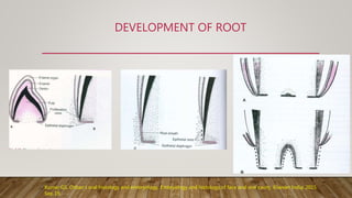
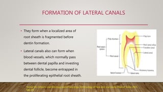
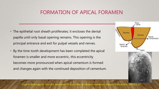
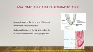
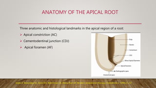
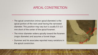
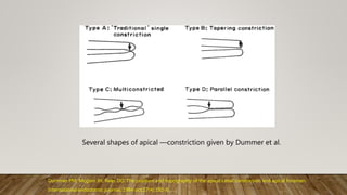
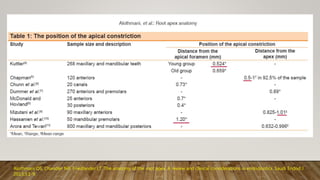
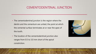
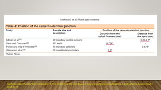
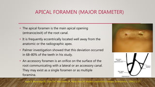
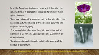
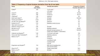
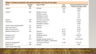
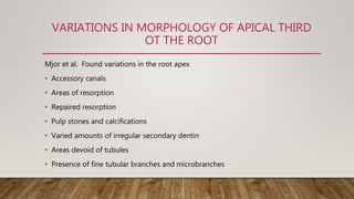
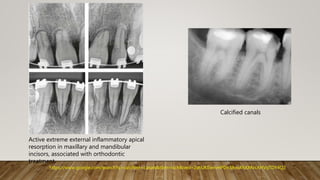
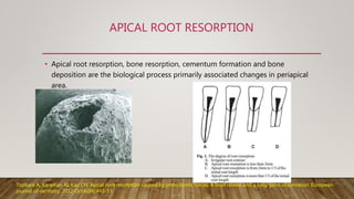
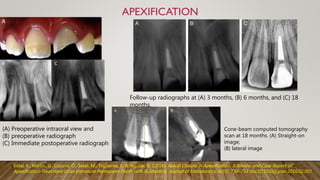
STRUCTURE OF
ROOT APEX
Presented by,
Swapnika.G.
(1 MDS)
Conservative Dentistry and Endodontics
CONTENTS
• Introduction
• Development of the structures in the root apex
• Histology of root apex
• Anatomy of the apical third of the root
• Variations in morphology of apical third
• Clinical significance
• Conclusion
• References
INTRODUCTION
• Anatomy is the foundation of art and science of healing.
• Of all the phases of anatomic study in human system, one of the most
complex is root canal system.
• The terminal part of root canal is the center of most activity and concern in
the treatment and filling of root canal.
Akashi CH, Geeta A, Girish P, Rrakesh V, Manjit K.Significance of Apical third.Scholars Journal of Applied Medical Sciences: 2014;
2(5B):1613-1617.
DEVELOPMENT OF ROOT
Kumar GS. Orban's oral histology and embryology. Embryology and histology of face and oral cavity. Elsevier India; 2015
Sep 19.
FORMATION OF LATERAL CANALS
• They form when a localized area of
root sheath is fragmented before
dentin formation.
• Lateral canals also can form when
blood vessels, which normally pass
between dental papilla and investing
dental follicle, become entrapped in
the proliferating epithelial root sheath.
Kumar GS. Orban's oral histology and embryology. Embryology of face and oral cavity. Elsevier India; 2015
Sep 19.
FORMATION OF ACCESSORY CANALS
Defect in the epithelial root
sheath
Failure in the induction of
dentinogenesis
Presence of blood
vessels
Ingle JI, Beveridge EE, Glick DH, Weichman JA, Abou-Rass M. Modern endodontic therapy. Endodontics. 1994;2:1-57.
FORMATION OF APICAL FORAMEN
• The epithelial root sheath proliferates; it encloses the dental
papilla until only basal opening remains. This opening is the
principal entrance and exit for pulpal vessels and nerves.
• By the time tooth development has been completed the apical
foramen is smaller and more eccentric, this eccentricity
becomes more pronounced when apical cementum is formed
and changes again with the continued deposition of cementum.
Ingle JI, Beveridge EE, Glick DH, Weichman JA, Abou-Rass M. Modern endodontic therapy. Endodontics. 1994;2:1-57.
Root completion (apical 1/3)
1year
ERUPTION
Canine 6-12 months
incisor 4-6 months
crown completion
ROOT COMPLETION
3 years
ERUPTION
3 years
crown completion
Time required for
development of primary
teeth
Time requirement for
development of
permanent teeth
Allen KL. Wheeler's Dental Anatomy, Physiology, and Occlusion. New York State Dental Journal. 2003 Nov 1;69(9):58.
HISTOLOGY OF ROOT APEX
• Primary dentinal tubules were found less frequently than in coronal dentine and were more
or less irregular in direction and density, some areas were completely devoid of tubules.
• Fine tubular branches (300-700 um in diameter) which run at 450 to the main tubules and
microtubules (25-200 um diameter) which run at 900 to the main tubules were frequently
present.
• This variable structure in the apical region presents challenges for root canal therapy.
Obturation techniques which rely on the penetration of adhesives into dentinal tubules may
not be successful.
ANATOMIC APEX AND RADIOGRAPHIC APEX
• Anatomic apex is the tip or end of the root
determined morphologically.
• Radiographic apex is the tip and end of the
of the root determined radio- graphically.
https://www.dentistryiq.com/home/contact/16373666/allan-s-deutsch-dmdRoot canal treatment: Where does the apex end?
ANATOMY OF THE APICAL ROOT
Three anatomic and histological landmarks in the apical region of a root:
 Apical constriction (AC)
 Cementodentinal junction (CDJ)
 Apical foramen (AF)
Ingle JI, Beveridge EE, Glick DH, Weichman JA, Abou-Rass M. Modern endodontic therapy. Endodontics. 1994;2:1-57.
APICAL CONSTRICTION
• The apical constriction (minor apical diameter) is the
apical portion of the root canal having the narrowest
diameter. This position may vary but is usually 0.5–1.0
mm short of the center of the apical foramen.
• The minor diameter widens apically toward the foramen
(major diameter) and assumes a funnel shape.
• Dummer and his associates reported many variations in
the apical constriction.
Deutsch AS. Endodontic insight: Where should the gutta-percha point end for optimal endodontic success?; Jan 7th 2017.
Several shapes of apical ―constriction given by Dummer et al.
Dummer PM, Mcginn JH, Rees DG. The position and topography of the apical canal constriction and apical foramen.
International endodontic journal. 1984 oct;17(4):192-8.
Alothmani OS, Chandler NP, Friedlander LT. The anatomy of the root apex: A review and clinical considerations in endodontics. Saudi Endod J
2013;3:1-9.
CEMENTODENTINAL JUNCTION
• The cementodentinal junction is the region where the
dentin and the cementum are united, the point at which
the cemental surface terminates at or near the apex of
the tooth.
• The location of the cementodentinal junction also
ranges from 0.5 to 3.0 mm short of the apical
constriction.
Yamamoto T, Domon T, Takahashi S, Islam H, Suzuki F, Wakita M. The structure and function of the cemento—dentinal junction in
human teeth. Journal of periodontal research. 1999 Jul;34(5):261-8.
Alothmani OS, Chandler NP, Friedlander LT. The anatomy of the root apex: A review and clinical considerations in endodontics. Saudi Endod J
2013;3:1-9.
APICAL FORAMEN (MAJOR DIAMETER)
• The apical foramen is the main apical opening
(entrance/exit) of the root canal.
• It is frequently eccentrically located well away from the
anatomic or the radiographic apex.
• Palmer investigation showed that this deviation occurred
in 68–80% of the teeth in his study.
• An accessory foramen is an orifice on the surface of the
root communicating with a lateral or an accessory canal.
They may exist as a single foramen or as multiple
foramina.
Ingle JI, Beveridge EE, Glick DH, Weichman JA, Abou-Rass M. Modern endodontic therapy. Endodontics. 1994;2:1-57.
• From the Apical constriction or minor apical diameter, the
canal widens as it approaches the apical foramen or major
apical diameter.
• The space between the major and minor diameters has been
described as funnel shaped or hyperbolic or as having the
shape of a morning glory.
• The mean distance between the major and minor apical
diameters is 0.5 mm in a young person and 0.67 mm in an
older individual.
• The distance is greater in older individuals because of the
buildup of cementum.
Ingle JI, Beveridge EE, Glick DH, Weichman JA, Abou-Rass M. Modern endodontic therapy. Endodontics. 1994;2:1-57.
Alothmani OS, Chandler NP, Friedlander LT. The anatomy of the root apex: A review and clinical considerations in endodontics. Saudi Endod J
2013;3:1-9.
APICAL DELTA
• Describes the primary or
secondary canals that terminates
short of apex with lateral canals
fanning out from the point to the
end of the root surface.
Gao, Xianhua & Tay, Frank & Gutmann, James & Fan, Wei & Xu, Ting & Fan, Bing. (2016). Micro-CT evaluation of apical delta morphologies in
human teeth. Scientific Reports. 6. 10.1038/srep36501.
VARIATIONS IN MORPHOLOGY OF APICAL THIRD
OT THE ROOT
Mjor et al. Found variations in the root apex
• Accessory canals
• Areas of resorption
• Repaired resorption
• Pulp stones and calcifications
• Varied amounts of irregular secondary dentin
• Areas devoid of tubules
• Presence of fine tubular branches and microbranches
Active extreme external inflammatory apical
resorption in maxillary and mandibular
incisors, associated with orthodontic
treatment.
Calcified canals
https://www.google.com/search?q=calcified+canals&tbm=isch&ved=2ahUKEwinmPDn38voAhXXKrcAHVqTDY4Q2
Weine’s classification
Vertucci's classification
Vertucci FJ. Root canal morphology and its relationship to endodontic procedures. Endodontic topics. 2005 Mar;10(1):3-29.
ISTHMUS
Kim et al. identified five types of isthmi that can be
found on a beveled root surface.
Type 1
Type 2
Type 3
Type 4
Type 5
Kim SY, Kim BS, Woo J, Kim Y. Morphology of mandibular first molars analyzed by cone-beam computed tomography in a Korean
population: variations in the number of roots and canals. J Endod. 2013;39:1516-21.
CLINICAL SIGNIFICANCE
• Working length determination
• Apical gauging
• Apical patency
• Apical root resection
• Apical third fracture
• Apical root resorption
• Apexification & apexogenesis
WORKING LENGTH DETERMINATION
Serene T, Krasny R, Ziegler P, et al. Principles of Preclinical Endodontics. Dubuque, IA: Kendall/Hunt Publishing; 1974.
Ingle JI, Beveridge EE, Glick DH, Weichman JA, Abou-Rass M. Modern endodontic therapy. Endodontics. 1994;2:1-57.
Dilacerated roots towards the buccal or lingual directions were determined by
evaluating the appearance of a round opaque area with a dark shadow in its central
region, the apical foramen in which the root canal gave a 'bulls-eye' appearance.
APICAL GAUGING
• Apical gauging is a technique to best determine the
size of the apical constriction and the taper of the
apical portion closest to the foramen.
• Apical gauging helps with:
 Choosing the best master cone that closely matches canal
length and taper.
 Achieving true tug back
 Minimizing GP extrusions during obturation, especially
with warm vertical compaction/ condensation.
Deutsch AS. Endodontic insight: Where should the gutta-percha point end for optimal endodontic success?; Jan 7th
APICAL PATENCY
Apical patency is leaving the apical foramen accessible, free from dentin chips, pulp
fragments, and other debris, as well as bacteria.
• Advantages of patency filing
 Establishment and Maintenance of Glide path
 Provides the clinician with knowledge of the anatomy of the apical root curvature
 Facilitates Length Determination
 To improve the efficiency of irrigation at the apical third level
 Minimizes apical blockage and loss of length
Khatavkar, roheet & hegde, vivek. (2010). Importance of patency in endodontics. Endodontology. 22. 85-91.
APICAL ROOT RESECTION
Color Atlas of Microsurgery in Endodontics, by S. Kim with G. Pecora and R. Rubinstein. W.B. Saunders Co., A Harcourt Health
Sciences Company, 2001.
APICAL THIRD FRACTURE
• A tooth whose root is fractured in its apical third has an excellent
prognosis because the pulp in the apical fragment usually remains vital,
and the tooth may remain firm in its socket.
• Mobile tooth should be ligated if the pulp in the coronal fragment
remains vital and the tooth is stable, with or without ligation, then no
additional treatment will be indicated.
• In the event that the pulp in the coronal fragment dies, then endodontic
treatment can be done, preferably limited to the coronal fragment.
• If the tooth fails to recover, the apical root fragment can be removed
surgically.Andreasen JO, Andreasen FM, Andersson L, editors. Textbook and color atlas of traumatic injuries to the teeth. John Wiley & Sons;
2018 Dec 17; 2nd ed, chapter 32 ,pages 842-3.
APICAL ROOT RESORPTION
• Apical root resorption, bone resorption, cementum formation and bone
deposition are the biological process primarily associated changes in periapical
area.
Topkara A, Karaman AI, Kau CH. Apical root resorption caused by orthodontic forces: A brief review and a long-term observation. European
journal of dentistry. 2012 Oct;6(04):445-53.
APEXOGENESIS
Forghani M, Parisay I, Maghsoudlou A. Apexogenesis and revascularization treatment procedures for two traumatized immature
permanent maxillary incisors: a case report. Restorative dentistry & endodontics. 2013 Aug 1;38(3):178-81.
Cone-beam computed tomography
scan at 18 months. (A) Straight-on
image;
(B) lateral image
Follow-up radiographs at (A) 3 months, (B) 6 months, and (C) 18
months.
c
(A) Preoperative intraoral view and
(B) preoperative radiograph
(C) Immediate postoperative radiograph
Vidal, K., Martin, G., Lozano, O., Salas, M., Trigueros, J., & Aguilar, G. (2016). Apical Closure in Apexification: A Review and Case Report of
Apexification Treatment of an Immature Permanent Tooth with Biodentine. Journal of Endodontics, 42(5), 730–734. doi:10.1016/j.joen.2016.02.007
CONCLUSION
• Outcomes of non-surgical and surgical endodontic procedures are highly influenced
by variable anatomic structures.
• Therefore clinicians must be aware of complex root canal structures & iatrogenic
alterations of canal anatomy.
• Careful interpretation of angled radiographs, proper access preparation and a detailed
exploration of the interior of the tooth, ideally under magnification, are essential
prerequisites for a successful treatment outcome.
REFERENCES
• Ingle JI, Beveridge EE, Glick DH, Weichman JA, Abou-Rass M. Modern endodontic therapy.
Endodontics. 1994;2:1-57.
• Weine FS, Kelly RF, Lio PJ. The effect of preparation procedures on original canal shape and on
apical foramen shape. Journal of endodontics. 1975 Aug 1;1(8):255-62.
• Berman LH, Hargreaves KM. Cohen's Pathways of the Pulp Expert Consult-E-Book. Cleaning and
shaping of root canal system. Elsevier Health Sciences; 2015 Sep 23.
• Vidal, K., Martin, G., Lozano, O., Salas, M., Trigueros, J., & Aguilar, G. (2016). Apical Closure in
Apexification: A Review and Case Report of Apexification Treatment of an Immature Permanent
Tooth with Biodentine. Journal of Endodontics, 42(5), 730–734. doi:10.1016/j.joen.2016.02.007
• Allen KL. Wheeler's Dental Anatomy, Physiology, and Occlusion. New York State Dental Journal.
2003 Nov 1;69(9):58.
• Forghani M, Parisay I, Maghsoudlou A. Apexogenesis and revascularization treatment procedures for
two traumatized immature permanent maxillary incisors: a case report. Restorative dentistry &
endodontics. 2013 Aug 1;38(3):178-81.
• Deutsch AS. Endodontic insight: Where should the gutta-percha point end for optimal endodontic
success?; Jan 7th 2017.
• Alothmani OS, Chandler NP, Friedlander LT. The anatomy of the root apex: A review and clinical
considerations in endodontics. Saudi Endod J 2013;3:1-9.
• Gao, Xianhua & Tay, Frank & Gutmann, James & Fan, Wei & Xu, Ting & Fan, Bing. (2016). Micro-CT
evaluation of apical delta morphologies in human teeth. Scientific Reports. 6. 10.1038/srep36501.
• Serene T, Krasny R, Ziegler P, et al. Principles of Preclinical Endodontics. Dubuque, IA: Kendall/Hunt
Publishing; 1974.
• Color Atlas of Microsurgery in Endodontics, by S. Kim with G. Pecora and R. Rubinstein. W.B.
Saunders Co., A Harcourt Health Sciences Company, 2001.
• Khatavkar, roheet & hegde, vivek. (2010). Importance of patency in endodontics. Endodontology.
22. 85-91.
• Dummer PM, Mcginn JH, Rees DG. The position and topography of the apical canal constriction
and apical foramen. International endodontic journal. 1984 oct;17(4):192-8.
• Vertucci FJ. Root canal morphology and its relationship to endodontic procedures. Endodontic
topics. 2005 Mar;10(1):3-29.
• Kim SY, Kim BS, Woo J, Kim Y. Morphology of mandibular first molars analyzed by cone-beam
computed tomography in a Korean population: variations in the number of roots and canals. J
Endod. 2013;39:1516-21.
• Andreasen JO, Andreasen FM, Andersson L, editors. Textbook and color atlas of traumatic injuries
to the teeth. John Wiley & Sons; 2018 Dec 17; 2nd ed, chapter 32 ,pages 842-3.
• Topkara A, Karaman AI, Kau CH. Apical root resorption caused by orthodontic forces: A brief
review and a long-term observation. European journal of dentistry. 2012 Oct;6(04):445-53.
• Kumar GS. Orban's oral histology and embryology. Embryology of face and oral cavity. Elsevier
• Yamamoto T, Domon T, Takahashi S, Islam H, Suzuki F, Wakita M. The structure and function
of the cemento—dentinal junction in human teeth. Journal of periodontal research. 1999
Jul;34(5):261-8.
• Kamble S, Survana P, Upendra H, Sayali Y. Apical preparation size in endodontics: A review.
International journal of Current Research, 10(06):70208-70211.
• Akashi CH, Geeta A, Girish P, Rrakesh V, Manjit K.Significance of Apical third.Scholars Journal
of Applied Medical Sciences: 2014; 2(5B):1613-1617.
structure of root apex

structure of root apex

  • 1.
    STRUCTURE OF ROOT APEX Presentedby, Swapnika.G. (1 MDS) Conservative Dentistry and Endodontics
  • 2.
    CONTENTS • Introduction • Developmentof the structures in the root apex • Histology of root apex • Anatomy of the apical third of the root • Variations in morphology of apical third • Clinical significance • Conclusion • References
  • 3.
    INTRODUCTION • Anatomy isthe foundation of art and science of healing. • Of all the phases of anatomic study in human system, one of the most complex is root canal system. • The terminal part of root canal is the center of most activity and concern in the treatment and filling of root canal. Akashi CH, Geeta A, Girish P, Rrakesh V, Manjit K.Significance of Apical third.Scholars Journal of Applied Medical Sciences: 2014; 2(5B):1613-1617.
  • 4.
    DEVELOPMENT OF ROOT KumarGS. Orban's oral histology and embryology. Embryology and histology of face and oral cavity. Elsevier India; 2015 Sep 19.
  • 5.
    FORMATION OF LATERALCANALS • They form when a localized area of root sheath is fragmented before dentin formation. • Lateral canals also can form when blood vessels, which normally pass between dental papilla and investing dental follicle, become entrapped in the proliferating epithelial root sheath. Kumar GS. Orban's oral histology and embryology. Embryology of face and oral cavity. Elsevier India; 2015 Sep 19.
  • 6.
    FORMATION OF ACCESSORYCANALS Defect in the epithelial root sheath Failure in the induction of dentinogenesis Presence of blood vessels Ingle JI, Beveridge EE, Glick DH, Weichman JA, Abou-Rass M. Modern endodontic therapy. Endodontics. 1994;2:1-57.
  • 7.
    FORMATION OF APICALFORAMEN • The epithelial root sheath proliferates; it encloses the dental papilla until only basal opening remains. This opening is the principal entrance and exit for pulpal vessels and nerves. • By the time tooth development has been completed the apical foramen is smaller and more eccentric, this eccentricity becomes more pronounced when apical cementum is formed and changes again with the continued deposition of cementum. Ingle JI, Beveridge EE, Glick DH, Weichman JA, Abou-Rass M. Modern endodontic therapy. Endodontics. 1994;2:1-57.
  • 8.
    Root completion (apical1/3) 1year ERUPTION Canine 6-12 months incisor 4-6 months crown completion ROOT COMPLETION 3 years ERUPTION 3 years crown completion Time required for development of primary teeth Time requirement for development of permanent teeth Allen KL. Wheeler's Dental Anatomy, Physiology, and Occlusion. New York State Dental Journal. 2003 Nov 1;69(9):58.
  • 9.
    HISTOLOGY OF ROOTAPEX • Primary dentinal tubules were found less frequently than in coronal dentine and were more or less irregular in direction and density, some areas were completely devoid of tubules. • Fine tubular branches (300-700 um in diameter) which run at 450 to the main tubules and microtubules (25-200 um diameter) which run at 900 to the main tubules were frequently present. • This variable structure in the apical region presents challenges for root canal therapy. Obturation techniques which rely on the penetration of adhesives into dentinal tubules may not be successful.
  • 10.
    ANATOMIC APEX ANDRADIOGRAPHIC APEX • Anatomic apex is the tip or end of the root determined morphologically. • Radiographic apex is the tip and end of the of the root determined radio- graphically. https://www.dentistryiq.com/home/contact/16373666/allan-s-deutsch-dmdRoot canal treatment: Where does the apex end?
  • 11.
    ANATOMY OF THEAPICAL ROOT Three anatomic and histological landmarks in the apical region of a root:  Apical constriction (AC)  Cementodentinal junction (CDJ)  Apical foramen (AF) Ingle JI, Beveridge EE, Glick DH, Weichman JA, Abou-Rass M. Modern endodontic therapy. Endodontics. 1994;2:1-57.
  • 12.
    APICAL CONSTRICTION • Theapical constriction (minor apical diameter) is the apical portion of the root canal having the narrowest diameter. This position may vary but is usually 0.5–1.0 mm short of the center of the apical foramen. • The minor diameter widens apically toward the foramen (major diameter) and assumes a funnel shape. • Dummer and his associates reported many variations in the apical constriction. Deutsch AS. Endodontic insight: Where should the gutta-percha point end for optimal endodontic success?; Jan 7th 2017.
  • 13.
    Several shapes ofapical ―constriction given by Dummer et al. Dummer PM, Mcginn JH, Rees DG. The position and topography of the apical canal constriction and apical foramen. International endodontic journal. 1984 oct;17(4):192-8.
  • 14.
    Alothmani OS, ChandlerNP, Friedlander LT. The anatomy of the root apex: A review and clinical considerations in endodontics. Saudi Endod J 2013;3:1-9.
  • 15.
    CEMENTODENTINAL JUNCTION • Thecementodentinal junction is the region where the dentin and the cementum are united, the point at which the cemental surface terminates at or near the apex of the tooth. • The location of the cementodentinal junction also ranges from 0.5 to 3.0 mm short of the apical constriction. Yamamoto T, Domon T, Takahashi S, Islam H, Suzuki F, Wakita M. The structure and function of the cemento—dentinal junction in human teeth. Journal of periodontal research. 1999 Jul;34(5):261-8.
  • 16.
    Alothmani OS, ChandlerNP, Friedlander LT. The anatomy of the root apex: A review and clinical considerations in endodontics. Saudi Endod J 2013;3:1-9.
  • 17.
    APICAL FORAMEN (MAJORDIAMETER) • The apical foramen is the main apical opening (entrance/exit) of the root canal. • It is frequently eccentrically located well away from the anatomic or the radiographic apex. • Palmer investigation showed that this deviation occurred in 68–80% of the teeth in his study. • An accessory foramen is an orifice on the surface of the root communicating with a lateral or an accessory canal. They may exist as a single foramen or as multiple foramina. Ingle JI, Beveridge EE, Glick DH, Weichman JA, Abou-Rass M. Modern endodontic therapy. Endodontics. 1994;2:1-57.
  • 18.
    • From theApical constriction or minor apical diameter, the canal widens as it approaches the apical foramen or major apical diameter. • The space between the major and minor diameters has been described as funnel shaped or hyperbolic or as having the shape of a morning glory. • The mean distance between the major and minor apical diameters is 0.5 mm in a young person and 0.67 mm in an older individual. • The distance is greater in older individuals because of the buildup of cementum. Ingle JI, Beveridge EE, Glick DH, Weichman JA, Abou-Rass M. Modern endodontic therapy. Endodontics. 1994;2:1-57.
  • 19.
    Alothmani OS, ChandlerNP, Friedlander LT. The anatomy of the root apex: A review and clinical considerations in endodontics. Saudi Endod J
  • 20.
    Alothmani OS, ChandlerNP, Friedlander LT. The anatomy of the root apex: A review and clinical considerations in endodontics. Saudi Endod J 2013;3:1-9.
  • 21.
    APICAL DELTA • Describesthe primary or secondary canals that terminates short of apex with lateral canals fanning out from the point to the end of the root surface. Gao, Xianhua & Tay, Frank & Gutmann, James & Fan, Wei & Xu, Ting & Fan, Bing. (2016). Micro-CT evaluation of apical delta morphologies in human teeth. Scientific Reports. 6. 10.1038/srep36501.
  • 22.
    VARIATIONS IN MORPHOLOGYOF APICAL THIRD OT THE ROOT Mjor et al. Found variations in the root apex • Accessory canals • Areas of resorption • Repaired resorption • Pulp stones and calcifications • Varied amounts of irregular secondary dentin • Areas devoid of tubules • Presence of fine tubular branches and microbranches
  • 23.
    Active extreme externalinflammatory apical resorption in maxillary and mandibular incisors, associated with orthodontic treatment. Calcified canals https://www.google.com/search?q=calcified+canals&tbm=isch&ved=2ahUKEwinmPDn38voAhXXKrcAHVqTDY4Q2
  • 24.
    Weine’s classification Vertucci's classification VertucciFJ. Root canal morphology and its relationship to endodontic procedures. Endodontic topics. 2005 Mar;10(1):3-29.
  • 25.
    ISTHMUS Kim et al.identified five types of isthmi that can be found on a beveled root surface. Type 1 Type 2 Type 3 Type 4 Type 5 Kim SY, Kim BS, Woo J, Kim Y. Morphology of mandibular first molars analyzed by cone-beam computed tomography in a Korean population: variations in the number of roots and canals. J Endod. 2013;39:1516-21.
  • 26.
    CLINICAL SIGNIFICANCE • Workinglength determination • Apical gauging • Apical patency • Apical root resection • Apical third fracture • Apical root resorption • Apexification & apexogenesis
  • 27.
    WORKING LENGTH DETERMINATION SereneT, Krasny R, Ziegler P, et al. Principles of Preclinical Endodontics. Dubuque, IA: Kendall/Hunt Publishing; 1974. Ingle JI, Beveridge EE, Glick DH, Weichman JA, Abou-Rass M. Modern endodontic therapy. Endodontics. 1994;2:1-57.
  • 28.
    Dilacerated roots towardsthe buccal or lingual directions were determined by evaluating the appearance of a round opaque area with a dark shadow in its central region, the apical foramen in which the root canal gave a 'bulls-eye' appearance.
  • 29.
    APICAL GAUGING • Apicalgauging is a technique to best determine the size of the apical constriction and the taper of the apical portion closest to the foramen. • Apical gauging helps with:  Choosing the best master cone that closely matches canal length and taper.  Achieving true tug back  Minimizing GP extrusions during obturation, especially with warm vertical compaction/ condensation. Deutsch AS. Endodontic insight: Where should the gutta-percha point end for optimal endodontic success?; Jan 7th
  • 30.
    APICAL PATENCY Apical patencyis leaving the apical foramen accessible, free from dentin chips, pulp fragments, and other debris, as well as bacteria. • Advantages of patency filing  Establishment and Maintenance of Glide path  Provides the clinician with knowledge of the anatomy of the apical root curvature  Facilitates Length Determination  To improve the efficiency of irrigation at the apical third level  Minimizes apical blockage and loss of length Khatavkar, roheet & hegde, vivek. (2010). Importance of patency in endodontics. Endodontology. 22. 85-91.
  • 31.
    APICAL ROOT RESECTION ColorAtlas of Microsurgery in Endodontics, by S. Kim with G. Pecora and R. Rubinstein. W.B. Saunders Co., A Harcourt Health Sciences Company, 2001.
  • 32.
    APICAL THIRD FRACTURE •A tooth whose root is fractured in its apical third has an excellent prognosis because the pulp in the apical fragment usually remains vital, and the tooth may remain firm in its socket. • Mobile tooth should be ligated if the pulp in the coronal fragment remains vital and the tooth is stable, with or without ligation, then no additional treatment will be indicated. • In the event that the pulp in the coronal fragment dies, then endodontic treatment can be done, preferably limited to the coronal fragment. • If the tooth fails to recover, the apical root fragment can be removed surgically.Andreasen JO, Andreasen FM, Andersson L, editors. Textbook and color atlas of traumatic injuries to the teeth. John Wiley & Sons; 2018 Dec 17; 2nd ed, chapter 32 ,pages 842-3.
  • 33.
    APICAL ROOT RESORPTION •Apical root resorption, bone resorption, cementum formation and bone deposition are the biological process primarily associated changes in periapical area. Topkara A, Karaman AI, Kau CH. Apical root resorption caused by orthodontic forces: A brief review and a long-term observation. European journal of dentistry. 2012 Oct;6(04):445-53.
  • 34.
    APEXOGENESIS Forghani M, ParisayI, Maghsoudlou A. Apexogenesis and revascularization treatment procedures for two traumatized immature permanent maxillary incisors: a case report. Restorative dentistry & endodontics. 2013 Aug 1;38(3):178-81.
  • 35.
    Cone-beam computed tomography scanat 18 months. (A) Straight-on image; (B) lateral image Follow-up radiographs at (A) 3 months, (B) 6 months, and (C) 18 months. c (A) Preoperative intraoral view and (B) preoperative radiograph (C) Immediate postoperative radiograph Vidal, K., Martin, G., Lozano, O., Salas, M., Trigueros, J., & Aguilar, G. (2016). Apical Closure in Apexification: A Review and Case Report of Apexification Treatment of an Immature Permanent Tooth with Biodentine. Journal of Endodontics, 42(5), 730–734. doi:10.1016/j.joen.2016.02.007
  • 36.
    CONCLUSION • Outcomes ofnon-surgical and surgical endodontic procedures are highly influenced by variable anatomic structures. • Therefore clinicians must be aware of complex root canal structures & iatrogenic alterations of canal anatomy. • Careful interpretation of angled radiographs, proper access preparation and a detailed exploration of the interior of the tooth, ideally under magnification, are essential prerequisites for a successful treatment outcome.
  • 37.
    REFERENCES • Ingle JI,Beveridge EE, Glick DH, Weichman JA, Abou-Rass M. Modern endodontic therapy. Endodontics. 1994;2:1-57. • Weine FS, Kelly RF, Lio PJ. The effect of preparation procedures on original canal shape and on apical foramen shape. Journal of endodontics. 1975 Aug 1;1(8):255-62. • Berman LH, Hargreaves KM. Cohen's Pathways of the Pulp Expert Consult-E-Book. Cleaning and shaping of root canal system. Elsevier Health Sciences; 2015 Sep 23. • Vidal, K., Martin, G., Lozano, O., Salas, M., Trigueros, J., & Aguilar, G. (2016). Apical Closure in Apexification: A Review and Case Report of Apexification Treatment of an Immature Permanent Tooth with Biodentine. Journal of Endodontics, 42(5), 730–734. doi:10.1016/j.joen.2016.02.007 • Allen KL. Wheeler's Dental Anatomy, Physiology, and Occlusion. New York State Dental Journal. 2003 Nov 1;69(9):58.
  • 38.
    • Forghani M,Parisay I, Maghsoudlou A. Apexogenesis and revascularization treatment procedures for two traumatized immature permanent maxillary incisors: a case report. Restorative dentistry & endodontics. 2013 Aug 1;38(3):178-81. • Deutsch AS. Endodontic insight: Where should the gutta-percha point end for optimal endodontic success?; Jan 7th 2017. • Alothmani OS, Chandler NP, Friedlander LT. The anatomy of the root apex: A review and clinical considerations in endodontics. Saudi Endod J 2013;3:1-9. • Gao, Xianhua & Tay, Frank & Gutmann, James & Fan, Wei & Xu, Ting & Fan, Bing. (2016). Micro-CT evaluation of apical delta morphologies in human teeth. Scientific Reports. 6. 10.1038/srep36501. • Serene T, Krasny R, Ziegler P, et al. Principles of Preclinical Endodontics. Dubuque, IA: Kendall/Hunt Publishing; 1974. • Color Atlas of Microsurgery in Endodontics, by S. Kim with G. Pecora and R. Rubinstein. W.B. Saunders Co., A Harcourt Health Sciences Company, 2001.
  • 39.
    • Khatavkar, roheet& hegde, vivek. (2010). Importance of patency in endodontics. Endodontology. 22. 85-91. • Dummer PM, Mcginn JH, Rees DG. The position and topography of the apical canal constriction and apical foramen. International endodontic journal. 1984 oct;17(4):192-8. • Vertucci FJ. Root canal morphology and its relationship to endodontic procedures. Endodontic topics. 2005 Mar;10(1):3-29. • Kim SY, Kim BS, Woo J, Kim Y. Morphology of mandibular first molars analyzed by cone-beam computed tomography in a Korean population: variations in the number of roots and canals. J Endod. 2013;39:1516-21. • Andreasen JO, Andreasen FM, Andersson L, editors. Textbook and color atlas of traumatic injuries to the teeth. John Wiley & Sons; 2018 Dec 17; 2nd ed, chapter 32 ,pages 842-3. • Topkara A, Karaman AI, Kau CH. Apical root resorption caused by orthodontic forces: A brief review and a long-term observation. European journal of dentistry. 2012 Oct;6(04):445-53. • Kumar GS. Orban's oral histology and embryology. Embryology of face and oral cavity. Elsevier
  • 40.
    • Yamamoto T,Domon T, Takahashi S, Islam H, Suzuki F, Wakita M. The structure and function of the cemento—dentinal junction in human teeth. Journal of periodontal research. 1999 Jul;34(5):261-8. • Kamble S, Survana P, Upendra H, Sayali Y. Apical preparation size in endodontics: A review. International journal of Current Research, 10(06):70208-70211. • Akashi CH, Geeta A, Girish P, Rrakesh V, Manjit K.Significance of Apical third.Scholars Journal of Applied Medical Sciences: 2014; 2(5B):1613-1617.