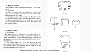
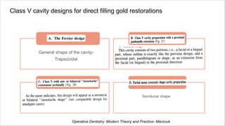
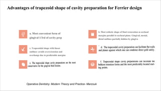
Dr. Sheetal Kotni
CLASS V
CAVITY
DESIGNS
CONTENTS
Summitt's Fundamentals of Operative Dentistry-4th ed.
Operating
Field
Gingival
retraction with
gingival
retraction cord
No. 212
retainer
Rubber dam
isolation
Sturdevant's Art and Science of Operative Dentistry, 6th ed.
01 Class V cavity designs for Amalgam restorations
Class V cavity designs for Amalgam restorations
01 0 1
• Cervical and root caries
• Incipient, smooth-surface enamel caries appears as a
chalky white line on facial surface
Indications
0 3
• Stronger
• Easier to place
• Less expensive
• Easily distinguished from the surrounding tooth structure
• Easier to finish and polish without damage to the adjacent
surfaces.
Advantages
0 2 • Esthetically important areas
Contraindications
0 4
Disadvantages
• Metallic and unesthetic.
• 90-degree cavosurface margins and specific axial depths
result in a less conservative preparation
Sturdevant's Art and Science of Operative Dentistry, 6th ed.
Outline form and Initial depth
Cavosurface margins in sound tooth structure
Limited axial depth-
0.5 mm inside the DEJ,
0.75 mm inside cementum (when on the root surface)
A tapered fissure bur of suitable size (e.g., No. 271)- Initial
entry into caries/ restoration.
The edge of the end of the bur > the flat end of the bur-
Reducing the bur’s “crawling.”
The bur orientation is adjusted to ensure that all external
walls are perpendicular to the external tooth surface and
parallel to the enamel rods
Alternatively, an appropriate carbide bur (usually No. 2 or
No. 4) for the initial tooth preparation. Round
burs are indicated in areas inaccessible to a fissure bur
that is
held perpendicular to the tooth surface. Smaller
round burs define the internal angles enhancing proper
placement of the retention grooves.
The axial wall is convex.
Depth of axial wall:
Incisal wall - more enamel ( 1-1.25 mm ) > gingival wall-
little or no enamel ( 0.75-1 mm)
Helps in pulp protection by increasing RDT.
Initial tooth preparation
Sturdevant's Art and Science of Operative Dentistry, 6th ed.
Removing any remaining
infected dentin with a No. 2 or
No. 4 round bur; pulp
protection; retention form;
finishing external walls; and
cleaning, inspecting, and
desensitizing.
• Final tooth
preparation
(1) No clinical or radiographic
evidence of recurrent caries
exists,
(2) The periphery of the base
and liner is intact
(3) The tooth is asymptomatic.
• Any old restorative
material (including
base and liner)
remaining may be
left if:
Because the walls of the
tooth preparation are
perpendicular to the external
tooth surface, they usually
diverge facially.
Consequently, no inherent
retention
•Retention form must
be provided because
the primary retention
form for an
amalgam restoration
is macromechanical.
Sturdevant's Art and Science of Operative Dentistry, 6th ed.
Sturdevant's Art and Science of Operative
Dentistry, 6th ed.
• The depth of the grooves - 0.25 mm (half of bur diameter).
• Adequate retention grooves - the only retention form to the preparation.
• In a large Class V amalgam preparation, extending the retention groove circumferentially around
all the internal line angles of the tooth preparation may enhance the retention form.
Sturdevant's Art and Science of Operative Dentistry, 6th ed.
Finally, the preparation is cleaned and inspected for completeness.
A desensitizer is applied.
• an angle-former chisel may be used to prepare the retention form.
• In addition, a No. 331/2 bur can be used
If access is inadequate for
use of the No. 1/4 round bur
• Amalgam can be condensed into rounded areas better than into sharp
areas, resulting in better adaptation of amalgam into the retention
grooves
The rounded retention form
placed with the No. 1/4 round
bur is generally preferred
• Suitable hand instruments (e.g., chisels, GMT) are used to plane the
enamel margins, verifying soundness and 90-degree cavosurface angles
If necessary
Sturdevant's Art and Science of Operative Dentistry, 6th ed.
Large Preparations That Include Line Angles
Sturdevant's Art and Science of Operative Dentistry, 6th ed.
• Because of the proximity of the coronoid process, access
to the facial surfaces of maxillary molars, particularly the
second molars, is often limited.
• Having the patient partially close and shift the mandible
toward the tooth being restored improves access and
visibility
• The previously placed amalgam serves as the distal wall
of the preparation.
• When proper treatment requires Class II and V amalgam
restorations on the same tooth, the Class II preparation
and restoration is completed before initiating the Class V
restoration.
• If the Class V restoration were done first, it might be
damaged by the matrix band and wedge needed for the
Class II restoration.
Sturdevant's Art and Science of Operative Dentistry, 6th ed.
Summitt's Fundamentals of Operative Dentistry-4th ed.
Operative Dentistry: Modern Theory and Practice- Marzouk
Class V cavity
designs for
Amalgam
restorations
ac. to Marzouk
Design 1
Design 2
Design 3
Design 4
Design 5
Operative Dentistry: Modern Theory and Practice- Marzouk
02 Class V cavity designs for direct filling gold
restorations
Class V cavity designs for direct filling gold restorations
Enter title
General shape of the cavity-
Trapezoidal
Enter title
Click here to add content of the text,
and briefly explain your point of view
Enter title
Click here to add content of the text,
and briefly explain your point of view
Enter title
Semilunar shape
Operative Dentistry: Modern Theory and Practice- Marzouk
Advantages of trapezoid shape of cavity preparation for Ferrier design
a. Most convenient form of
gingival 1/3rd of cavity prep
b. Most esthetic shape of final restoration as occlusal
margins parallel to occlusal plane. Gingival, mesial,
distal outlines partially hidden by gingiva
c. Trapezoidal shape with linear
outlines- avoids overextension and
overhangs due to predictable margins
Operative Dentistry: Modern Theory and Practice- Marzouk
Convex axial wall following
tooth contour
2 planed mesial, distal walls.
4 planed gingival, occlusal walls
Operative Dentistry: Modern Theory and Practice- Marzouk
No. 331/2 inverted
cone bur, flat
bladed plastic
instrument to
protect dam
Straight and
smooth gingival
wall at right
angles to other
walls
Planing occlusal
wall with
wedelstaedt
chisel
900 Cavosurface
angle
900 axioocclusal
angle
Other walls
refined with
small
monoangled
chisel
Principles and Practice of Operative Dentistry- Charbeneau 3rd ed.
Operative Dentistry: Modern Theory and Practice- Marzouk
• Indications: Apical location of height of contour
• No clear demarcation between mesial, distal, gingival walls
• Limited inciso-gingival width
Textbook of Operative Dentistry: Vimal K Sikri, 4th ed.
03 Class V cavity designs for Direct composite
restorations
For small or moderate
lesions that don't
extend onto the root
surface
Decalcified
enamel lesion having a broken,
rough surface
extending mesially or distally
from the cavitated lesion
Aberrant Smooth
Surface Pit Fault
With bilateral
extension occlusally
With unilateral
extension occlusally
Class V Abrasion or
Erosion Area
Large Lesions or
Defects that Extend
onto the Root Surface
Designs of Class V cavity preparation for Composite
Restorations based on extent of lesion
Textbook of Operative Dentistry: Vimal K Sikri, 4th ed. Sturdevant's Art and Science of Operative Dentistry, 6th ed
Class V Tooth Preparation for Small or Moderate Lesions or Defects That Do
Not Extend Onto the Root Surface
Class V (E and F) initial composite restorations (primary caries).
Restoring as
conservatively
No butt joints,
no groove
retention
Lesion is
scooped out
Divergent walls,
axial wall not
uniform in
depth
Initial tooth prep- round
diamond or carbide
bur eliminating the entire
enamel lesion
Dentin extension
only if necessary
Results in a
slightly bevelled
enamel margin
Sturdevant's Art and Science of Operative Dentistry, 6th ed.
Ideal for small enamel defects or
small primary caries lesions
(Fig. 9-25, A). These include
decalcified and hypoplastic
areas located in the cervical third
of the teeth
Sturdevant's Art and Science of Operative Dentistry, 6th ed.
A path of a decalcified
enamel lesion having a broken,
rough surface
extends mesially or distally from the
cavitated lesion (or
failing existing restoration). After
preparation of the cavitated
lesion (or failing restoration), the
margins of the preparation
are extended to include these areas
of decalcification by using
a round diamond or bur to prepare
the cavosurface margin in
the form of a chamfer, extended in
the enamel only to a depth
that removes the defect
Sturdevant's Art and Science of Operative Dentistry, 6th ed.
Textbook of Operative Dentistry: Vimal K Sikri, 4th ed.
Tooth Preparation for A Class V Abrasion or Erosion Area
Roughening of internal walls, bevelling of enamel margins, If necessary, the root surface cavosurface
margins should be prepared to approximately 90 degrees
Sturdevant's Art and Science of Operative Dentistry, 6th ed.
Class V Tooth Preparation for Large Lesions or Defects that Extend
onto the Root Surface
When a tapered fissure bur(271) or diamond is used, the handpiece is maneuvered to maintain the bur’s
long axis perpendicular to the external surface of the tooth during preparation of the outline form, which
should result in 90-degree cavosurface margins.
• Bevel-
Flame-
shaped
diamond
• Angle-
450
• Width-
atleast
0.5mm
Sturdevant's Art and Science of Operative Dentistry, 6th ed.
Tooth Preparation for Aberrant Smooth Surface Pit Fault
• The outline form (extensions and depth) is dictated by the extent of the fault or caries lesion.
• Faults existing entirely in enamel are prepared with an appropriately-sized round diamond instrument by merely
eliminating the defect (see Fig. 9-31, B).
• Adequate retention is obtained by bonding.
• When the defect includes carious dentin, the infected portion is removed also, leaving a flared enamel margin
Sturdevant's Art and Science of Operative Dentistry, 6th ed.
Flowable resin composite
• As the tooth flexes, the less rigid restoration might be able to accommodate the change in
cavity shape and therefore be more difficult to dislodge. Not supported by clinical trials.
• The use of a flowable resin composite as a liner has not been shown to improve clinical
performance
Reduced filler
particle
loading
Lower elastic
modulus
Higher
polymerization
shrinkage
Higher COTE
Lower fracture
toughness
relative to
traditional
resin
composites
04 Class V cavity designs for GIC restorations
Glass Ionomer Restorations for Class V cavities
Indications
• GIC- Anticariogenic -
Material of choice for
restoring root-surface
caries in patients with
high caries activity,
esthetics is not as
critical.
• Notched cervical
defects of idiopathic
erosion or abrasion
orgin
• Gingival recession
leading to caries
Preparation GI sandwich technique Compomer
• Similar to dental
amalgam without the
mechanical retention
• Cavosurface bevels
are not recommended
because GIC is a
brittle material that
requires bulk for
strength.
• A 90-degree butt joint
approximately 1 mm
deep is a reasonable
minimum thickness.
• GI- replaces the missing
dentin, reduce leakage
improve the potential for
tissue attachment for
subgingival restorations, and
potentially increase retention.
• A veneer of resin composite
is placed to enhance
esthetics, increase color
stability, improve marginal
performance, provide a
smoother surface, and
increase abrasion resistance
• Restore teeth that have
carious cervical lesions
and NCCLs
• Pro- lack of “stickiness”
has brought them ready
acceptance in the
marketplace.
• Cons- the marginal
integrity of compomers
has been worse than that
of resin composites in
long-term clinical trials.
Summitt's Fundamentals of Operative
Dentistry-4th ed.
Summitt's Fundamentals of Operative Dentistry-4thed
THANK
YOU

Class 5 cavity designs

  • 1.
    Dr. Sheetal Kotni CLASSV CAVITY DESIGNS
  • 2.
  • 3.
    Summitt's Fundamentals ofOperative Dentistry-4th ed.
  • 4.
    Operating Field Gingival retraction with gingival retraction cord No.212 retainer Rubber dam isolation Sturdevant's Art and Science of Operative Dentistry, 6th ed.
  • 5.
    01 Class Vcavity designs for Amalgam restorations
  • 6.
    Class V cavitydesigns for Amalgam restorations 01 0 1 • Cervical and root caries • Incipient, smooth-surface enamel caries appears as a chalky white line on facial surface Indications 0 3 • Stronger • Easier to place • Less expensive • Easily distinguished from the surrounding tooth structure • Easier to finish and polish without damage to the adjacent surfaces. Advantages 0 2 • Esthetically important areas Contraindications 0 4 Disadvantages • Metallic and unesthetic. • 90-degree cavosurface margins and specific axial depths result in a less conservative preparation Sturdevant's Art and Science of Operative Dentistry, 6th ed.
  • 7.
    Outline form andInitial depth Cavosurface margins in sound tooth structure Limited axial depth- 0.5 mm inside the DEJ, 0.75 mm inside cementum (when on the root surface) A tapered fissure bur of suitable size (e.g., No. 271)- Initial entry into caries/ restoration. The edge of the end of the bur > the flat end of the bur- Reducing the bur’s “crawling.” The bur orientation is adjusted to ensure that all external walls are perpendicular to the external tooth surface and parallel to the enamel rods Alternatively, an appropriate carbide bur (usually No. 2 or No. 4) for the initial tooth preparation. Round burs are indicated in areas inaccessible to a fissure bur that is held perpendicular to the tooth surface. Smaller round burs define the internal angles enhancing proper placement of the retention grooves. The axial wall is convex. Depth of axial wall: Incisal wall - more enamel ( 1-1.25 mm ) > gingival wall- little or no enamel ( 0.75-1 mm) Helps in pulp protection by increasing RDT. Initial tooth preparation
  • 8.
    Sturdevant's Art andScience of Operative Dentistry, 6th ed.
  • 9.
    Removing any remaining infecteddentin with a No. 2 or No. 4 round bur; pulp protection; retention form; finishing external walls; and cleaning, inspecting, and desensitizing. • Final tooth preparation (1) No clinical or radiographic evidence of recurrent caries exists, (2) The periphery of the base and liner is intact (3) The tooth is asymptomatic. • Any old restorative material (including base and liner) remaining may be left if: Because the walls of the tooth preparation are perpendicular to the external tooth surface, they usually diverge facially. Consequently, no inherent retention •Retention form must be provided because the primary retention form for an amalgam restoration is macromechanical. Sturdevant's Art and Science of Operative Dentistry, 6th ed.
  • 10.
    Sturdevant's Art andScience of Operative Dentistry, 6th ed.
  • 11.
    • The depthof the grooves - 0.25 mm (half of bur diameter). • Adequate retention grooves - the only retention form to the preparation. • In a large Class V amalgam preparation, extending the retention groove circumferentially around all the internal line angles of the tooth preparation may enhance the retention form. Sturdevant's Art and Science of Operative Dentistry, 6th ed.
  • 12.
    Finally, the preparationis cleaned and inspected for completeness. A desensitizer is applied. • an angle-former chisel may be used to prepare the retention form. • In addition, a No. 331/2 bur can be used If access is inadequate for use of the No. 1/4 round bur • Amalgam can be condensed into rounded areas better than into sharp areas, resulting in better adaptation of amalgam into the retention grooves The rounded retention form placed with the No. 1/4 round bur is generally preferred • Suitable hand instruments (e.g., chisels, GMT) are used to plane the enamel margins, verifying soundness and 90-degree cavosurface angles If necessary Sturdevant's Art and Science of Operative Dentistry, 6th ed.
  • 13.
    Large Preparations ThatInclude Line Angles Sturdevant's Art and Science of Operative Dentistry, 6th ed.
  • 14.
    • Because ofthe proximity of the coronoid process, access to the facial surfaces of maxillary molars, particularly the second molars, is often limited. • Having the patient partially close and shift the mandible toward the tooth being restored improves access and visibility • The previously placed amalgam serves as the distal wall of the preparation. • When proper treatment requires Class II and V amalgam restorations on the same tooth, the Class II preparation and restoration is completed before initiating the Class V restoration. • If the Class V restoration were done first, it might be damaged by the matrix band and wedge needed for the Class II restoration. Sturdevant's Art and Science of Operative Dentistry, 6th ed.
  • 15.
    Summitt's Fundamentals ofOperative Dentistry-4th ed.
  • 16.
    Operative Dentistry: ModernTheory and Practice- Marzouk
  • 17.
    Class V cavity designsfor Amalgam restorations ac. to Marzouk Design 1 Design 2 Design 3 Design 4 Design 5
  • 18.
    Operative Dentistry: ModernTheory and Practice- Marzouk
  • 19.
    Operative Dentistry: ModernTheory and Practice- Marzouk
  • 20.
    Operative Dentistry: ModernTheory and Practice- Marzouk
  • 21.
    Operative Dentistry: ModernTheory and Practice- Marzouk
  • 22.
    02 Class Vcavity designs for direct filling gold restorations
  • 23.
    Class V cavitydesigns for direct filling gold restorations Enter title General shape of the cavity- Trapezoidal Enter title Click here to add content of the text, and briefly explain your point of view Enter title Click here to add content of the text, and briefly explain your point of view Enter title Semilunar shape Operative Dentistry: Modern Theory and Practice- Marzouk
  • 24.
    Advantages of trapezoidshape of cavity preparation for Ferrier design a. Most convenient form of gingival 1/3rd of cavity prep b. Most esthetic shape of final restoration as occlusal margins parallel to occlusal plane. Gingival, mesial, distal outlines partially hidden by gingiva c. Trapezoidal shape with linear outlines- avoids overextension and overhangs due to predictable margins Operative Dentistry: Modern Theory and Practice- Marzouk
  • 25.
    Convex axial wallfollowing tooth contour 2 planed mesial, distal walls. 4 planed gingival, occlusal walls Operative Dentistry: Modern Theory and Practice- Marzouk
  • 26.
    No. 331/2 inverted conebur, flat bladed plastic instrument to protect dam Straight and smooth gingival wall at right angles to other walls Planing occlusal wall with wedelstaedt chisel 900 Cavosurface angle 900 axioocclusal angle Other walls refined with small monoangled chisel Principles and Practice of Operative Dentistry- Charbeneau 3rd ed.
  • 27.
    Operative Dentistry: ModernTheory and Practice- Marzouk
  • 28.
    Operative Dentistry: ModernTheory and Practice- Marzouk
  • 29.
    • Indications: Apicallocation of height of contour • No clear demarcation between mesial, distal, gingival walls • Limited inciso-gingival width
  • 30.
    Textbook of OperativeDentistry: Vimal K Sikri, 4th ed.
  • 31.
    03 Class Vcavity designs for Direct composite restorations
  • 32.
    For small ormoderate lesions that don't extend onto the root surface Decalcified enamel lesion having a broken, rough surface extending mesially or distally from the cavitated lesion Aberrant Smooth Surface Pit Fault With bilateral extension occlusally With unilateral extension occlusally Class V Abrasion or Erosion Area Large Lesions or Defects that Extend onto the Root Surface Designs of Class V cavity preparation for Composite Restorations based on extent of lesion Textbook of Operative Dentistry: Vimal K Sikri, 4th ed. Sturdevant's Art and Science of Operative Dentistry, 6th ed
  • 33.
    Class V ToothPreparation for Small or Moderate Lesions or Defects That Do Not Extend Onto the Root Surface Class V (E and F) initial composite restorations (primary caries). Restoring as conservatively No butt joints, no groove retention Lesion is scooped out Divergent walls, axial wall not uniform in depth Initial tooth prep- round diamond or carbide bur eliminating the entire enamel lesion Dentin extension only if necessary Results in a slightly bevelled enamel margin Sturdevant's Art and Science of Operative Dentistry, 6th ed.
  • 34.
    Ideal for smallenamel defects or small primary caries lesions (Fig. 9-25, A). These include decalcified and hypoplastic areas located in the cervical third of the teeth Sturdevant's Art and Science of Operative Dentistry, 6th ed.
  • 35.
    A path ofa decalcified enamel lesion having a broken, rough surface extends mesially or distally from the cavitated lesion (or failing existing restoration). After preparation of the cavitated lesion (or failing restoration), the margins of the preparation are extended to include these areas of decalcification by using a round diamond or bur to prepare the cavosurface margin in the form of a chamfer, extended in the enamel only to a depth that removes the defect Sturdevant's Art and Science of Operative Dentistry, 6th ed.
  • 36.
    Textbook of OperativeDentistry: Vimal K Sikri, 4th ed.
  • 37.
    Tooth Preparation forA Class V Abrasion or Erosion Area Roughening of internal walls, bevelling of enamel margins, If necessary, the root surface cavosurface margins should be prepared to approximately 90 degrees Sturdevant's Art and Science of Operative Dentistry, 6th ed.
  • 38.
    Class V ToothPreparation for Large Lesions or Defects that Extend onto the Root Surface When a tapered fissure bur(271) or diamond is used, the handpiece is maneuvered to maintain the bur’s long axis perpendicular to the external surface of the tooth during preparation of the outline form, which should result in 90-degree cavosurface margins. • Bevel- Flame- shaped diamond • Angle- 450 • Width- atleast 0.5mm Sturdevant's Art and Science of Operative Dentistry, 6th ed.
  • 39.
    Sturdevant's Art andScience of Operative Dentistry, 6th ed.
  • 40.
    Tooth Preparation forAberrant Smooth Surface Pit Fault • The outline form (extensions and depth) is dictated by the extent of the fault or caries lesion. • Faults existing entirely in enamel are prepared with an appropriately-sized round diamond instrument by merely eliminating the defect (see Fig. 9-31, B). • Adequate retention is obtained by bonding. • When the defect includes carious dentin, the infected portion is removed also, leaving a flared enamel margin Sturdevant's Art and Science of Operative Dentistry, 6th ed.
  • 41.
    Flowable resin composite •As the tooth flexes, the less rigid restoration might be able to accommodate the change in cavity shape and therefore be more difficult to dislodge. Not supported by clinical trials. • The use of a flowable resin composite as a liner has not been shown to improve clinical performance Reduced filler particle loading Lower elastic modulus Higher polymerization shrinkage Higher COTE Lower fracture toughness relative to traditional resin composites
  • 42.
    04 Class Vcavity designs for GIC restorations
  • 43.
    Glass Ionomer Restorationsfor Class V cavities Indications • GIC- Anticariogenic - Material of choice for restoring root-surface caries in patients with high caries activity, esthetics is not as critical. • Notched cervical defects of idiopathic erosion or abrasion orgin • Gingival recession leading to caries Preparation GI sandwich technique Compomer • Similar to dental amalgam without the mechanical retention • Cavosurface bevels are not recommended because GIC is a brittle material that requires bulk for strength. • A 90-degree butt joint approximately 1 mm deep is a reasonable minimum thickness. • GI- replaces the missing dentin, reduce leakage improve the potential for tissue attachment for subgingival restorations, and potentially increase retention. • A veneer of resin composite is placed to enhance esthetics, increase color stability, improve marginal performance, provide a smoother surface, and increase abrasion resistance • Restore teeth that have carious cervical lesions and NCCLs • Pro- lack of “stickiness” has brought them ready acceptance in the marketplace. • Cons- the marginal integrity of compomers has been worse than that of resin composites in long-term clinical trials. Summitt's Fundamentals of Operative Dentistry-4th ed.
  • 44.
    Summitt's Fundamentals ofOperative Dentistry-4thed
  • 47.