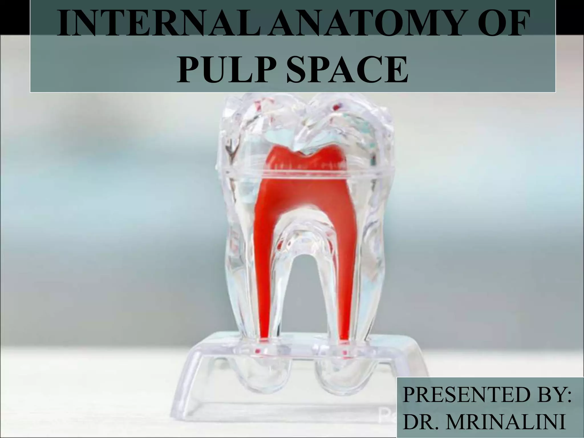
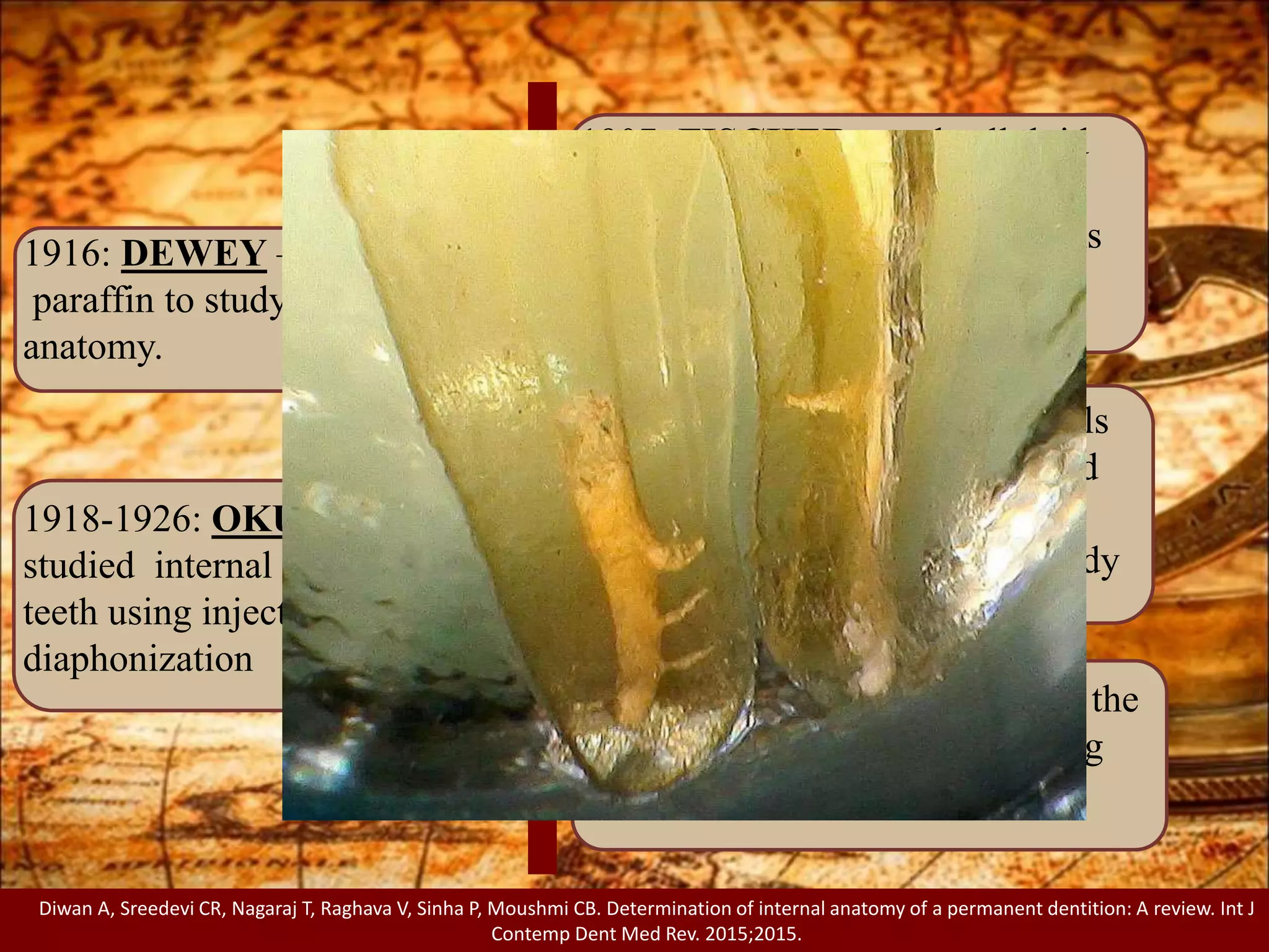
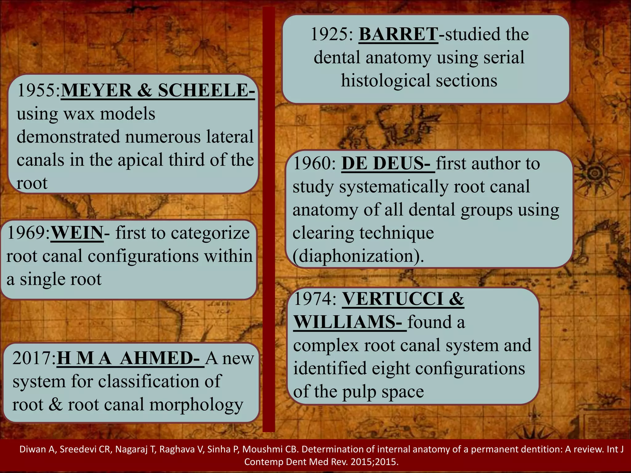
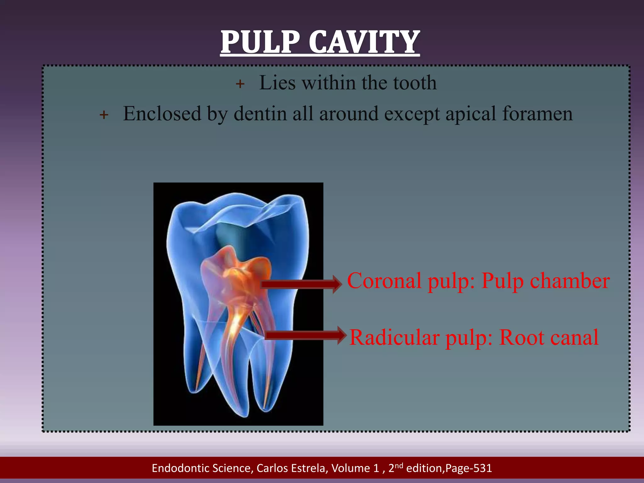
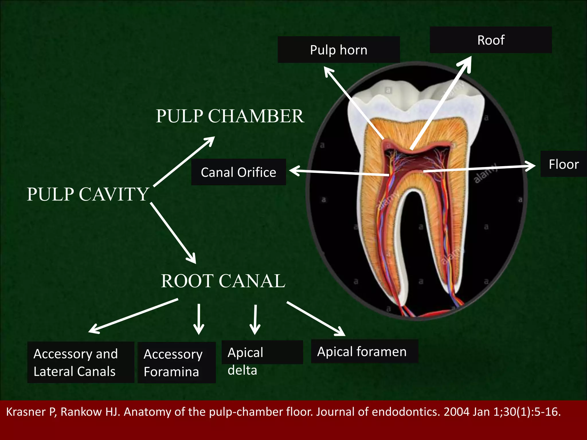
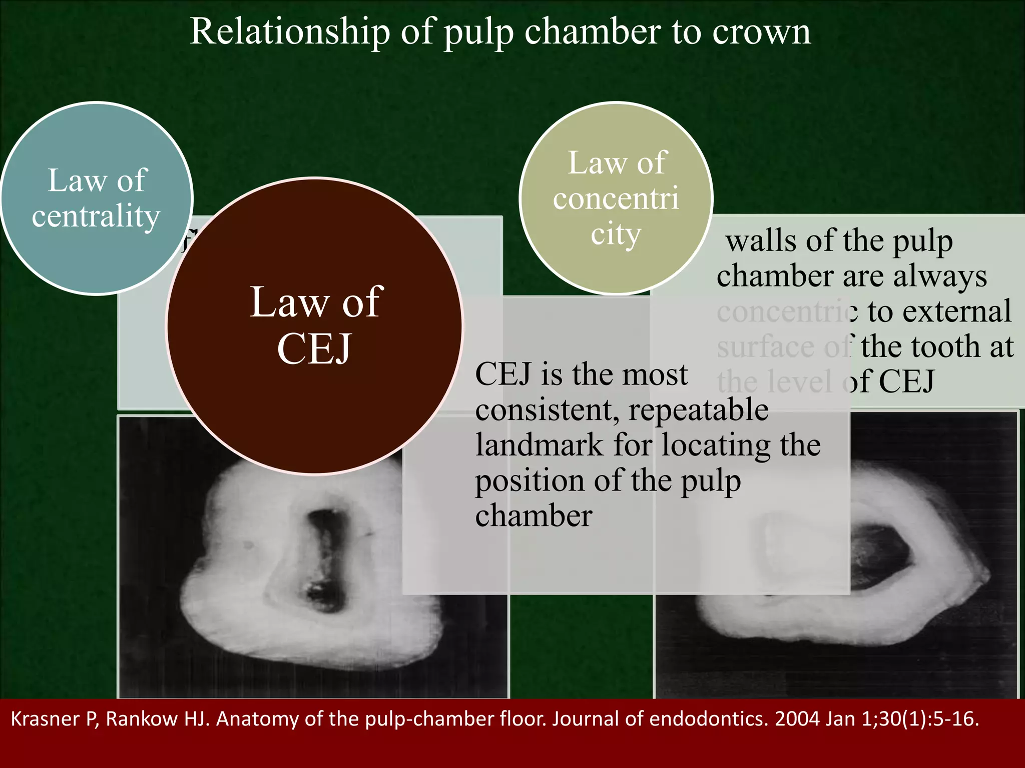
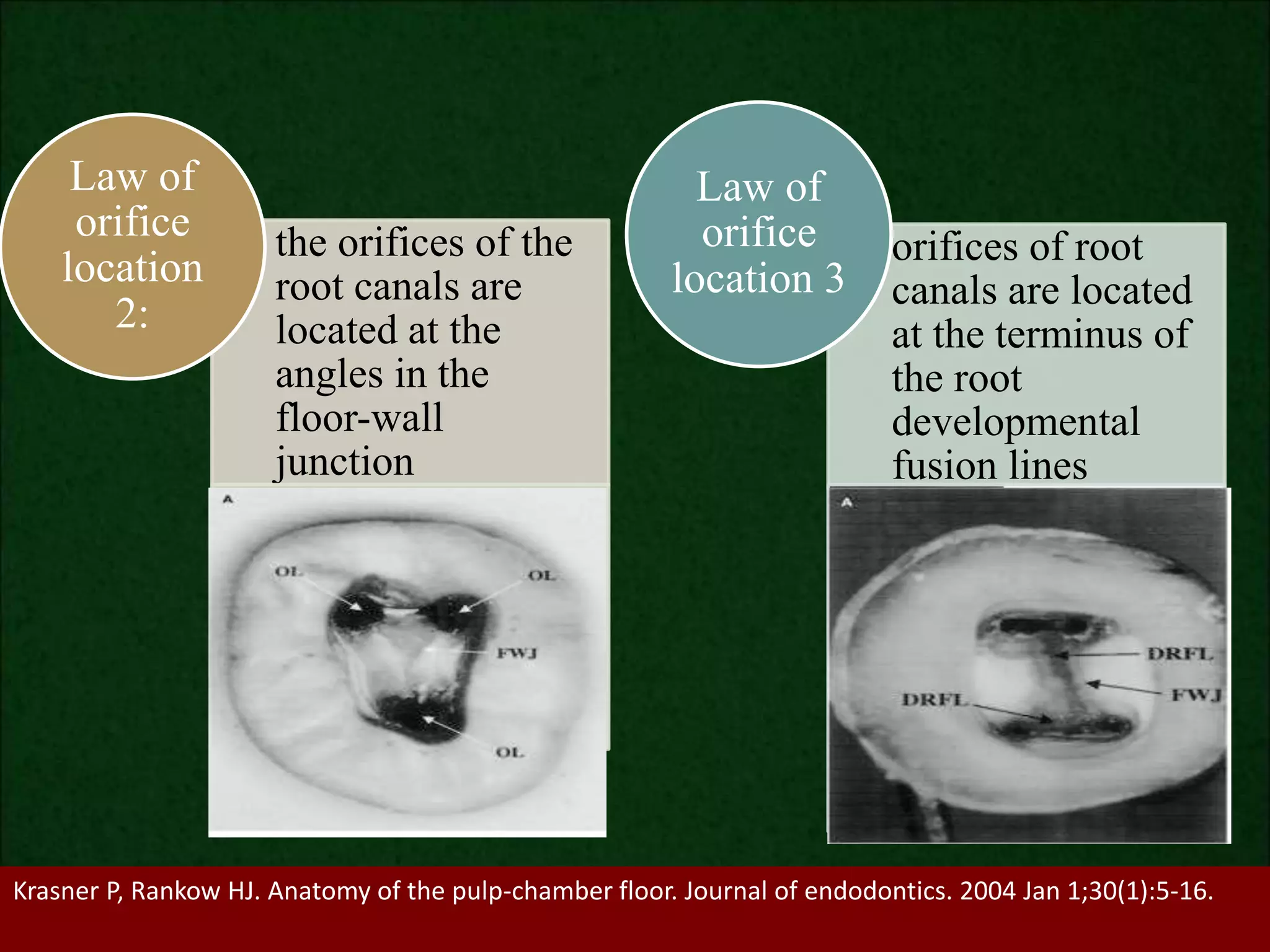
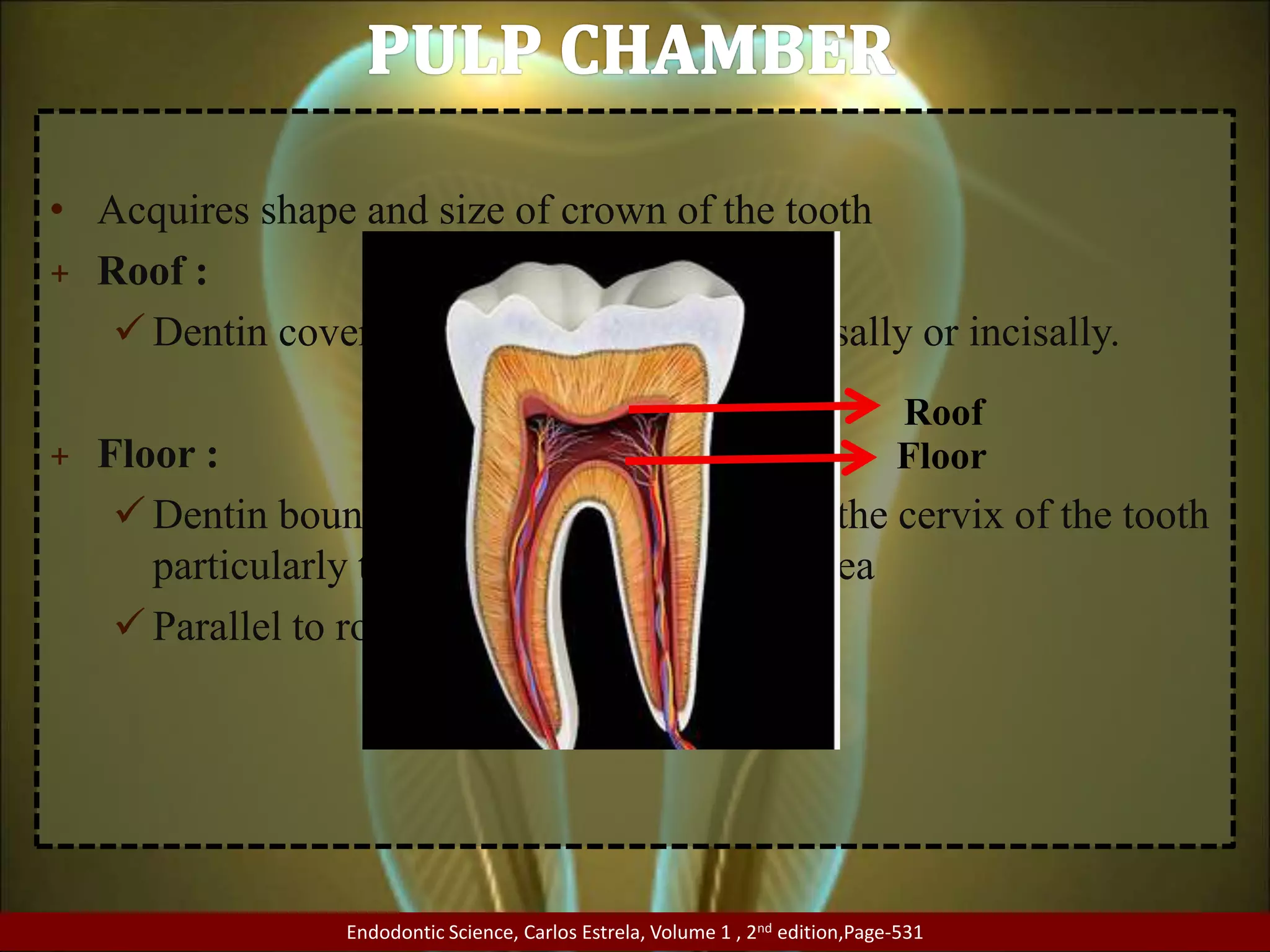
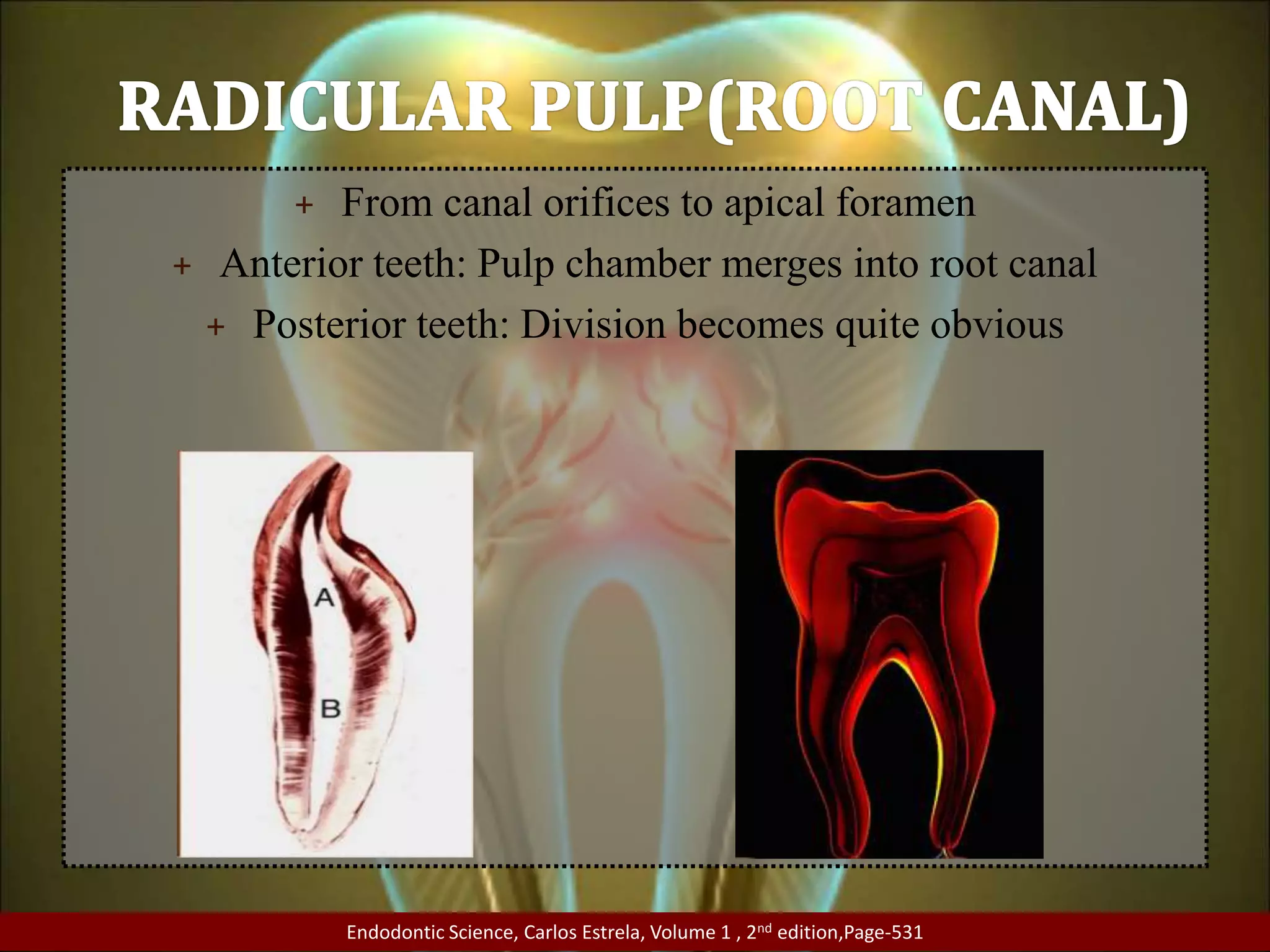
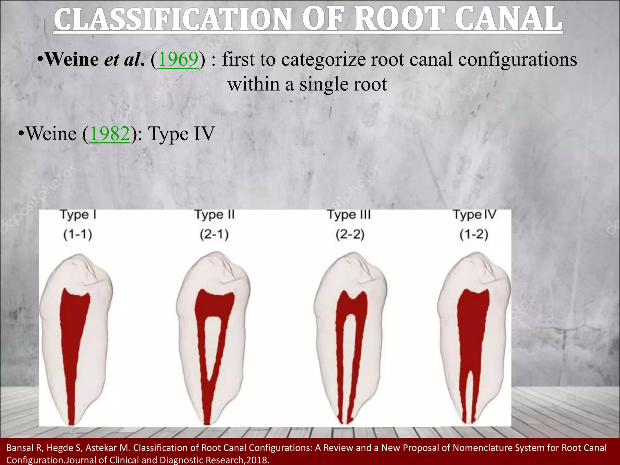
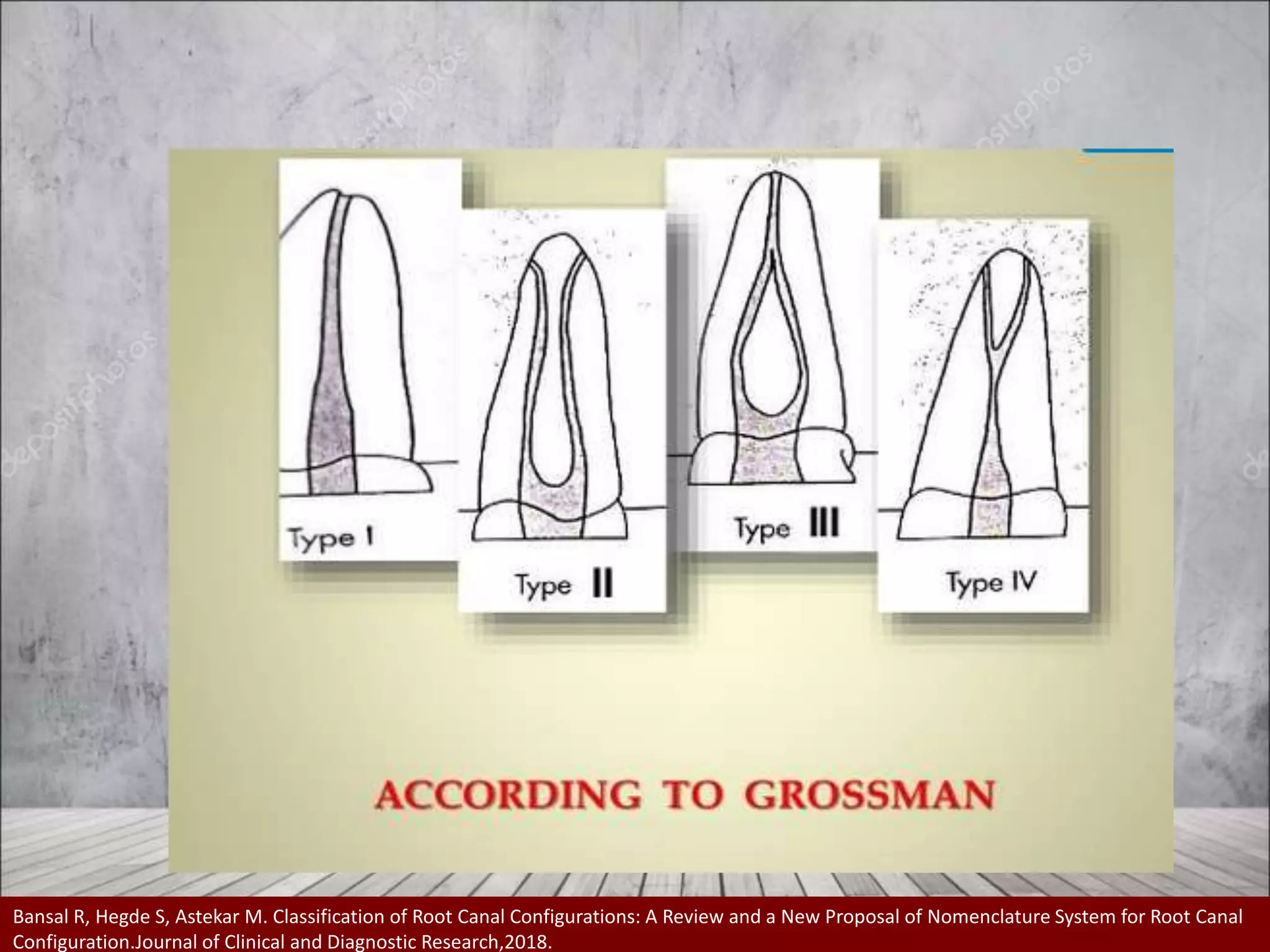
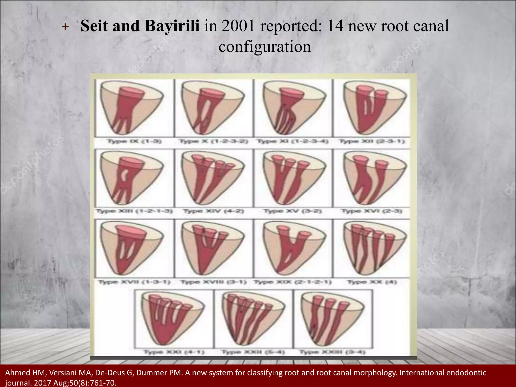
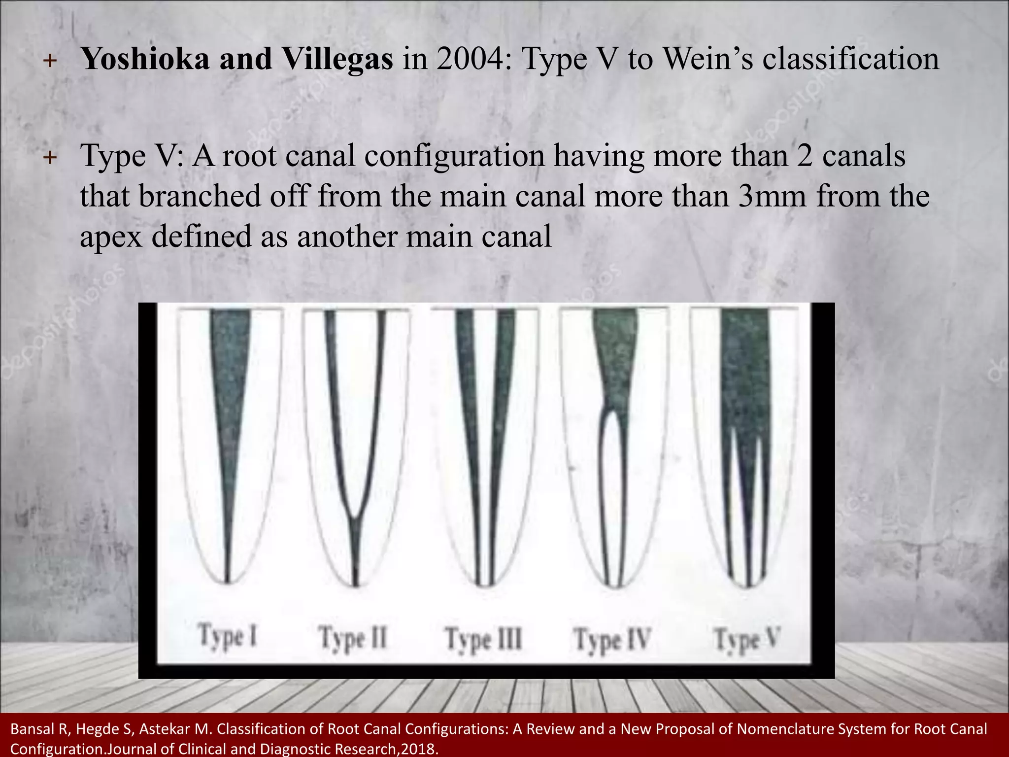
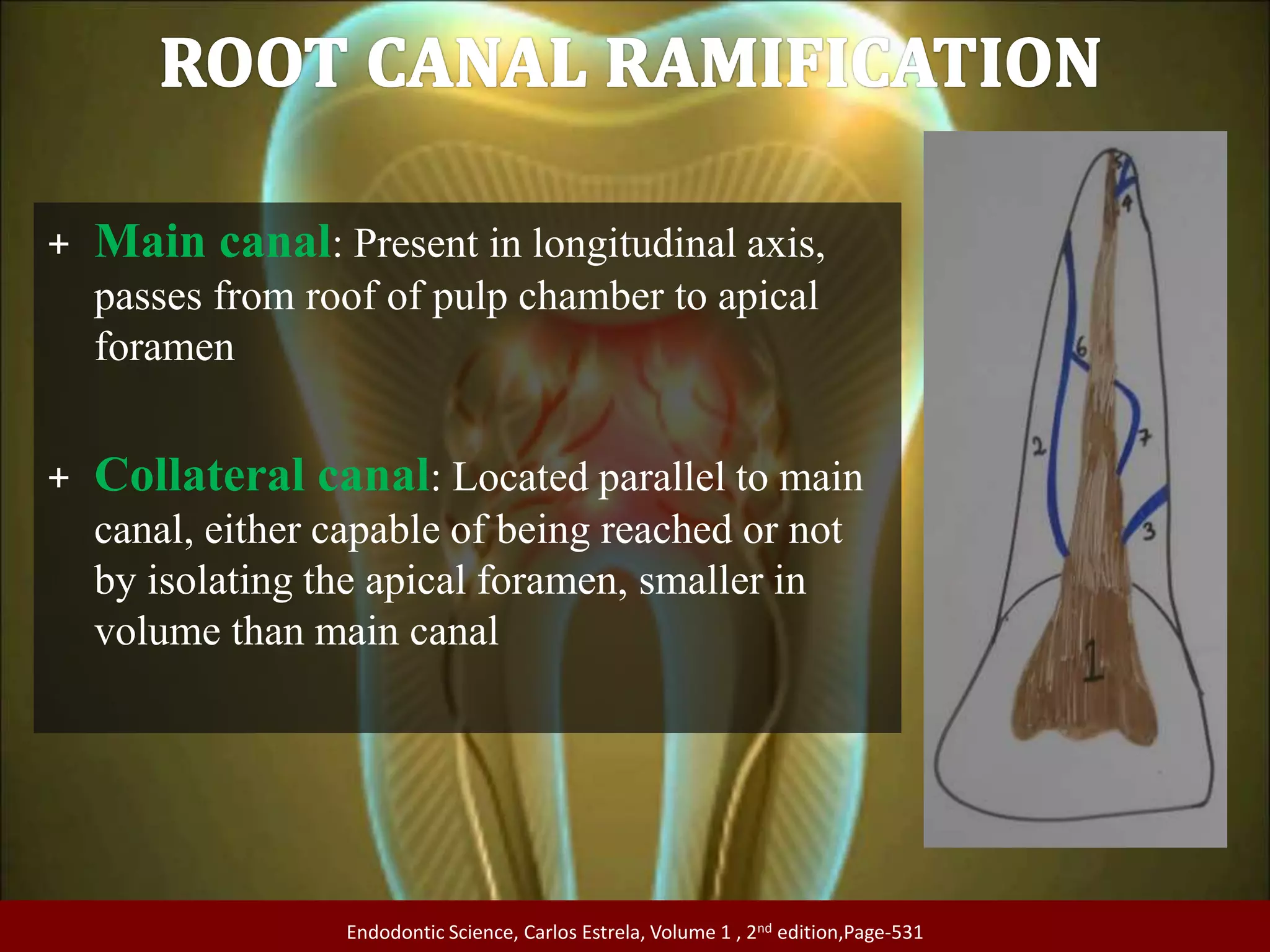
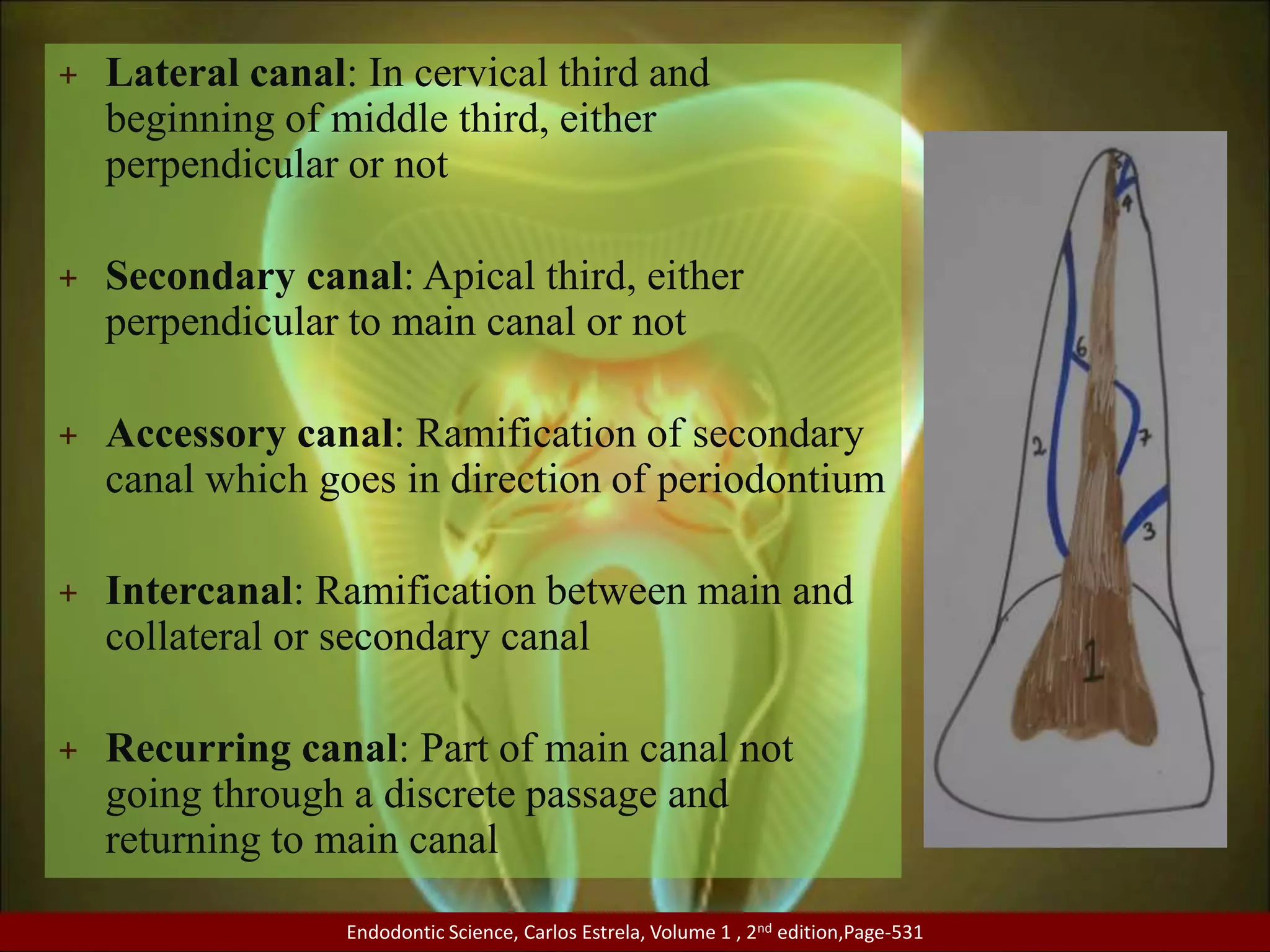
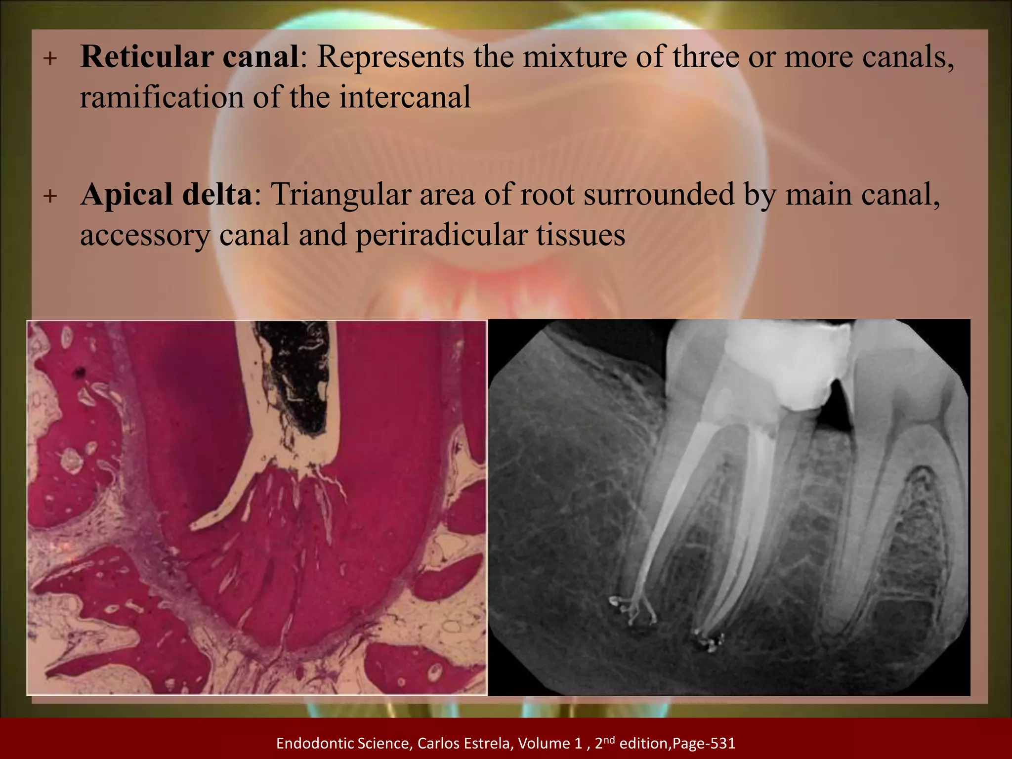
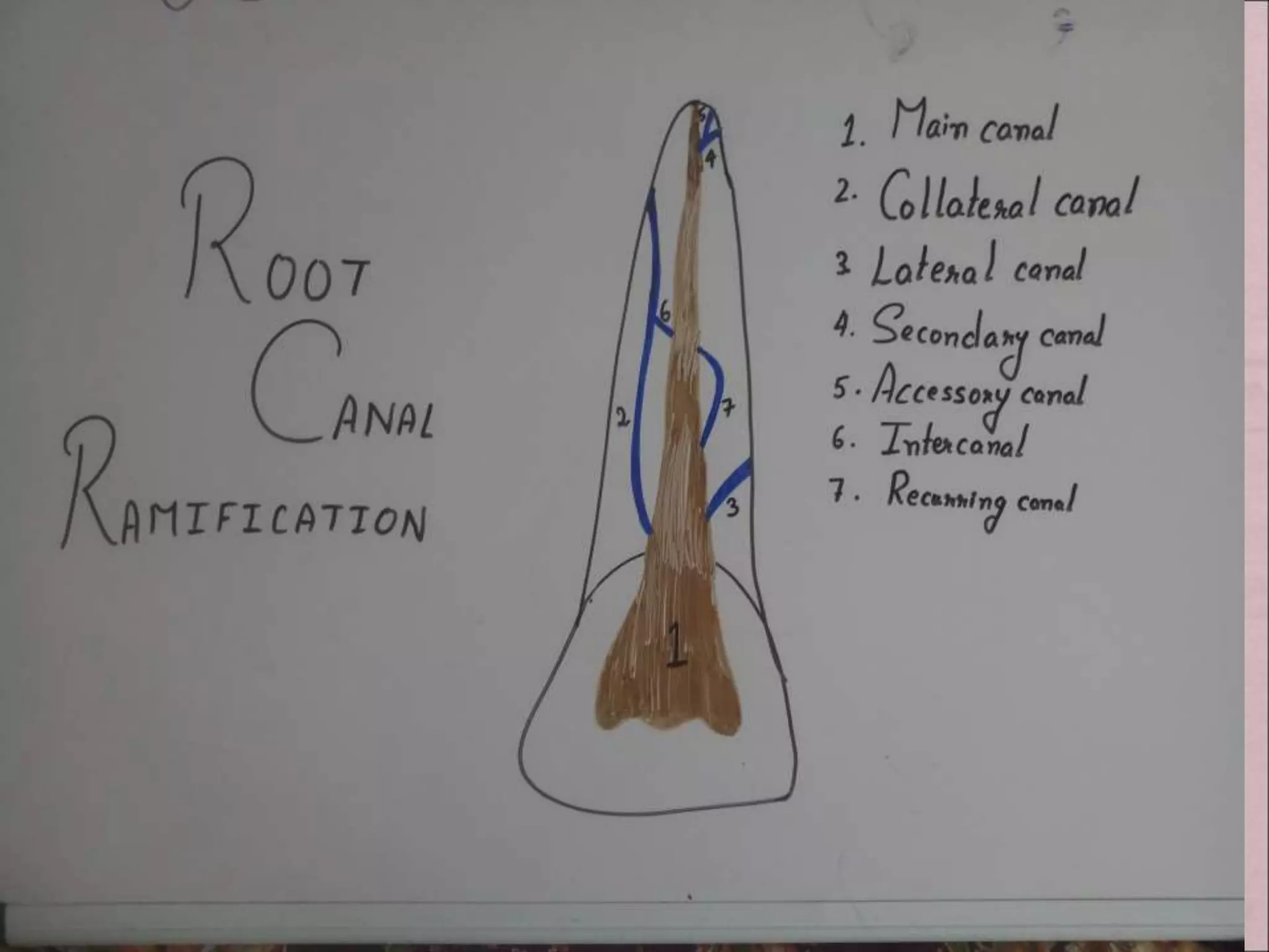
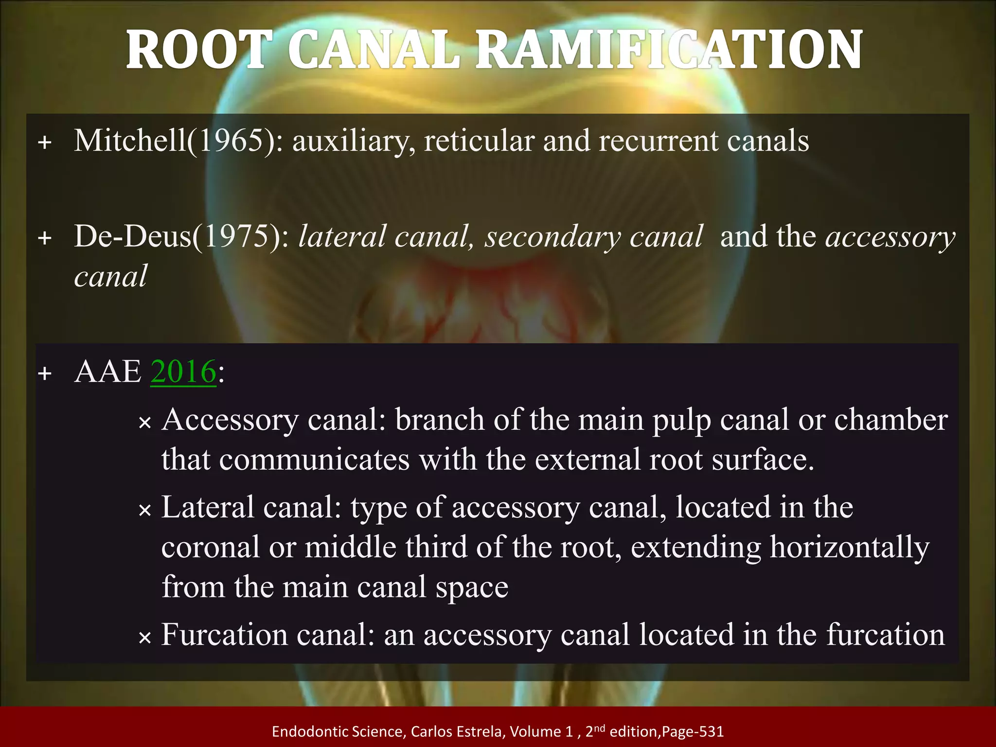
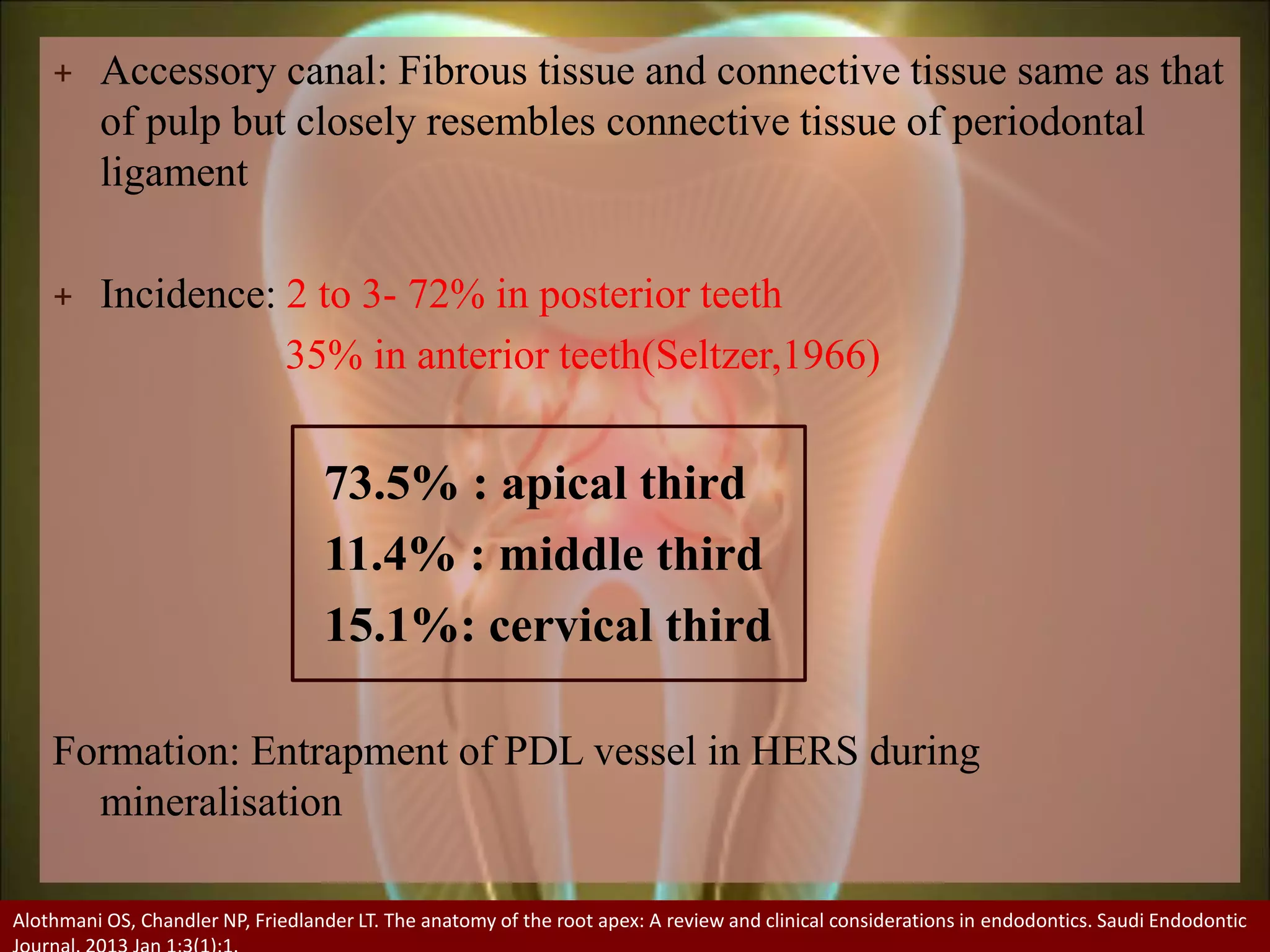
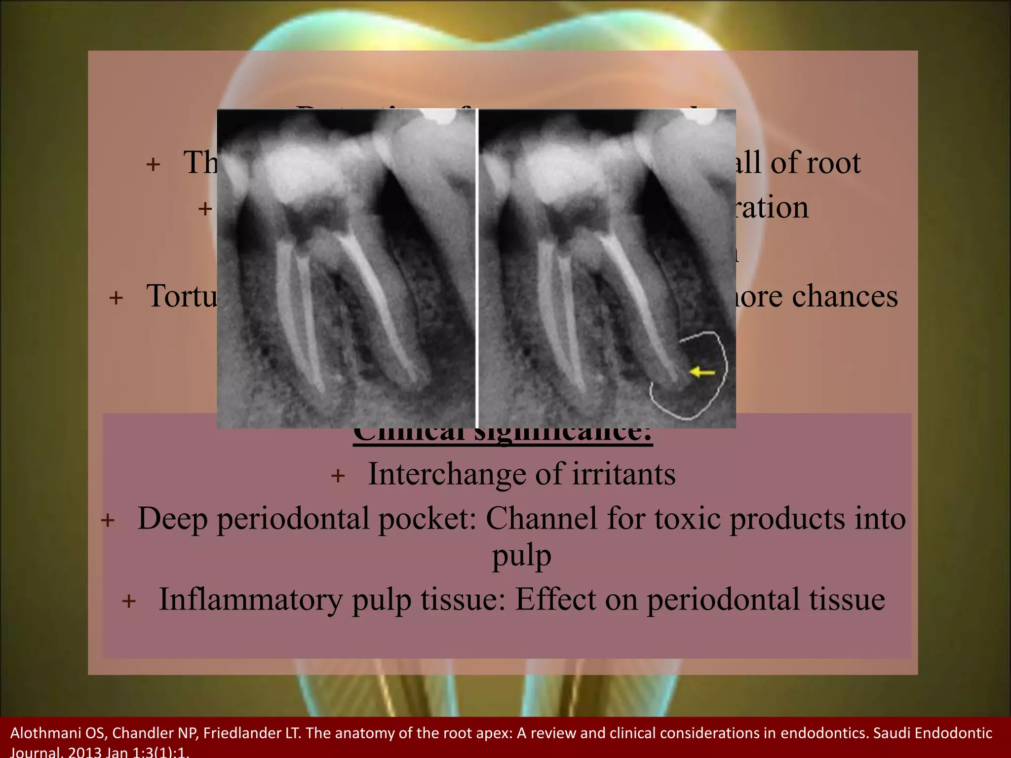
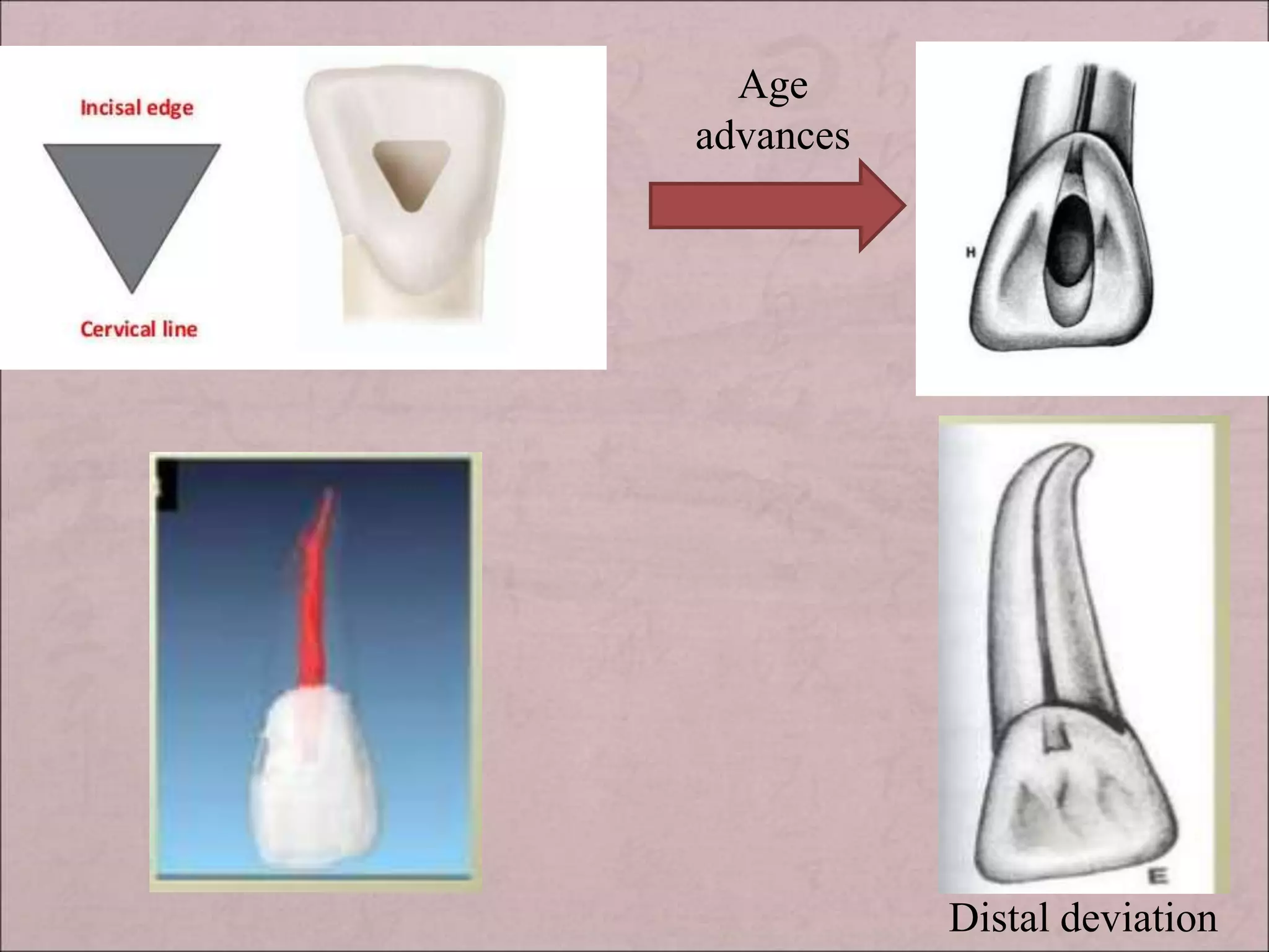
Internal anatomy of pulp space. Includes history, development of pulp, classification of the root canal, isthmus, root canal ramifications, regressive changes, apical root anatomy, variations in pulpal anatomy, methods of determining anatomy, pulp space anatomy of permanent teeth.
This document provides an overview of regenerative endodontics, which aims to regenerate dental tissues through biologically-based procedures using stem cells, growth factors, and scaffolds. Key elements for pulp regeneration include reliable stem cell sources, such as dental pulp stem cells, growth factors to stimulate cell proliferation and differentiation, and appropriate scaffolds. Potential regenerative therapies include revascularization, stem cell therapy, scaffold implantation, and gene delivery. Measuring clinical outcomes and further applications are areas of future focus to develop regenerative endodontic therapies.
This document discusses apexogenesis and apexification procedures for teeth with open apices. Apexogenesis refers to treatments that maintain pulp vitality and allow continued root development and apex closure. These include indirect pulp capping, direct pulp capping, and apical closure pulpotomy using materials like calcium hydroxide or MTA. Apexification induces artificial root end closure when the pulp is necrotic, using repeated dressings of calcium hydroxide or MTA to encourage hard tissue deposition. Clinical success depends on factors like the treatment material and maintaining a bacteria-tight seal during root development.
The document discusses the benefits of exercise for mental health. Regular physical activity can help reduce anxiety and depression and improve mood and cognitive function. Exercise causes chemical changes in the brain that may help protect against mental illness and improve symptoms for those who already suffer from conditions like anxiety and depression.
This document provides an overview of working length determination in endodontics. It discusses the historical perspectives on working length, important definitions like working length and anatomical structures at the root apex. Methods of determining working length are also covered, including both radiographic and non-radiographic techniques. Factors that influence working length like root canal anatomy and methods to prevent loss of working length are described. The importance of accurately determining working length for treatment success is emphasized.
1. Access cavity preparation is the first and most important phase of root canal treatment, with the goals of achieving straight line access to the apical foramen, locating all root canal orifices, and conserving sound tooth structure.
2. The principles of access cavity preparation include establishing an outline form based on pulp chamber size and shape and the number/direction of root canals, providing a convenience form for improved visibility and instrumentation, and removing caries/defective restorations and debris from the pulp chamber.
3. Key steps in access cavity preparation depend on the specific tooth but involve using burs and instruments to locate and prepare access to all root canal orifices while avoiding errors like
This document discusses various obturation techniques. It introduces lateral compaction and warm vertical compaction as two primary methods. Warm vertical compaction involves vertically compacting gutta-percha into the prepared canal with heated pluggers. Variations include single/continuous wave compaction. Warm lateral compaction uses an electrically heated spreader to laterally condense softened gutta-percha. The document provides details on the procedure, advantages, and limitations of different obturation methods.
Tooth hypersensitivity is a common problem encountered in everyday life and clinical practice. This presentation clearly shows causes, methods of prevention and treatment in such cases.
The document discusses the anatomy of the pulp cavity. It begins by introducing the importance of understanding pulp anatomy for endodontic therapy. The pulp cavity is divided into the coronal pulp chamber and radicular root canal. The pulp chamber occupies the crown and merges with the root canal. Multi-rooted teeth have a single chamber and 3 or more canals. Anatomical structures like the roof, floor, canal orifices and isthmus are described. Root canals extend from the orifice to the apical foramen. Classification systems and methods to study pulp anatomy are also outlined. Variations in shape, number of canals and pathological changes are discussed.
The document discusses various endodontic mishaps that can occur during root canal treatment. It describes mishaps related to access preparation, instrumentation, and obturation. Access-related mishaps include treating the wrong tooth, missing canals, damaging existing restorations, perforating the access cavity, and crown fractures. Instrumentation mishaps include ledge formation, perforating the root, and separated instruments. Obturation mishaps include overfilling or underfilling the canal. The document provides details on the causes, recognition, correction, prevention and prognosis of several common endodontic mishaps.
The document discusses the anatomy and root canal morphology of maxillary central incisors. It notes that maxillary central incisors typically have:
- A single root with a single root canal system
- A triangular/ovoid root cross-section that tapers lingually
- Midroot and apical lateral canals that are common
- The root apex and apical foramen located distolabially on average
- A triangularly-shaped pulp chamber located in the center of the crown that follows the contours of the crown and has three pulp horns
This document discusses root canal curvatures and methods for determining their degree. It begins with an introduction on the importance of understanding root canal anatomy for successful endodontic treatment. It then covers causes of canal curvatures, various classification systems, and techniques for assessing curvature using periapical radiographs, cone-beam computed tomography, and angular measurement methods. The relationship between curvature degree and ledge formation risk is also addressed, as are canal characteristics often invisible on routine radiographs. The document concludes by emphasizing the challenges of treating severely curved canals and the need for accurate preoperative assessment and proper instrumentation.
This document discusses apexification and indirect pulp capping procedures. Apexification is used to induce calcification at the open apex of an immature tooth to allow for root canal treatment. Materials used include calcium hydroxide, tricalcium phosphate, and MTA. Calcium hydroxide is placed in the canal and replaced every 3 months until radiographic evidence of apical barrier formation. MTA can also be used by placing it at the apex. Indirect pulp capping covers the deepest carious dentin with a biocompatible material like calcium hydroxide to prevent pulpal exposure while removing infected dentin, allowing for pulp preservation.
Burs, mirrors, probes and excavators are used to prepare the access cavity and remove pulp tissue. K-files, reamers, and Hedstrom files are used to shape the root canal in a twisting motion. NiTi files are used to initially flare canals. Gates Glidden drills and Peeso reamers further widen canals. Irrigation, paper points and gutta percha are used to clean canals and conduct root canal fillings. Spreaders and pluggers are used to condense gutta percha during filling.
This document provides an overview of root canal anatomy and discusses various considerations for endodontic treatment. It describes the components of the root canal system, including the pulp chamber and root canals. It also discusses accessory canals and various classifications of root canal configurations. Additionally, it outlines objectives and guidelines for access cavity preparation and clinical determinations of root canal configurations based on coronal, mid-root, and apical considerations. Specific anatomical features and clinical considerations are also described for different types of anterior and posterior teeth.
This document discusses irrigation in endodontics. It provides an introduction to irrigation solutions and devices used, challenges of irrigation, and recent advances. It describes the ideal characteristics of endodontic irrigants and commonly used solutions such as sodium hypochlorite. Sodium hypochlorite is the current irrigant of choice and its properties, concentrations, effects on dentin, and safety considerations are discussed in detail. The document concludes by emphasizing the importance of irrigation in endodontic treatment.
Radiographic Assessment of the Prevalence of Pulp Stones in Malaysians
Kannan et al.
JOE — Volume 41, Number 3, March 2015
Pulp stones are discrete calcified bodies found in the dental pulp.
They have calcium phosphorous ratios similar to dentin and can be seen in healthy, diseased, or even unerupted teeth
Radiographically, pulp stones appear as radiopaque structures in the pulp space that frequently act as an impediment during endodontic treatment
The document discusses tooth preparation for dental restorations. It describes the objectives of tooth preparation as removing defects, protecting the pulp, extending restorations conservatively, and allowing functional and esthetic placement of restorative materials. Factors that affect tooth preparation include pulpal and periodontal status, dental anatomy, occlusion, patient factors, affected dentin, and restorative materials. Different types of cavity preparations are also described, including simple, compound, and complex cavities as well as Class I, Class II, and modified cavity preparations for both amalgam and composite restorations. Techniques for cavity preparation are provided for both conventional and modified designs depending on the restoration needs.
This document discusses the anatomy of root canals and related structures. It begins with an introduction and overview, then discusses topics like pulp chamber anatomy, root canal classification systems, the apical foramen, accessory foramina, apical constriction, isthmuses, C-shaped canals, and anatomy of individual teeth. It also touches on developmental disturbances and references other research. The overall document provides an in-depth look at root canal anatomy, variations, related structures, and classification systems.
This document provides an overview of anterior composite restorations. It discusses the indications, contraindications, advantages, and disadvantages of composite resins. It also describes the different types of composites and their composition. The document outlines techniques for cavity preparations for Class III, IV, and V lesions and the steps for placing composite restorations, including acid etching, bonding, matrix placement, increment placement, and finishing/polishing. Composite resins are presented as esthetic restorative materials that conserve tooth structure when used for anterior restorations according to the guidelines provided.
This document provides an overview of root canal anatomy, including the divisions of the pulp cavity, landmarks in the apical region, classifications of canal configurations, and detailed descriptions of canal morphology for different tooth types. Key points include:
- The pulp cavity is divided into the coronal pulp chamber and radicular root canals.
- Important apical landmarks include the apical constriction, foramen, cementodentinal junction, and accessory canals.
- Classification systems describe common canal morphologies, such as single versus multiple canals.
- Tooth-specific details are given for maxillary incisors, canines, premolars, and molars, including average canal numbers, lengths
This document discusses common errors that can occur during endodontic treatment and ways to prevent or manage them. It covers errors related to accessing the pulp space like treating the wrong tooth, incomplete caries removal, or perforating through a full coverage restoration. It also discusses errors during canal cleaning and shaping such as ledge formation, canal deviations, or instrument separation. Finally, it addresses procedural errors during obturation like underfilling or overfilling the canal with gutta percha. Throughout, it provides tips for preventing errors like using small instruments sequentially, maintaining canal patency, and taking pre-operative radiographs to understand anatomy.
This document discusses the cleaning and shaping of root canals. It defines cleaning as the removal of pathogenic contents from the root canal and shaping as creating a 3D tapered shape that is widest coronally and narrowest apically. The objectives of shaping are outlined as both mechanical and biological to remove debris without forcing it periapically and create sufficient space for obturation. Various techniques are described such as step-back, crown-down, and hybrid techniques. Considerations like instrument movements, irrigation methods, and the goals of apical enlargement are also covered.
An inlay may cap none, or may cap all but one cusp.
Sturdevant’s 4th ed. page579
Inlays may be used as single-tooth restorations for proximo-occlusal or gingival lesions with minimal to moderate extensions
Shillingburg page 1
An inlay may be defined as a restoration which has been constructed out of mouth from gold, porcelain, or other material & then cemented into the prepared cavity of a tooth.
William McGehee pg410
The cast metal restoration is versatile and is especially applicable to Class II onlay preparations. The process has many steps, involves many dental materials, and requires meticulous attention to prepration.
Tooth treatment planned to be restored with an intracoronal restoration, but the decay or fracture is so extensive that a direct restoration, such as amalgam or composite, would not be able to sustain or bear forces.
Additionally, when decay or fracture incorporate areas of cusp or remaining tooth structure that undermines perimeter walls of a tooth, an onlay might be indicated.
Anatomy of apical third /certified fixed orthodontic courses by Indian dental...Indian dental academy
The Indian Dental Academy is the Leader in continuing dental education , training dentists in all aspects of dentistry and offering a wide range of dental certified courses in different formats.
Indian dental academy provides dental crown & Bridge,rotary endodontics,fixed orthodontics,
Dental implants courses.for details pls visit www.indiandentalacademy.com ,or call
0091-9248678078
Anatomy of apical third /certified fixed orthodontic courses by Indian dental...Indian dental academy
Welcome to Indian Dental Academy
The Indian Dental Academy is the Leader in continuing dental education , training dentists in all aspects of dentistry and offering a wide range of dental certified courses in different formats.
Indian dental academy has a unique training program & curriculum that provides students with exceptional clinical skills and enabling them to return to their office with high level confidence and start treating patients
State of the art comprehensive training-Faculty of world wide repute &Very affordable.
Clinical Approach of a Tooth with Radix Entomolaris and Five Root CanalsAbu-Hussein Muhamad
The endodontic treatment of a mandibular molar with aberrant canal configuration can be diagnostically and technically challenging. Radix Entomolaris (RE) is one such aberration where an extra root is present on the distolingual aspect of mandibular first molar . This article presents a case report of mandibular first molar with five root canals.
This document provides an overview of the history and evolution of dental implants from ancient times to the modern era. It discusses early attempts at implant dentistry dating back thousands of years, including the use of animal teeth, carved ivory, and other materials as implants. The document then outlines several key periods in the more recent history and development of dental implants, including pioneers who advanced implant techniques and materials in the 18th century through the early 20th century. It focuses on the foundational work done in the late 1930s and 1940s that marked the beginning of modern implant dentistry.
This document summarizes a study examining the anatomy of pulp chambers in extracted teeth. The researchers observed 500 teeth and identified consistent anatomical patterns in the location of pulp chambers and root canal orifices. They proposed several "laws" based on their observations, such as the pulp chamber always being located in the center of the tooth at the cementoenamel junction level. Understanding these anatomical patterns can help clinicians more systematically locate pulp chambers and root canal orifices during root canal procedures.
This document discusses pontics, which are the artificial tooth components of fixed partial dentures that replace missing natural teeth. It defines pontics and outlines their functions and requirements, including classifications based on shape and materials used. The document reviews literature on the development of pontic designs and materials over time. It discusses factors to consider for ideal pontic form, such as the dental material, design, and shape of the edentulous ridge. The document also covers pontic modifications, esthetic considerations, and the importance of pontic design for maintaining oral health and hygiene.
The document discusses the anatomy and histology of the root apex. It describes the development of root structures including the apical constriction, cementodentinal junction, and apical foramen. Variations in root apex morphology are discussed including accessory canals, resorption, and calcifications. The clinical significance of understanding root apex anatomy for procedures like working length determination, apical resection, and apexification are summarized.
The first comprehensive studies on root canal anatomy were published in the 1840s, with further contributions in the late 19th century using techniques like sectioning teeth and injecting materials into canals. In the early 20th century, improved methods like injecting celluloid or paraffin allowed for more detailed studies of variations. Landmark publications in the 1950s and 1960s further characterized the complex and variable nature of root canal anatomy using wax models and clearing techniques.
This document outlines the basics of root canal treatment through a lecture given by Dr. Syed Mukhtar-un- Nisar Andrabi. It begins with definitions of endodontics and discusses root canal anatomy, microbiology, and the step-by-step root canal treatment procedure. The treatment procedure involves access preparation, shaping and cleaning, irrigation, obturation, and post endodontic restoration. Case examples are also provided to demonstrate successful root canal treatments. The goal of root canal treatment is to eliminate infection and prevent reinfection to allow healing of periapical tissues. Proper diagnosis, instrumentation, obturation and restoration are essential for optimal treatment outcomes.
anatomy of pulp cavity and access opening.pptxadityabhagat62
The document describes the anatomy of the pulp cavity and access openings for various teeth. It discusses the pulp cavity, which consists of the pulp chamber and root canals. The pulp chamber roof and floor are described along with the location of canal orifices. Guidelines are provided for access cavity preparation, including removal of carious tooth structure and de-roofing the pulp chamber. The document then reviews the anatomy and access openings for various individual teeth.
The document discusses the junctional epithelium (JE), providing definitions, historical concepts, and details on its structure and function. Some key points:
- JE is the non-keratinized stratified squamous epithelium that forms a collar around the cervical portion of the tooth below the cementoenamel junction.
- There has been debate around its attachment to the tooth surface, but transmission electron microscopy showed it is attached via hemidesmosomes to the internal basal lamina on the tooth surface.
- JE develops as the tooth erupts, with the reduced enamel epithelium transforming into JE over 1-2 years in a coronal to apical direction via cell changes.
- It plays a
The document discusses the pathogenesis and classification of apical periodontitis. It begins by explaining that pulpal involvement usually precedes periapical pathology. Acute apical periodontitis is characterized by neutrophil infiltration and causes pain and sensitivity in the affected tooth. Chronic apical periodontitis is a long-standing inflammation characterized by lymphocytes, plasma cells and macrophages that can form granulomas or cysts. The document also examines different classification systems for apical periodontitis from organizations like the WHO and AAE.
This document provides an introduction and history of orthodontics. It discusses early concepts and treatments from ancient civilizations through the 14th-17th centuries. Key figures who advanced orthodontic understanding and techniques are highlighted from the 18th century onward, including Fauchard, Hunter, Greenwood, Kingsley, Harris, Farrar, Angell, Baker, and Angle. Angle is described as revolutionizing orthodontics in the late 19th/early 20th century by developing his classification system and innovative appliances like the edgewise bracket. The document traces the evolution of orthodontics to become a recognized specialty.
This document provides a history of endodontics from the 17th century to present day. It describes how endodontic procedures have evolved from early attempts to relieve pain by removing pulp tissue, to modern advancements like digital radiography, rotary instruments, ultrasonic irrigation and new filling materials like mineral trioxide aggregate. The document also discusses important figures who advanced the field through the introduction of gutta-percha, rubber dams, x-rays and the concept of aseptic technique. Overall it traces the progression of endodontic treatment and technology.
The branch of dentistry which – in the interests of justice – deals with the proper handling and examination of dental evidence and with the proper evaluation and presentation of dental findings. - Keiser Neilsen - 1970
Furcation involvement is a common sequela of severe chronic periodontal disease. Its effective management has a profound influence on the outcome of periodontal therapy.
One of the main goals in orthodontics is to attain excellence in treatment with comfortable and esthetic appliances. From the esthetic perspective, lingual orthodontics provides the best option.
This presentation covers the history and evolution of lingual brackets and the various methods of lingual bonding in orthodontics
The document provides a history of dental implants from ancient times to the modern era. Some key points discussed include:
- Ancient implants dating back to 400 BC made of materials like ivory and animal teeth.
- Medieval period saw allotransplantation of animal teeth and use of materials like bone and ivory.
- Foundational period in the 1800s included early modern style root-form implants made of gold and other materials.
- Premodern era from 1910-1930 saw advances like Payne's silver capsule implant and Greenfield's hollow basket implant design.
- Dawn of modern era from 1935 included developments like Strock's threaded vitallium implant and subperiosteal implant frames
This document discusses the complex anatomy of teeth, with a focus on the anatomy of the root apex. It provides background on early classification systems for tooth anatomy and describes various anatomical features of the root apex, including the apical constriction, apical foramen, types of apical constrictions, root apex shapes, canal morphologies, and root canal classifications. Understanding the detailed anatomy and variations of the root apex is important for effective endodontic treatment and procedures.
This document provides a timeline of important events and discoveries in dentistry and related fields from 1901 to 1972. Some key highlights include:
- In 1901, William Rollins published over 200 articles warning about the potential dangers of x-rays and advocated for precautions when using them.
- In 1937, Bernard Fantus opened the world's first blood bank and coined the term.
- In 1955, Michael Buonocore described the acid etch technique, laying the foundation for dental bonding.
- The first commercial electric toothbrush was introduced in 1960.
- In 1971, Hall and Jako described tissue reactions to laser light and wound healing, advancing the use of lasers in dentistry.
Journal club: Lidocaine and Sodium hypochlorite interactionMrinaliniDr
(1) When lidocaine hydrochloride is used for intrapulpal injections and sodium hypochlorite is subsequently used as an irrigant, a precipitate forms that coats the dentin at all levels of the root canal.
(2) Conventional chemomechanical instrumentation does not fully remove this precipitate from the dentin surface.
(3) Using lidocaine hydrochloride with sterile water irrigation avoids precipitate formation and results in patent dentinal tubules.
David H Pashley: Clinical considerations of microleakageMrinaliniDr
Microleakage and its effect. Includes factors affecting microleakage, the role of smear layer, cytotoxicity of dental materials, blood flow, lymphatics in pulp
The study compared the healing of surgically exposed dental pulps in germ-free and conventional rats with and without corticosteroid medication. In germ-free rats, dentinal bridging began at 14 days and was complete by 21-28 days regardless of exposure severity or medication. In conventional rats, pulps deteriorated from inflammation to necrosis and abscess formation by 36 days. Corticosteroid medication had no effect on germ-free pulp healing but did not prevent necrosis in conventional pulps, demonstrating that presence or absence of microbes is the major determinant in exposed pulp healing.
Techniques of direct composite restorationMrinaliniDr
Techniques of the direct composite restoration. Includes different instruments, matrix system, wedges, bevel, etching, bonding, and placement of composite along with finishing and polishing and clinical management
Management of biofilm in endodontics. History, Classification, use of sodium hypochlorite, EDTA, Chlorhexidine, recent irrigants, LASERS, Ultrasonics, Natural agents, Nanoparticles and probiotics
Hand instruments in operative dentistryMrinaliniDr
Hand instruments in operative dentistry. Includes definition, history, classification, instrument design, instrument formula, parts, nomenclature, grasps, rests and guards, sharpening of instruments and recent advances
Includes definition, classification, history, formation, salient features, gene transfer( conjugation, transformation, transduction), antibiotic resistance, nutritional influence, quorum sensing, role in pathogenesis, and controversies.
How to Setup Warehouse & Location in Odoo 17 InventoryCeline George
In this slide, we'll explore how to set up warehouses and locations in Odoo 17 Inventory. This will help us manage our stock effectively, track inventory levels, and streamline warehouse operations.
Temple of Asclepius in Thrace. Excavation resultsKrassimira Luka
The temple and the sanctuary around were dedicated to Asklepios Zmidrenus. This name has been known since 1875 when an inscription dedicated to him was discovered in Rome. The inscription is dated in 227 AD and was left by soldiers originating from the city of Philippopolis (modern Plovdiv).
Philippine Edukasyong Pantahanan at Pangkabuhayan (EPP) CurriculumMJDuyan
(𝐓𝐋𝐄 𝟏𝟎𝟎) (𝐋𝐞𝐬𝐬𝐨𝐧 𝟏)-𝐏𝐫𝐞𝐥𝐢𝐦𝐬
𝐃𝐢𝐬𝐜𝐮𝐬𝐬 𝐭𝐡𝐞 𝐄𝐏𝐏 𝐂𝐮𝐫𝐫𝐢𝐜𝐮𝐥𝐮𝐦 𝐢𝐧 𝐭𝐡𝐞 𝐏𝐡𝐢𝐥𝐢𝐩𝐩𝐢𝐧𝐞𝐬:
- Understand the goals and objectives of the Edukasyong Pantahanan at Pangkabuhayan (EPP) curriculum, recognizing its importance in fostering practical life skills and values among students. Students will also be able to identify the key components and subjects covered, such as agriculture, home economics, industrial arts, and information and communication technology.
𝐄𝐱𝐩𝐥𝐚𝐢𝐧 𝐭𝐡𝐞 𝐍𝐚𝐭𝐮𝐫𝐞 𝐚𝐧𝐝 𝐒𝐜𝐨𝐩𝐞 𝐨𝐟 𝐚𝐧 𝐄𝐧𝐭𝐫𝐞𝐩𝐫𝐞𝐧𝐞𝐮𝐫:
-Define entrepreneurship, distinguishing it from general business activities by emphasizing its focus on innovation, risk-taking, and value creation. Students will describe the characteristics and traits of successful entrepreneurs, including their roles and responsibilities, and discuss the broader economic and social impacts of entrepreneurial activities on both local and global scales.
This document provides an overview of wound healing, its functions, stages, mechanisms, factors affecting it, and complications.
A wound is a break in the integrity of the skin or tissues, which may be associated with disruption of the structure and function.
Healing is the body’s response to injury in an attempt to restore normal structure and functions.
Healing can occur in two ways: Regeneration and Repair
There are 4 phases of wound healing: hemostasis, inflammation, proliferation, and remodeling. This document also describes the mechanism of wound healing. Factors that affect healing include infection, uncontrolled diabetes, poor nutrition, age, anemia, the presence of foreign bodies, etc.
Complications of wound healing like infection, hyperpigmentation of scar, contractures, and keloid formation.
it describes the bony anatomy including the femoral head , acetabulum, labrum . also discusses the capsule , ligaments . muscle that act on the hip joint and the range of motion are outlined. factors affecting hip joint stability and weight transmission through the joint are summarized.
Beyond Degrees - Empowering the Workforce in the Context of Skills-First.pptxEduSkills OECD
Iván Bornacelly, Policy Analyst at the OECD Centre for Skills, OECD, presents at the webinar 'Tackling job market gaps with a skills-first approach' on 12 June 2024
2. + Introduction
+ History
+ Development of pulp
+ Pulp cavity
Laws Of Pulp Cavity
Coronal pulp(Pulp chamber)
Roof and Floor
Pulp horns
Canal orifices
Radicular pulp(Root canal)
Classification of root canals
Weine’s classification(1969,1982)
Vertucci’s classification(1974)
Grossman’s classification
3. Gulabiwala and Coworkers(2001)
Seit and Bayirili(2001)
Yoshioka and Villegas(2004)
Other classifications
o Classification by H M AAhmed(2017)
o Classification by Rashmi Bansal et al.(2018)
Isthmus
o Identification
o Classification
o Clinical Significance
Root canal ramification
Terminologies
Classification of root canal ramification
Accessory Canal
o Incidence
4. o Formation
o Classification of accessory canal
o Clinical significance
Concept of radius of curvature and angle of curvature
Classification of root canal curvature
Ingle and Taintor(1980) and Pucci and Reig(1986)
Zeidle’s classification of root canal system(1986)
Schneider’s classification(1986)
Wein’s classification
Csaba Dobo Negi et al(1995)
Relationship between degree of curvature and ledge
formation
Management of apical curvature
Management of curvature in middle third
+ Regressive changes in anatomy of root canal
5. + Apical root anatomy
Minor Constriction
o Introduction
o Topography
o Position
Major constriction
o Introduction
o Location
o Associated studies
Cementodentinal Junction
Radiographic apex
Significance of apical third
Optimal working length
Apical tissue
6. + Variations in the pulpal anatomy of teeth
Variations in development
C-shaped
oIntroduction
oIncidence
oClassification(Melton’s and Fan’s classification
for C-shaped canal)
oExternal root anatomy of C shaped canal
configuration molar
oSignificance
oManagement of C shaped canal
Access cavity preparation
Biomechanical preparation
Obturation
Post endodontic restoration
8. Variations in shape of pulp cavity
Gradual curve
Apical curve
C shaped canal
Bayonet shaped
Dilaceration
Sickle shaped
Variations in pulp cavity due to pathology
Pulp stones
Calcifications
Internal resorption
External resorption
Variations in apical third
Different locations of apex
Accessory and lateral canals
Open apex
9. + Methods of determining pulp anatomy
Clinical methods
Diagnostic method
Anatomic studies
Radiographs
Radiovisiography
Cone beam computed tomography
Dental operating microscope
Fiberoptic endoscope
Magnetic resonance imaging
Visualisation endogram
In vitro methods
Sectioning of teeth
Use of dyes
Filling and Clearing of teeth
10. Contrasting media
Radiography
Scanning electron microscopic analysis
+ Factors affecting internal anatomy
– Age
– Irritant
– Calcification
– Resorption
+ Pulp space anatomy of permanent teeth
Maxillary central
Maxillary lateral incisor
Maxillary canine
Maxillary first premolar
Maxillary second premolar
Maxillary first molar
11. Maxillary second molar
Maxillary third molar
Mandibular central
Mandibular lateral incisor
Mandibular canine
Mandibular premolars
Mandibular first molar
Mandibular second molar
Mandibular third molar
+ Difference from primary teeth
+ Conclusion
+ Previously asked questions
12. Attempting to treat the root-canal system
without detailed anatomic description
would be equivalent of a physician
looking for an appendix without ever
having read Gray’s Anatomy.
-Paul Krasner
13. INTRODUCTION
+ Of all the phases of anatomic study in the human system, one of
the most complex is the pulpal morphology.
+ For succcess of endodontic therapy, knowledge of pulp anatomy
cannot be ruled out.
+ It is essential to have the knowledge of normal and usual
configuration of the pulp cavity along with variations.
14. 1842:Investigation of tooth
anatomy
CARABELLI: published
drawings of sectioned teeth
detailing the root canal system
1870: MUHLREITER -first
one to investigate root canal
anatomy, sectioned teeth in all
planes & described the internal
anatomy with details 1890: G. V. BLACK-contributed
with the study of the root canal
anatomy in the 1st edition of his
book
1892: ALFRED GYSI-
presented pictures of
histological sections of the
tooth showing the complexity
of the internal anatomy.
1901:PREISWERCK -injected
molten metal within the pulp
followed by complete
decalcification of tooth and obtained
a metal model of internal anatomy
Diwan A, Sreedevi CR, Nagaraj T, Raghava V, Sinha P, Moushmi CB. Determination of internal anatomy of a permanent dentition: A review. Int J
Contemp Dent Med Rev. 2015;2015.
HISTORY
15. 1907: FISCHER- used celluloid
instead of metal
Better results, small ramifications
of the replicas broke easily as
celluloid was fragile.
1916: DEWEY –injected
paraffin to study the root canal
anatomy.
1917: HESS- injected root canals
with vulcanized rubber, removed
hard tissue by decalcification.
Material:still valuable to the study
of the root canal anatomy.
1918-1926: OKUMARA-
studied internal anatomy of
teeth using injection of dye &
diaphonization
1923: CLYDE DAVIS- studied the
anatomy of the apical third using
ground sections of the tooth.
Diwan A, Sreedevi CR, Nagaraj T, Raghava V, Sinha P, Moushmi CB. Determination of internal anatomy of a permanent dentition: A review. Int J
Contemp Dent Med Rev. 2015;2015.
16. 1960: DE DEUS- first author to
study systematically root canal
anatomy of all dental groups using
clearing technique
(diaphonization).
1925: BARRET-studied the
dental anatomy using serial
histological sections
1955:MEYER & SCHEELE-
using wax models
demonstrated numerous lateral
canals in the apical third of the
root
1974: VERTUCCI &
WILLIAMS- found a
complex root canal system and
identified eight configurations
of the pulp space
Diwan A, Sreedevi CR, Nagaraj T, Raghava V, Sinha P, Moushmi CB. Determination of internal anatomy of a permanent dentition: A review. Int J
Contemp Dent Med Rev. 2015;2015.
1969:WEIN- first to categorize
root canal configurations within
a single root
2017:H M A AHMED- A new
system for classification of
root & root canal morphology
17. + Begins at 8th week of intrauterine life
CEMENTUM
18. + Lies within the tooth
+ Enclosed by dentin all around except apical foramen
Endodontic Science, Carlos Estrela, Volume 1 , 2nd edition,Page-531
Coronal pulp: Pulp chamber
Radicular pulp: Root canal
19. PULP CAVITY
PULP CHAMBER
ROOT CANAL
Roof
Floor
Pulp horn
Canal Orifice
Accessory and
Lateral Canals
Accessory
Foramina
Apical
delta
Apical foramen
Krasner P, Rankow HJ. Anatomy of the pulp-chamber floor. Journal of endodontics. 2004 Jan 1;30(1):5-16.
20. Krasner and Rankow: studies pulp chamber of 500 extracted teeth
Krasner P, Rankow HJ. Anatomy of the pulp-chamber floor. Journal of endodontics. 2004 Jan 1;30(1):5-16.
Anatomic Laws
1. Relationship of pulp
chamber to crown
2. Relationship of root canal
orifice to pulp chamber floor
21. floor of the pulp
chamber is always
located in center of
the tooth at the level
of the CEJ
Law of
centrality
walls of the pulp
chamber are always
concentric to external
surface of the tooth at
the level of CEJ
Law of
concentri
city
Relationship of pulp chamber to crown
CEJ is the most
consistent, repeatable
landmark for locating the
position of the pulp
chamber
Law of
CEJ
Krasner P, Rankow HJ. Anatomy of the pulp-chamber floor. Journal of endodontics. 2004 Jan 1;30(1):5-16.
22. Law of symmetry 1:
except for maxillary
molars, orifices of
canals are equidistant from
a line drawn in a mesial
distal direction through the
pulp-chamber floor.
Law of symmetry 2:
except for the maxillary
molars, orifices
of canals lie on a line
perpendicular to a line
drawn in a mesial-distal
direction across the
center of the floor of the
pulp chamber
Krasner P, Rankow HJ. Anatomy of the pulp-chamber floor. Journal of endodontics. 2004 Jan 1;30(1):5-16.
Relationship of root canal orifice to pulp chamber
floor
23. the color of the
pulp-chamber
floor is always
darker than the
walls
Law of
Color
Change:
orifices of root
canals are always
located at the
junction of the
walls and the
floor
Law of
orifice
location 1:
Krasner P, Rankow HJ. Anatomy of the pulp-chamber floor. Journal of endodontics. 2004 Jan 1;30(1):5-16.
24. the orifices of the
root canals are
located at the
angles in the
floor-wall
junction
Law of
orifice
location
2:
orifices of root
canals are located
at the terminus of
the root
developmental
fusion lines
Law of
orifice
location 3
Krasner P, Rankow HJ. Anatomy of the pulp-chamber floor. Journal of endodontics. 2004 Jan 1;30(1):5-16.
25. • Acquires shape and size of crown of the tooth
+ Roof :
Dentin covering the pulp chamber occlusally or incisally.
+ Floor :
Dentin bounding the pulp chamber near the cervix of the tooth
particularly that forming the furcation area
Parallel to roof
Endodontic Science, Carlos Estrela, Volume 1 , 2nd edition,Page-531
Roof
Floor
26. + Walls and angles :
Walls: correspond to respective walls of the tooth surface.
Angles correspond to the respective angles formed from the
walls of pulp chamber.
+ Pulp horns:
Between occlusal and pulp chamber
Accentuation of roof of pulp chamber directly under a cusp or
developmental lobe
+ Canal orifices:
Openings in the floor of pulp chamber leading to root canals
Continuous with pulp chamber and root canal
Endodontic Science, Carlos Estrela, Volume 1 , 2nd edition,Page-531
27. + From canal orifices to apical foramen
+ Anterior teeth: Pulp chamber merges into root canal
+ Posterior teeth: Division becomes quite obvious
Endodontic Science, Carlos Estrela, Volume 1 , 2nd edition,Page-531
28. Bansal R, Hegde S, Astekar M. Classification of Root Canal Configurations: A Review and a New Proposal of Nomenclature System for Root Canal
Configuration.Journal of Clinical and Diagnostic Research,2018.
•Weine et al. (1969) : first to categorize root canal configurations
within a single root
•Weine (1982): Type IV
29. Bansal R, Hegde S, Astekar M. Classification of Root Canal Configurations: A Review and a New Proposal of Nomenclature System for Root Canal
Configuration.Journal of Clinical and Diagnostic Research,2018.
30. Vertucci et al. (1974): based on evaluation of 200 cleared maxillary
2nd premolars in which the pulp cavities were stained with dye
Bansal R, Hegde S, Astekar M. Classification of Root Canal Configurations: A Review and a New Proposal of Nomenclature System for Root Canal
Configuration.Journal of Clinical and Diagnostic Research,2018.
31. Bansal R, Hegde S, Astekar M. Classification of Root Canal Configurations: A Review and a New Proposal of Nomenclature System for Root Canal
Configuration.Journal of Clinical and Diagnostic Research,2018.
32. + Seit and Bayirili in 2001 reported: 14 new root canal
configuration
Bansal R, Hegde S, Astekar M. Classification of Root Canal Configurations: A Review and a New Proposal of Nomenclature System for Root Canal
Configuration.Journal of Clinical and Diagnostic Research,2018.
Ahmed HM, Versiani MA, De‐Deus G, Dummer PM. A new system for classifying root and root canal morphology. International endodontic
journal. 2017 Aug;50(8):761-70.
33. + Yoshioka and Villegas in 2004: Type V to Wein’s classification
+ Type V: A root canal configuration having more than 2 canals
that branched off from the main canal more than 3mm from the
apex defined as another main canal
Bansal R, Hegde S, Astekar M. Classification of Root Canal Configurations: A Review and a New Proposal of Nomenclature System for Root Canal
Configuration.Journal of Clinical and Diagnostic Research,2018.
34. + Christie wt al(1991), Carlsen & Alexandersen (2000), Baratto‐Filho
et al.( 2002), Versiani et al (2012): Maxillary molars with four roots
+ Carlsen & Alexandersen (2000), Baratto‐Filho et al. (2002), Versiani
et al. (2012): maxillary premolars with three canals
+ Belizzi & Hartwell (1981), Ahmed & Cheung (2012): the middle
mesial canal
+ Pomeranz et al. (1981): distolingual root in mandibular molars
+ Kottoor et al. (2012) and Albuquerque et al. (2012) suggested a new
nomenclature to classify root canal anatomy in maxillary and
mandibular molars, respectively.
Ahmed HM, Versiani MA, De‐Deus G, Dummer PM. A new system for classifying root and root canal morphology. International endodontic
35. + Simple, accurate and useful :information on root and root
canal anatomy.
+ Does not address the degree of root and root canal curvature,
degree of root/canal separation, exact level of bifurcation of
canals/roots, accessory canals
+ Codes for three separate components: the tooth number, the
number of roots and their configuration, and the root canal
configuration
Ahmed HM, Versiani MA, De‐Deus G, Dummer PM. A new system for classifying root and root canal morphology. International endodontic
36. Ahmed HM, Versiani MA, De‐Deus G, Dummer PM. A new system for classifying root and root canal morphology. International endodontic
37. Ahmed HM, Versiani MA, De‐Deus G, Dummer PM. A new system for classifying root and root canal morphology. International endodontic
38. Tooth number:FDI
Root nomenclature:
right side
Course of canal:
bracket
Foramen through
which canal is
exiting at the apex:
after slash
Anatomic
Variations: Left eg.
C shaped canal-
C,Taurodont-T
Single root as R in
the right side,
Bansal R, Hegde S, Astekar M. Classification of Root Canal Configurations: A Review and a New Proposal of Nomenclature System for Root Canal
Configuration.Journal of Clinical and Diagnostic Research,2018.
39. + Narrow ribbon shaped communication between the root canals
containing pulp or pulpally derived tissues is called isthmus
Endodontic Science, Carlos Estrela, Volume 1 , 2nd edition,Page-531
41. + Nidus for recurrent infection
+ Highest incidence: Mesial root of mandibular 1st molar
+ Cambruzzi & Marshall: Use of methylene blue dye for
visualisation
+ Microscope: for identification
+ Ultrasonic :tips for preparation and filling
Endodontic Science, Carlos Estrela, Volume 1 , 2nd edition,Page-531
42. + Main canal: Present in longitudinal axis,
passes from roof of pulp chamber to apical
foramen
+ Collateral canal: Located parallel to main
canal, either capable of being reached or not
by isolating the apical foramen, smaller in
volume than main canal
Endodontic Science, Carlos Estrela, Volume 1 , 2nd edition,Page-531
43. + Lateral canal: In cervical third and
beginning of middle third, either
perpendicular or not
+ Secondary canal: Apical third, either
perpendicular to main canal or not
+ Accessory canal: Ramification of secondary
canal which goes in direction of periodontium
+ Intercanal: Ramification between main and
collateral or secondary canal
+ Recurring canal: Part of main canal not
going through a discrete passage and
returning to main canal
Endodontic Science, Carlos Estrela, Volume 1 , 2nd edition,Page-531
44. + Reticular canal: Represents the mixture of three or more canals,
ramification of the intercanal
+ Apical delta: Triangular area of root surrounded by main canal,
accessory canal and periradicular tissues
Endodontic Science, Carlos Estrela, Volume 1 , 2nd edition,Page-531
45.
46. + Mitchell(1965): auxiliary, reticular and recurrent canals
+ De-Deus(1975): lateral canal, secondary canal and the accessory
canal
+ AAE 2016:
Accessory canal: branch of the main pulp canal or chamber
that communicates with the external root surface.
Lateral canal: type of accessory canal, located in the
coronal or middle third of the root, extending horizontally
from the main canal space
Furcation canal: an accessory canal located in the furcation
Endodontic Science, Carlos Estrela, Volume 1 , 2nd edition,Page-531
47. + Accessory canal: Fibrous tissue and connective tissue same as that
of pulp but closely resembles connective tissue of periodontal
ligament
+ Incidence: 2 to 3- 72% in posterior teeth
35% in anterior teeth(Seltzer,1966)
73.5% : apical third
11.4% : middle third
15.1%: cervical third
Formation: Entrapment of PDL vessel in HERS during
mineralisation
Alothmani OS, Chandler NP, Friedlander LT. The anatomy of the root apex: A review and clinical considerations in endodontics. Saudi Endodontic
48. Detection of accessory canal:
+ Thickening of PDL or lesion in lateral wall of root
+ Usually becomes noticeble post obturation
+ Bulbous root: more ramification
+ Tortuous root canal or sharp bend in root: more chances
Clinical significance:
+ Interchange of irritants
+ Deep periodontal pocket: Channel for toxic products into
pulp
+ Inflammatory pulp tissue: Effect on periodontal tissue
Alothmani OS, Chandler NP, Friedlander LT. The anatomy of the root apex: A review and clinical considerations in endodontics. Saudi Endodontic
49. + Yoshiuchi et al. (1972): staining and clearing method
+ Based on the region of the root: Kasahara et al. (1990),
Miyashita et al. (1997), Adorno et al. (2010)
Accessory canal at 5/10–9/10, 4/10–2/10, 1/10
or less of the root length: cervical, middle or
apical location, respectively
Ahmed HM, Neelakantan P, Dummer PM. A new system for classifying accessory canal morphology. International endodontic journal. 2018 Feb
50. Ahmed HM, Neelakantan P, Dummer PM. A new system for classifying accessory canal morphology. International endodontic journal. 2018 Feb
52. Ingle and Taintor(1980) and Pucci and Reig(1986)
+ Apical curve
+ Gradual curve
+ Sickle shaped
+ Dilaceration
+ Bayonet
Balani P, Niazi F, Rashid H. A brief review of the methods used to determine the curvature of root canals. Journal of Restorative Dentistry. 2015
Sep 1;3(3):57.
53. Zeidle’s classification of root canal system(1986)
+ Severe curve
+ Dilacerated curve
+ Bayonet curve
+ Apical bifurcation
+ Apical curve
+ Additional canals
+ Lateral and Accessory canals
Balani P, Niazi F, Rashid H. A brief review of the methods used to determine the curvature of root canals. Journal of Restorative Dentistry. 2015
Sep 1;3(3):57.
54. •a mid-point marked on the file
at the level of canal orifice
•straight line drawn parallel to
the image and that point is
labeled as point A
•second point is marked where
the flare starts to deviate that is
labeled point B
•third point is marked at the
apical foramen and is termed
point C and the angle formed by
the intersection of these lines is
measured
Easy: straight and curved less
than 5 degree
Average: curved more than 10
less than 25
Difficult: curved more than 25
Schneider’s classification(1986): Based on degree of curvature in root
canal, measured using protactor
Balani P, Niazi F, Rashid H. A brief review of the methods used to determine the curvature of root canals. Journal of Restorative Dentistry. 2015
Sep 1;3(3):57.
55. Balani P, Niazi F, Rashid H. A brief review of the methods used to determine the curvature of root canals. Journal of Restorative Dentistry. 2015
Sep 1;3(3):57.
Point A: at the center of the canal
orifices
Point B: 2 mm below the orifices in the
long axis of the canal
Primary line: Point A and Point B
Point C: 1 mm coronal to the apical
foramen
Point D: At apical foramen
Secondary line: Point C and Point D
56. Weine’s classification:
+ Curvature of 30 to 45 degree
+ Curvature of 45 to 60 degree
+ Curvature of 60 to 90 degree
+ Curvature more than 90 degree
+ Bayonet shaped curve
+ Backman et al(1976) and Southard et al(1990) : Based on
radius quotient(angle divided by radius)
+ Dabo Negi et al: Schnieder’s angle and radius of circle
superimposed on curved part of root canal
Balani P, Niazi F, Rashid H. A brief review of the methods used to determine the curvature of root canals. Journal of Restorative Dentistry. 2015
Sep 1;3(3):57.
57. Csaba Dobo Negi et al(1995)
+ Straight or ‘I form’
+ Apical curve or ‘J form’
+ Curved canal along its entire length or ‘C form’
+ Multicurved or ‘S form’
Balani P, Niazi F, Rashid H. A brief review of the methods used to determine the curvature of root canals. Journal of Restorative Dentistry. 2015
Sep 1;3(3):57.
58. Balani P, Niazi F, Rashid H. A brief review of the methods used to determine the curvature of root canals. Journal of Restorative Dentistry. 2015
Sep 1;3(3):57.
(More than 20O
59. Apical curvature:
+ Straight line access
+ Start : smaller diameter K file such as #08 or #10(precurved)
+ Chelating agent (EDTA) ,irrigation with sodium hypochlorite
+ Segal: reamer instead of K-file, more flexible .
– Once removed, describes the degree, type, location, and
direction of the curvature,
– Due to its flexibility may lead to canal transportation.
+ Stainless steel files of smaller diameter with light passive
movement ,diameter of glide path is then increased with nickel-
titanium (NiTi) hand files before the preparation of the canal with
rotary NiTi file
Balani P, Niazi F, Rashid H. A brief review of the methods used to determine the curvature of root canals. Journal of Restorative Dentistry. 2015
Sep 1;3(3):57.
60. Managing middle curvature
+ Adequate access and good coronal third preparation
+ Coronal third preparation followed by the mid-portion
preparation using precurved files
+ Precurved file: negotiating the canal and makes a glide path
before rotary NiTi files are introduced for cleaning and shaping
Balani P, Niazi F, Rashid H. A brief review of the methods used to determine the curvature of root canals. Journal of Restorative Dentistry. 2015
Sep 1;3(3):57.
61. + Receded pulp horns
+ Shorter and smaller pulp chamber
+ Narrower root canals( due to secondary or reparative
dentin deposition)
+ Narrower minor diameter, wider major diameter
+ Reduced no. of accessory foramina(due to calcification of
contained soft tissue)
+ Narrower or obliterated dentinal tubules
Receded pulp horns
Shorter and smaller pulp chamber
Narrower root canals( due to
secondary or reparative dentin
deposition)
Narrower minor diameter and
wider major diameter
Reduced no. of accessory foramina(due
to calcification of contained soft tissue)
Narrower or obliterated dentinal
tubules
62. + Apical constriction(minor
diameter/physiological foramen):
Apical part of root canal having
narrowest diameter short of apical
foramina or radiographic apex
May or may not coincide with CDJ
Histologically: at the junction
between pulpal connective tissue and
interstitial loose connective tissue of
periodontal ligament
Alothmani OS, Chandler NP, Friedlander LT. The anatomy of the root apex: A review and clinical considerations in endodontics. Saudi Endodontic
Journal. 2013 Jan 1;3(1):1.
63. Dummer et al:
+ Type A: Single constriction
+ Type B: Tapering
constriction with narrowest
portion of canal very near
to actual apex
+ Type C: Number of
constrictions present
+ Type D: Constriction
followed by narrow,
parallel portion of canal
+ 5th type: canal completely
blocked with secondary
dentin or cementum
Alothmani OS, Chandler NP, Friedlander LT. The anatomy of the root apex: A review and clinical considerations in endodontics. Saudi Endodontic
Journal. 2013 Jan 1;3(1):1.
64. The distance between the AC and AF ranged between 0.4-1.2 mm, while its
reported location in relation to the root apex ranged between 0.5-1.01 mm
Alothmani OS, Chandler NP, Friedlander LT. The anatomy of the root apex: A review and clinical considerations in endodontics. Saudi Endodontic
Journal. 2013 Jan 1;3(1):1.
65. + Apical foramen(major diameter):
Main apical opening on surface of
root canal through which blood
vessels enter
Diameter: almost double the apical
constriction, funnel shaped described
as morning glory or hyperbolic
+
Alothmani OS, Chandler NP, Friedlander LT. The anatomy of the root apex: A review and clinical considerations in endodontics. Saudi Endodontic
Journal. 2013 Jan 1;3(1):1.
66. + Changes as a result of functional influence on the teeth
+ Mesial migration or tipping: apex tilt to opposite side
+ Tissues entering pulp exert pressure on one wall of foramen :
resorption and cementum deposition on opposing wall
+ Shifts with: Aging, mesial migration, occlusal drift and
continuous cementum deposition
Alothmani OS, Chandler NP, Friedlander LT. The anatomy of the root apex: A review and clinical considerations in endodontics. Saudi Endodontic
Journal. 2013 Jan 1;3(1):1.
67. Deviation of the AF from the root apex is common, with a reported frequency
ranging from 17-100%
Alothmani OS, Chandler NP, Friedlander LT. The anatomy of the root apex: A review and clinical considerations in endodontics. Saudi Endodontic
Journal. 2013 Jan 1;3(1):1.
68. Green’s study(1955,1956 and 1960): Major apical foramen
situated directly at apex more frequently in:
+ Maxillary first premolar and mandibular second premolar
+ Maxillary central and lateral incisor
+ Maxillary molars and all mandibular teeth with exception of
2nd premolar: main apical foramina coincides with apices less
frequently
Green D.A stereo-binocular microscopic study of the root apices and surrounding areas of 100 mandibular molars.Oral Surg Oral Med Oral Pathol
1955;8:1298–1304.
Green D.A stereomicroscopic study of the root apices of 400 maxillary and mandibular anterior teeth. Oral Surg Oral Med Oral Pathol 1956;9:1224–
32.
Green D. Stereomicroscopic study of 700 root apices of maxillary and mandibular posterior teeth. Oral Surg Oral Med Oral Pathol 1960;13:728–33.
69. + Mean distance between major and minor diameter
+ Increased length in older individual: increased cementum
+ Cementodentinal junction: Usually lies 0.1mm from the apical
foramen
+ Tooth apex: Radiographic apex
Young person: 0.5mm
Older person: 0.7mm
Alothmani OS, Chandler NP, Friedlander LT. The anatomy of the root apex: A review and clinical considerations in endodontics. Saudi Endodontic
Journal. 2013 Jan 1;3(1):1.
70.
71. SIGNIFICANCE OF APICAL THIRD
+ Great degree of variation in shape and size: problem during
endodontic procedure
+ Presence of accessory canal, pulp stones, areas of resorption,
irregular secondary dentin: alter root canal therapy
+ Most of the curvature occurs in this area
+ Obturation should end at apical constriction
+ Apical 3mm is resected during endodontic surgery to eliminate
canal abberations
Alothmani OS, Chandler NP, Friedlander LT. The anatomy of the root apex: A review and clinical considerations in endodontics. Saudi Endodontic
Journal. 2013 Jan 1;3(1):1.
72. Several apical reference points
CDJ:
•Prevent microbial escape into periapical tissues & block
entry of tissue fluids into canal space (theoretically)
•Histological point: cannot be located clinically and its
appearance varies from tooth to tooth
•Few teeth: located inside the root canal
Apical foramen:
•Cleaning and shaping short of AF: entire procedure is
performed within root canal regardless of the position or
existence of AC
•Accurate location of the AF is only possible histologically
Alothmani OS, Chandler NP, Friedlander LT. The anatomy of the root apex: A review and clinical considerations in endodontics. Saudi Endodontic
Journal. 2013 Jan 1;3(1):1.
73. Apical constriction:
•Result in least amount of tissue damage
•Quality guidelines of European Society of Endodontology
(2006) :working length determination should be as close as
possible to the AC.
•Divergent shape of canal apical to AC: difficult to adequately clean.
•Most favorable histological response at the periapical region:
instrumentation and filling ended at the level of the AC
•Method of identifying AC not clear, teeth prepared 1 mm short of
radiographic apex if the AF could not be identified radiographically
Alothmani OS, Chandler NP, Friedlander LT. The anatomy of the root apex: A review and clinical considerations in endodontics. Saudi Endodontic
Journal. 2013 Jan 1;3(1):1.
74. Apical constriction:
•Instrumentation at level of AC: better treatment outcomes.
•Kuttler: all root canal procedures should terminate 0.5 mm short of
AF(nearest to AC)
•Risks: leaving diseased tissue apical to AC.
•Histologically not identified in many teeth.
•Clinically: setting WL 1 mm short of radiographic apex may position
the file exactly at AC in 22%, 35% and 11% of anteriors, premolars &
molars respectively
•Cementum deposition: alters relation of radiographic apex to AC
Alothmani OS, Chandler NP, Friedlander LT. The anatomy of the root apex: A review and clinical considerations in endodontics. Saudi Endodontic
Journal. 2013 Jan 1;3(1):1.
75. Radiographic apex
•Include all apical ramifications in the disinfection and root
filling procedures
•Simon: suggested instrumentation to the radiographic apex and
then stepping back to create an apical stop for the root filling
•Results in under- or over-instrumentation as AF is usually not
located at the radiographic apex.
•in vitro:50% of the teeth had files extending beyond the AF
when inserted till radiographic apex.
•in vivo:extended beyond the AF in most cases
Alothmani OS, Chandler NP, Friedlander LT. The anatomy of the root apex: A review and clinical considerations in endodontics. Saudi Endodontic
Journal. 2013 Jan 1;3(1):1.
76. Alothmani OS, Chandler NP, Friedlander LT. The anatomy of the root apex: A review and clinical considerations in endodontics. Saudi Endodontic
Journal. 2013 Jan 1;3(1):1.
Normal periapical tissue: Working length 1mm short of
radiographic apex
Bone resorption: 1.5 mm short of apex
Bone and apex resorption: 2mm short of apex
77. Alothmani OS, Chandler NP, Friedlander LT. The anatomy of the root apex: A review and clinical considerations in endodontics. Saudi Endodontic
Journal. 2013 Jan 1;3(1):1.
More fibrous, fewer cells
Histologically (Yamashi et al,1986): larger concentration of
glycogen, a condition compatible for presence of anaerobic
environment
Gross appearance: Collagenous tissue white in colour
Fibrous tissue: acts as barrier against apical progression of
pulpal inflammation
79. + Root and their root canals with their cross-sectional morphology C-
shaped are called C-shaped canals
+ First documented in endodontic literature : Cooke and Cox in 1979
+ Fusion of mesial and distal roots on either buccal or lingual root surface
or due to failure of HERS to fuse on buccal or lingual root surface
+ Most common: Mandibular 2nd molars
+ May also be seen in: Mandibular 1st molar, Maxillary 1st and 2nd molar
+ Common in Asians and Caucasians
Cooke HG, Cox FL. C-shaped canal configurations in mandibular molars. The Journal of the American Dental Association. 1979 Nov 1;99(5):836-9.
80.
81. •High prevalence in mandibular
second molars
(2.7%-45.5%).
•Incidence studies in mandibular
premolars have been reported in
Chinese, Indian and Iranian
population, with the highest
frequency being reported in the
Chinese population (29.7%).
•Bilateral occurrence of C-shaped
canals: 70%-81%.
Fernandes M, De Ataide I, Wagle R. C-shaped root canal configuration: A review of literature. Journal of conservative dentistry: JCD. 2014
Jul;17(4):312.
82. Fernandes M, De Ataide I, Wagle R. C-shaped root canal configuration: A review of literature. Journal of conservative dentistry: JCD. 2014
Jul;17(4):312.
83. Fernandes M, De Ataide I, Wagle R. C-shaped root canal configuration: A review of literature. Journal of conservative dentistry: JCD. 2014
Jul;17(4):312.
84. Fernandes M, De Ataide I, Wagle R. C-shaped root canal configuration: A review of literature. Journal of conservative dentistry: JCD. 2014
Jul;17(4):312.
Type I: Canals merge
into one before exit
Type II: 2 Canals-
separate exit
Type III:1 canal
curved and
superimposed to
radiolucent line
85. + A conical or square configuration of roots
+ Roots: occluso-apical groove on the buccal or lingual surface,
(line of fusion between mesial and distal roots)
+ pulp chambers :greater apico-occlusal width with a low
bifurcation
+ root canal system: broad, fan-shaped communications from the
coronal to the apical third of the canal
Fernandes M, De Ataide I, Wagle R. C-shaped root canal configuration: A review of literature. Journal of conservative dentistry: JCD. 2014
Jul;17(4):312.
86. + four radiographic characteristics that can allow prediction of
the existence of this anatomical condition:
radicular fusion
radicular proximity
a large distal canal
blurred image of a third canal in between.
+ Crown morphology: does not present with any special features
that can aid in the diagnosis.
+ A longitudinal groove on lingual or buccal surface of the root
with a C-shaped anatomy may be present.
Fernandes M, De Ataide I, Wagle R. C-shaped root canal configuration: A review of literature. Journal of conservative dentistry: JCD. 2014
Jul;17(4):312.
87. + Continuous C-shape or arc like Mesiobuccal-Distal (MB-D)
+ Number of canals: one to three
+ Oval or flat orifice: one or two canal
+ Round orifice: usually only one canal
+ Continuous C-shape orifice: 3 initial files are inserted, one at
either end and one in the middle.
+ Oval orifice: two files inserted, one file at each end of the
orifice
+ Exploration: small size endodontic files,(no. 8, 10, 15 K-file)
with a small, abrupt apically placed curve, to ensure that
irregularities are not missed.
Fernandes M, De Ataide I, Wagle R. C-shaped root canal configuration: A review of literature. Journal of conservative dentistry: JCD. 2014
Jul;17(4):312.
ACCESS CAVITY PREPARATION
88. • Cleaning and shaping
• Orifice : widened with Gates Glidden drills.
+ C1 (continuous C type) & C2 (semicolon type) configurations
:always have a narrow isthmus, avoid perforation during their
preparation.
+ Narrow isthmus areas: GGdrills should not be used, cleaning
should be carried out using a size 25 instrument or smaller.
+ High risk of root perforation at the thinner lingual walls of C-
shaped canals during cleaning and shaping.
Fernandes M, De Ataide I, Wagle R. C-shaped root canal configuration: A review of literature. Journal of conservative dentistry: JCD. 2014
Jul;17(4):312.
89. + Nickel-titanium rotary instruments safe
+ Enlargement to an apical dimension greater than size 30 (0.06
taper): not recommended.
+ Self-adjusting file (SAF) system: more efficacious than the
protaper system for shaping of C-shaped canals.
Fernandes M, De Ataide I, Wagle R. C-shaped root canal configuration: A review of literature. Journal of conservative dentistry: JCD. 2014
Jul;17(4):312.
90. + Large canal space: intracanal
instruments reaching and
debriding the entire portion is
doubtful, irrigation procedures
more significant.
+ Cleaning of the C-shaped canal
system with rotary instruments:
assisted by ultrasonic irrigation.
+ Use of chemical agents for
disinfection: calcium hydroxide
as an intracanal medicament for a
period of 7-10 days.
Fernandes M, De Ataide I, Wagle R. C-shaped root canal configuration: A review of literature. Journal of conservative dentistry: JCD. 2014
Jul;17(4):312.
91. Obturation
Barnett technique:
Placing a large diameter file in the most distal portion of the canal
Seating the master cone in the mesial canal
File is withdrawn and the master cone of the distal canal is seated
Placement of accessory cones in the middle portion of the C-
shaped canal.
Fernandes M, De Ataide I, Wagle R. C-shaped root canal configuration: A review of literature. Journal of conservative dentistry: JCD. 2014
Jul;17(4):312.
92. + Following cleaning and shaping: RDT around canals usually
0.2 to 0.3 mm.
+ Resultant forces of compaction during obturation can exceed
the dentin canal resistance resulting in root fracture and
perforation of the root.
+ Thermoplasticized gutta-percha technique may prove to be
more beneficial.
+ Aim of this technique: move gutta-percha and sealer into root
canal system under hydraulic force.
Fernandes M, De Ataide I, Wagle R. C-shaped root canal configuration: A review of literature. Journal of conservative dentistry: JCD. 2014
Jul;17(4):312.
93. + C-shaped canals : hydraulic forces can dramatically decrease
and can seriously compromise the obturation quality due to:
+ (a) there are divergent areas that are frequently unshaped, which
may offer resistance to obturating material flow
+ (b) communications exist between the main canals of the C-
shape through which the entrapped filling materials that should
be captured between the apical tug back area and the level of
condensation may pass from one canal to another.
Fernandes M, De Ataide I, Wagle R. C-shaped root canal configuration: A review of literature. Journal of conservative dentistry: JCD. 2014
Jul;17(4):312.
94. To overcome these: Walid's technique
+ Placing the master points simultaneously in the C-shaped
canal
+ Large plugger is placed on one of the seared master points
while the other master point is down packed with a smaller
plugger.
+ This increases the resistance towards the passage of obturating
material from one canal to another.
+ The smaller plugger is then held in place while the other point
is down packed.
+ This offers backpressure on entrapped filling materials and
enhances the seal.
Fernandes M, De Ataide I, Wagle R. C-shaped root canal configuration: A review of literature. Journal of conservative dentistry: JCD. 2014
Jul;17(4):312.
95. Post endodontic restorations
Prefabricated or cast posts increase the risk of creating a strip
perforation.
No prefabricated post (circular or conical i.e. of a circular cross
section) would fit the C-shaped canals.
Since the floor of the pulp chamber is deep: provide ample
retention from the available undercuts.
Chamber-retained, bonded amalgam or composite: better
choice as the core or as the final restoration in these teeth.
Fernandes M, De Ataide I, Wagle R. C-shaped root canal configuration: A review of literature. Journal of conservative dentistry: JCD. 2014
Jul;17(4):312.
96. GEMINATION
Attempt at division of a single
tooth resulting in incomplete
formation of two teeth
Mahendra L, Govindarajan S, Jayanandan M, Shamsudeen SM, Kumar N, Madasamy R. Complete bilateral gemination of maxillary incisors with
97. Mahendra L, Govindarajan S, Jayanandan M, Shamsudeen SM, Kumar N, Madasamy R. Complete bilateral gemination of maxillary incisors with
Before treatment.
Study Cast
Intraoral periapical radiographs showing pre- and
postendodontic treatment.
Clinical photograph of split crowns
98. FUSION
Union of two normally separated tooth germ
Separate or fused pulp space
Chipashvili N, Vadachkoria D, Beshkenadze E. Gemination or fusion?-challenge for dental practitioners (case study). Georgian Med News. 2011
May;194:28-33.
99. + Localization and access to the canals might pose additional
difficulties.
+ Internal morphology varies and pulp chambers may be
together or separated.
+ Communication between pulp chambers of fused teeth:
common.
Chipashvili N, Vadachkoria D, Beshkenadze E. Gemination or fusion?-challenge for dental practitioners (case study). Georgian Med News. 2011
May;194:28-33.
100. Clinical view of the fused
teeth before treatment
Separated pulp chamber and two root canals.
Palatal view of endodontic access cavity.
Radiographic view of teeth after treatment.
Clinical view of resin composite veneer
restoration.
Radiographic view of teeth at the
end of one month.
101. CONCRESCENCE
Fusion after root formation
Joined by cementum only
Law L, Fishelberg G, Skribner JE, Lin LM. Endodontic treatment of mandibular molars with concrescence. Journal of Endodontics. 1994 Nov
102. Law L, Fishelberg G, Skribner JE, Lin LM. Endodontic treatment of mandibular molars with concrescence. Journal of Endodontics. 1994 Nov
103. TAURODONTISM
Body of tooth enlarged at expense of root(Bull like teeth)
Pulp chamber: extremely large(greater apicoocclusally)
Pulp: Lacks normal constriction at cervical region
Conditions: Klienfelter’s and Down’s syndrome
Jafarzadeh H, Azarpazhooh A, Mayhall JT. Taurodontism: a review of the condition and endodontic treatment challenges. International
endodontic journal. 2008 May;41(5):375-88.
104. + Wide variation in size and shape of pulp chamber
+ Varying degrees of obliteration and canal configuration
+ Apically positioned canal orifices and potential for additional root
canal systems
+ Shifman & Buchner (1976): access to root canal orifices can easily
obtained as floor of pulp chamber not affected by the formation of
reactional dentine as in normal teeth.
Jafarzadeh H, Azarpazhooh A, Mayhall JT. Taurodontism: a review of the condition and endodontic treatment challenges. International
endodontic journal. 2008 May;41(5):375-88.
105. + Durr et al. (1980): morphology could
hamper the location of the orifices, thus
difficulty in instrumentation and filling
+ Exploration of grooves between all
orifices, with magnification (Tsesis et
al. 2003): additional orifices and canals
+ Complete removal of necrotic pulp :
2.5% sodium hypochlorite initially as an
irrigant to digest pulp tissue(Prakash et
al. 2005).
+ Application of final ultrasonic
irrigation: ensure no pulp remains
(Prakash et al. 2005).
Jafarzadeh H, Azarpazhooh A, Mayhall JT. Taurodontism: a review of the condition and endodontic treatment challenges. International
endodontic journal. 2008 May;41(5):375-88.
106. + Modified filling technique: combined lateral compaction in apical
region with vertical compaction of elongated pulp chamber
(Tsesis et al. 2003).
+ Hypertaurodont: vital pulpotomy instead of pulpectomy-treatment
of choice (Shifman & Buchner 1976, Neville et al. 2002).
+ PRosthetic treatment: post-placement avoided for tooth
reconstruction less surface area of the tooth is embedded in the
alveolus (Tsesis et al. 2003).
Jafarzadeh H, Azarpazhooh A, Mayhall JT. Taurodontism: a review of the condition and endodontic treatment challenges. International
endodontic journal. 2008 May;41(5):375-88.
107. Chowdappa NS, Hegde MN, Shetty S, Bhat GT. " Management of taurodont right mandibular second molar tooth": A case
108. TALON’S CUSP
Resembles eagle’s talon
Projects lingually from cingulum area of maxillary or
mandibular incisor
varying extensions of pulp tissue, or maybe devoid of pulp
tissue
Shafer’s Oral Pathology, 7th edition
109. + DILACERATION
+ Extraordinary curving of root
Etiology: Trauma during root development
Jafarzadeh H, Abbott PV. Dilaceration: review of an endodontic challenge. Journal of endodontics. 2007 Sep 1;33(9):1025-30.
110. + “Scout file” : provide critical information regarding extent
and direction of root canal dilaceration
+ Greater incidence : blocking, ledging, apical cavitation like
transportation or zipping, perforation & instrument breakage
+ Precurvature of files: depends on curvature of the canal, size of
the instrument and depth at which instrument is to be used
Jafarzadeh H, Abbott PV. Dilaceration: review of an endodontic challenge. Journal of endodontics. 2007 Sep 1;33(9):1025-30.
111. + Severely curved canals:Instruments discarded after use(“single
use instruments”)
+ Multi-visit approach : interappointment intracanal
medicaments
+ Calcium hydroxide with glycerin rather than with sterile
water.
+ Glycerin : significantly superior to water in regards to the length
of filling and density in the apical third of curved canals
Jafarzadeh H, Abbott PV. Dilaceration: review of an endodontic challenge. Journal of endodontics. 2007 Sep 1;33(9):1025-30.
112. + DENTINOGENESIS IMPERFECTA
+ Defective formation of dentin
+ Partial or total obliteration of pulp chamber or root canal due to
continued formation of dentin
Yeh PY, Pai SF, Lee YY, Yang SF. Dentinogenesis imperfecta: a challenge for root canal treatment-case report. Journal of Dental Sciences. 2008
113. + Multiple-purpose probes , Surgical-length contra-angle burs &
Chelating agents: help gain access and improve the possibility
of negotiating calcified canals
+ Perforations and ledges are common
+ Chelating agents not advised: further softening of original
defective dentin
+ Periapical surgery: for a tooth with persistent apical pathosis
+ Rotary instruments : gentle force and as few times as required
Yeh PY, Pai SF, Lee YY, Yang SF. Dentinogenesis imperfecta: a challenge for root canal treatment-case report. Journal of Dental Sciences. 2008
114. + DENTIN DYSPLASIA
+ Characterized by formation of normal enamel, atypical dentin and
abnormal pulpal morphology, Obliterated canals
Diwan A, Sreedevi CR, Nagaraj T, Raghava V, Sinha P, Moushmi CB. Determination of internal anatomy of a permanent dentition: A review. Int J
Contemp Dent Med Rev. 2015;2015
115. + DENS INVAGINATUS(DENS IN DENTE)
+ Exaggeration of lingual pit
Invagination of enamel organ into the dental papilla before
calcification has occur
Most commonly: max lateral incisor
Tendency of plaque accumulation: predisposes to decay
Gallacher A, Ali R, Bhakta S. Dens invaginatus: diagnosis and management strategies. British dental journal. 2016 Oct;221(7):383.
116. Gallacher A, Ali R, Bhakta S. Dens invaginatus: diagnosis and management strategies. British dental journal. 2016 Oct;221(7):383.
117. + Two canal orifices: one regular and one invagination opening
+ May present with wide open or ‘blunderbuss’ open apices
+ Class II lesions(close proximity with pulp): the invagination
dressed with mineral trioxide aggregate (MTA), remaining
defect restored with composite resin.
Gallacher A, Ali R, Bhakta S. Dens invaginatus: diagnosis and management strategies. British dental journal. 2016 Oct;221(7):383.
118. + Ultrasonic alloy tips: debride the lesions
+ Irrigants :ultrasonically activated to maximise their efficacy
and ensure that they reach all parts of the anomaly.
+ Pulpal portion of the tooth: treated with endodontic files,
thorough irrigation of sodium hypochlorite
+ Thermoplastic gutta percha (to ensure that the complex
anatomy has been completely sealed)
Gallacher A, Ali R, Bhakta S. Dens invaginatus: diagnosis and management strategies. British dental journal. 2016 Oct;221(7):383.
119. Gallacher A, Ali R, Bhakta S. Dens invaginatus: diagnosis and management strategies. British dental journal. 2016 Oct;221(7):383.
120. + DENS EVAGINATUS
+ Anamolous tubercle or cusp on occlusal surface
Tubercle wears off fast: Early
exposure of accessory pulp horn
that extend into tubercle
May result in periradicular pathology in otherwise caries free
teeth
Common: Premolars
Ayer A, Vikram M, Suwal P. Dens evaginatus: a problem-based approach. Case reports in dentistry. 2015;2015.
121. + Usually contains pulp tissue
+ Trauma during mastication fracture of the tubercle
necrosis of pulp and periapical infection
+ Vital pulp: selective reduction of opposing occluding teeth
+ Fractured tubercle: it can be sealed with resin.
+ Pulp exposure(early phase of root development): mineral
trioxide aggregate (MTA) pulpotomy.
+ Necrotic pulp: MTA root end barrier(immature apex) and
conventional root canal treatment(mature tooth)
Ayer A, Vikram M, Suwal P. Dens evaginatus: a problem-based approach. Case reports in dentistry. 2015;2015.
122. + Gradual curve: Most common
+ Apical curve: Commonly seen in maxillary lateral incisor and
mesiobuccal root of maxillary molar
+ C-shaped canal: Common in mandibular molars
Diwan A, Sreedevi CR, Nagaraj T, Raghava V, Sinha P, Moushmi CB. Determination of internal anatomy of a permanent dentition: A review. Int J
123. + Bayonet shaped canal: Common in premolars
+ Sickle shaped canal: Common in mandibular molars, Canal: Ribbon
shaped
Sakkir N, Thaha KA, Nair MG, Joseph S, Christalin R. Management of Dilacerated and S-shaped Root Canals-An Endodontist’s Challenge.
Journal of clinical and diagnostic research: JCDR. 2014 Jun;8(6):ZD22.
124. + Strip perforation: very high.
+ Guttman: preflaring the
coronal 1/3rd of the
canal(reduce the angle of
curvature).
+ Precurving the file: A
precurved file traverses the
curve better than a straight file.
+ Precurving is done in two
ways:
– Placing a gradual curve for
the entire length of the file
– Placing a sharp curve of
nearly 45° near the apical
end of the instrument
Sakkir N, Thaha KA, Nair MG, Joseph S, Christalin R. Management of Dilacerated and S-shaped Root Canals-An Endodontist’s Challenge.
Journal of clinical and diagnostic research: JCDR. 2014 Jun;8(6):ZD22.
125. + Smaller number files :follow
canal curvature(flexibility).
+ Intermediate size files: allows
smoother transition of
instrument sizes to cause
smoother cutting in curved
canals (cutting 1 mm of No. 15
file makes it No. 17 file as there
is an increase of 0.02 mm of
diameter per mm of length).
+ Flexible files (NiTi files, Flex R
files): maintain shape of curve
& avoid procedural errors
(ledge, zipping).
Sakkir N, Thaha KA, Nair MG, Joseph S, Christalin R. Management of Dilacerated and S-shaped Root Canals-An Endodontist’s Challenge.
Journal of clinical and diagnostic research: JCDR. 2014 Jun;8(6):ZD22.
126. + Coronal pre-flaring and crown down technique.
+ Balanced force technique: less prone to cause iatrogenic
damage, decreases the extrusion of debris apically and
maintains the instruments centrally within the root canal
Sakkir N, Thaha KA, Nair MG, Joseph S, Christalin R. Management of Dilacerated and S-shaped Root Canals-An Endodontist’s Challenge.
Journal of clinical and diagnostic research: JCDR. 2014 Jun;8(6):ZD22.
127. + Pulp stones and calcification: Calcified masses present in
either coronal or radicular pulp or both
50% of teeth
Due to injury or normal phenomenon
Calcification sometimes obliterate the pulp
Shafer’s Oral Pathology, 7th edition
128.
129. + For locating calcified canals: LN bur
(Caulk/ Denstply), the Mueller bur
(Brasseler, Savannah) and thin
ultrasonic tips.
+ Orifice location: DG-16 explorer.
+ Small files(No. 8 or No. 10 K –
file):to negotiate the canal.
+ Alternative option: Canal
Pathfinder(reduced flute), Pathfinder
CS-greater shaft strength ( Kerr
Manufacturing Co.)
Kothari H. Calcified Canals–A Review.IOSR Journal of Dental and Medical Sciences.2014;13(5):38-43
LN BUR
MUELLER BUR
ORIFICE LOCATION
130. + Orifice not negotiated with a fine instrument: drill 1-2 mm into
the center of the orifice with a No.2 round bur on slow speed &
use the explorer to re-establish the canal orifice
+ Slow speed bur: remove whitish chips that accumulate in the
orifice.
+ Light stream of air blown into the chamber: chips appear as
white spots on dark floor of chamber and serve as markers for
exploration or further troughing
Kothari H. Calcified Canals–A Review.IOSR Journal of Dental and Medical Sciences.2014;13(5):38-43
131. + Grinding of floor: dark- colored dentin visible
+ Locating canals and initial penetration under the microscope is
also aided by fine instruments like the Micro- Orifice Opener
Kothari H. Calcified Canals–A Review.IOSR Journal of Dental and Medical Sciences.2014;13(5):38-43
132. Biomechanical Preparation
+ Coronal flaring: crown- down fashion .
+ Incremental instrumentation: new increments between
established widths by cutting off a portion of the file tip(wider
in diameter).
+ Extremely sclerotic canals: 0.5 mm segments trimmed (width
increases by 0.01mm )
+ size 10 into a size 11 (cutting shaft-flat tip, a metal nail file used
to smooth the end and reestablish a bevel)
Kothari H. Calcified Canals–A Review.IOSR Journal of Dental and Medical Sciences.2014;13(5):38-43
133. + Chelator preparations: adjuncts for root canal preparation,
especially in narrow and calcified root canals.
+ Apical dentin: more frequently sclerosed and more mineralized.
+ EDTA solution into the pulp chamber (pipette, cotton pellet): to
identify the entrance to calcified canals.
+ EDTA: not used initially , may lead to transportation due to
increased dentin permeability
Kothari H. Calcified Canals–A Review.IOSR Journal of Dental and Medical Sciences.2014;13(5):38-43
134. + Internal resorption: Resorption begins centrally within the tooth
Mostly initiated by: Inflammation of pulp
Oval shaped enlargement of root canal space
Common: Maxillary central incisor
R/F: Smooth widening of root canal wall
Mittal S, Kumar T, Mittal S, Sharma J. “Internal root resorption: An endodontic challenge”: A case series. Journal of conservative dentistry: JCD.
2014 Nov;17(6):590.
135. + Materials :
• MTA
• Glass ionomer cement
• Super EBA
• Hydrophilic plastic polymer (2-hydroxyethyl methacrylate
with barium salts)
• Zinc oxide eugenol
• Zinc acetate cement
• Amalgam alloy(not used)
• Composite resin(not used)
• Thermoplasticized gutta-percha(injection or condensation
techniques)
Mittal S, Kumar T, Mittal S, Sharma J. “Internal root resorption: An endodontic challenge”: A case series. Journal of conservative dentistry: JCD.
2014 Nov;17(6):590.
136. + Nonsurgical pulp space therapy with a calcium hydroxide
dressing: Andreasen.
+ MTA: repair material due to superior sealing ability,
biocompatibility and fibroblastic stimulation.
+ Obturating material cold filling gutta-percha system
(GuttaFlow®2) combines two products in one: Gutta-percha in
powder form with a particle size of less than 30 μm and sealer.
+ Good flow properties, low solubility and tight seal of the root
canal due to its slight expansion, hence, no forces exerted on the
weakened tooth structure as in comparison to thermomechanical
or cold lateral compaction
Hegde N, Hegde MN. Internal and external root resorption management: a report of two cases. International journal of clinical pediatric
dentistry. 2013 Jan;6(1):44.
137. + Different location of apical foramen
+ Accessory or lateral canals
+ Open apex(Blunderbass canal): Due to periapical pathology
before completion of root development or as a result of trauma
or injury causing pulpal exposure
138. 1. Diagnostic Measures:
Exploration: Analysing
anatomy by an experienced
clinician
Troughing Grooves - with
ultrasonic tips
Champagne Bubble Test: with
sodium hypochlorite, bubbles at
canal orifice due to
liberation of free oxygen
Diwan A, Sreedevi CR, Nagaraj T, Raghava V, Sinha P, Moushmi CB. Determination of internal anatomy of a permanent
139. 2.ANATOMIC STUDIES:
Basic anatomy and frequent variations can be studied before
endodontic procedure
3.RADIOGRAPHY: One of the most common methods of
analyzing pulp space by a clinician
Disadvantage: 2dimensional
Overlying canals: Clark rule or SLOB rule
Diwan A, Sreedevi CR, Nagaraj T, Raghava V, Sinha P, Moushmi CB. Determination of internal anatomy of a permanent dentition: A review. Int J
140. If canal suddenly stops in radicular region: bifurcated or
trifurcated.
To confirm this 2nd radiograph with 10-30 degree mesial
angulation should be taken (Fast break appearance).
Diwan A, Sreedevi CR, Nagaraj T, Raghava V, Sinha P, Moushmi CB. Determination of internal anatomy of a permanent dentition: A review. Int J
141. Lateral radiolucency: Lateral canals
Knob like image: Apex that curves towards or away
from the beam of the X-ray machine
Multiple vertical lines: Possibility of thin roots
Diwan A, Sreedevi CR, Nagaraj T, Raghava V, Sinha P, Moushmi CB. Determination of internal anatomy of a permanent dentition: A review. Int J
142. 4.RADIOVISIOGRAPHY: Mouyen et al(1989)
+ Provides additional visual information more easily
because of mapping effect of radiopaque measuring
instruments
+ Advantages: Less exposure to radiation
Elimination of chemical processing
Diwan A, Sreedevi CR, Nagaraj T, Raghava V, Sinha P, Moushmi CB. Determination of internal anatomy of a permanent dentition: A review. Int J
143. 5. CBCT
+ 3D imaging of root canals
+ Computer image processing.
+ Obtains up to 600 distinct
images by rotating around
patients head.
+ Model can be rotated in any
plane in space and analyzed
internally and externally, can
be sectioned transversally and
longitudinally.
+ Canal volume can also be
evaluated.
Diwan A, Sreedevi CR, Nagaraj T, Raghava V, Sinha P, Moushmi CB. Determination of internal anatomy of a permanent dentition: A review. Int J
144. 6. DENTAL OPERATING MICROSCOPE
+ Nylen(1922): first to develop a monocular microscope.
+ Apotheker(1978): developed the dentiscope, commercially
available for dental surgery and other procedures.
+ Enables to take photos of high quality and magnification
for documentation
Diwan A, Sreedevi CR, Nagaraj T, Raghava V, Sinha P, Moushmi CB. Determination of internal anatomy of a permanent dentition: A review. Int J
145. 7. FIBEROPTIC ENDOSCOPE(ORASCOPE)
Magnified intracanal visualisation
0.7mm flexible fiberoptic endoscope: canal morphology
Difference between an orascope and an endoscope: orascope made
of fiber optics and an endoscope made of glass rods
Infection control: placing disposable, optical-grade, plastic sheaths
over the distal end of the probe
Diwan A, Sreedevi CR, Nagaraj T, Raghava V, Sinha P, Moushmi CB. Determination of internal anatomy of a permanent dentition: A review. Int J
146. 8. HIGH RESOLUTION COMPUTED TOMOGRAPHY
3D imaging of root canals, area, perimeter of cross-section
and volume can be evaluated
Diwan A, Sreedevi CR, Nagaraj T, Raghava V, Sinha P, Moushmi CB. Determination of internal anatomy of a permanent dentition: A review. Int J
147. 9. MAGNETIC RESONANCE IMAGING:
Permits creation of two and three-dimensional reconstructions
that can be rotated and sectioned.
Diwan A, Sreedevi CR, Nagaraj T, Raghava V, Sinha P, Moushmi CB. Determination of internal anatomy of a permanent dentition: A review. Int J
148. + Non-destructive method
+ Tutton et al.(2002): determine the roots of multi
rooted teeth, smaller branches of the
neurovascular bundle could be clearly identified
entering apical foramina.
Diwan A, Sreedevi CR, Nagaraj T, Raghava V, Sinha P, Moushmi CB. Determination of internal anatomy of a permanent dentition: A review. Int J
149. 10.VISUALISATION ENDOGRAM: Utilises Ruddle’s solution.
+ Solution is passively injected in canal and radiograph is taken.
+ Advantage: Irrigation as well as visualization
COMPONENTS
Sodium hypochlorite: Dissolves organic
tissues
17% EDTA: dissolves inorganic part
Hypque: Iodine containing radiopaque
contrast medium
Diwan A, Sreedevi CR, Nagaraj T, Raghava V, Sinha P, Moushmi CB. Determination of internal anatomy of a permanent dentition: A review. Int J
150. Mechanism of action
+ Hypaque :visualize root canal system anatomy (water soluble and
radiopaque contrast solution).
+ Sodium hypochlorite : solvent.
+ EDTA: improved penetration
access cavity preparation
Injection of ruddle’s solution
Sodium hypochlorite dissolves the pulp and
eliminates the bacteria within the root canal system.
Iodine portion of the Ruddle’s solution flows into vacated
spaces which are cleared by the solvent action of the solution.
Diwan A, Sreedevi CR, Nagaraj T, Raghava V, Sinha P, Moushmi CB. Determination of internal anatomy of a permanent dentition: A review. Int J
151. IN-VITRO METHODS
1. TOOTH SECTIONING
Diwan A, Sreedevi CR, Nagaraj T, Raghava V, Sinha P, Moushmi CB. Determination of internal anatomy of a permanent dentition: A review. Int J
152. 2. DYES:
Methylene blue, Fluorescein sodium
Stains vital or dystrophic pulp tissue
Diwan A, Sreedevi CR, Nagaraj T, Raghava V, Sinha P, Moushmi CB. Determination of internal anatomy of a permanent dentition: A review. Int J
153. 3. FILLING AND CLEARING
Tooth decalcified under 5% nitric acid or 10% hydrochloric
acid
dehydrated with varying concentration of alcohol
immersed in clearing agents(xylene, benzene, methyl
salicylate etc.)
Tooth becomes transparent and pulp space can be visualised
4. RADIOGRAPHY
Diwan A, Sreedevi CR, Nagaraj T, Raghava V, Sinha P, Moushmi CB. Determination of internal anatomy of a permanent dentition: A review. Int J
154. 5. CONTRASTING MEDIA:
Iodine containing radiopaque contrast media could be
ionic(Hypaque, Ruddle’s solution) or non-ionic(Saigram,
Iopamido)
Endogram: Radiographic appearance of pulp space in
the tooth after receiving radiopaque contrasting
media
Diwan A, Sreedevi CR, Nagaraj T, Raghava V, Sinha P, Moushmi CB. Determination of internal anatomy of a permanent dentition: A review. Int J
155. 6. SEM ANALYSIS:
Advanced and sophisticated method
Determine number and size of apical foramen,
accesory foramen and their distance from anatomic
apex
Diwan A, Sreedevi CR, Nagaraj T, Raghava V, Sinha P, Moushmi CB. Determination of internal anatomy of a permanent dentition: A review. Int J
156. + Uses focused beam of electrons for scanning a sample to
produce an image.
+ Its a microscope that uses electron instead of light to form
an image.
+ Advantages:
– Large depth of field
– Achieve resolution better than 1 nm.
– Specimens can be observed in high vacuum, in low
vacuum, in wet conditions, and at a wide range of
cryogenic or elevated temperatures.
Diwan A, Sreedevi CR, Nagaraj T, Raghava V, Sinha P, Moushmi CB. Determination of internal anatomy of a permanent dentition: A review. Int J
157. PULP SPACE ANATOMY OF TEETH
+ 52 pulp organs:32(permanent)+ 20(primary)
+ Total pulp volume in permanent teeth: 0.38cc
+ Mean pulp volume: 0.02cc
+ Largest average pulp volume: Maxillary molar
+ Lowest pulp volume: Mandibular incisor
158. MAXILLARY
CENTRAL INCISOR
MAXILLARY
LATERAL INCISOR
Average tooth length 22.5 22 mm
Pulp chamber Equidistant from wall
3 pulp horns
Wider & ovoid
mesiodistally
Similar to maxillary CI
but smaller
2 or no pulp horn
Broader mesiodistally
Root canal Usually 1 root 1canal
(99.4%), 2 canal(0.6%)
Centrally located, conical
in shape
Lateral canal: 20%
Usually 1 root 1canal
De Deus(1992)- 3% with
2 root canal, Walvekar
(1997): 3 root canal
Lateral canal: 24%
Cross-section Cervical:
triangular(young),
oval(old),Middle: ovoid
Apical : round
Cervical:
ovoid(labiopalatally)
Middle: ovoid
Apical : round
159. MAXILLARY
CENTRAL INCISOR
MAXILLARY
LATERAL INCISOR
Others Root curvature: 75%-
straight
Distal -8%, Mesial -4%
Labial -9%, Lingual-4%
Root: deflected palatal &
distal(more than 50%)
Clinical significance Access cavity: Triangular
to slightly oval
Removal of lingual
shoulder
Access outline: more
oval as tooth matures
Labial Perforation:
common
Access cavity too far
palatally: Straight line
access difficult
Access cavity: Smaller
Gemination, fusion
concrescence, dens
invaginatus, talon’s cusp,
microdontia : common.
Removal of cervical
constriction
Lateral canals: more
common
Labial perforation: Most
common
166. Average tooth length: 20.8 mm
Largest pulp chamber
4 pulp horns: MB,DB,MP,DP
Pulpal roof: rhomboidal, floor: traingular
Palatal orifice: Largest, oval
MB orifice: Below MB cusp, long buccopalatally
DB orifice: Distal & Palatal to MB orifice, accessible
from mesial
Cohen’s Pathways of pulp, 8th edition
167. MB root: broad buccopalatally, distal
curve-78%
DB: small, almost round, 54% straight,
17% mesial, 19% distal & 10% S or
bayonet shaped
Palatal: largest & longest, flat ribbon
like wider mesiodistally, 40% straight,
55% buccally, lateral canal in 40%
Oswald(1979)- Curvature in palatal
canal is so common that it should be
assumed that the curve is present
unless proved otherwise
Cohen’s Pathways of pulp, 8th edition
168. Ingle(2002): 3 canal in 41.1%, 4 canal in 56.5% and 2 canal in
2.4%
MB: narrowest
MB2-51.5-95.2% in vitro studies & 18.6-77.2% in vivo studies
On avg. MB2: 1.8 mm away from MB canal in Palatomesial
direction
DB: single narrow taperiang, round at apical third
Palatal canal: Ovoid mesiodistally, at apex round
Buccal curvature: 85%, Curve to buccal and to palatal: 13%
Cohen’s Pathways of pulp, 8th edition
169. Access cavity: Traingular, MB2 canal- Cloverleaf appearance
or Shamrock preparation(Luebke)
MB2 canal(troughing & countersinking by ultrasonic tips):
from distopalatal angle as initial canal curvature is mesial
Buccal curvature of palatal canal: may not be visible on
radiograph
Isthmus: Between MB canals (sometimes)
Close to sinus floor
Cohen’s Pathways of pulp, 8th edition
170.
171. NON- NEGOTIABLE MB2 CANAL:
Narrow, Diffuse calcification, Pulp stones,Tortuous pathway
IDENTIFICATION
+ Piezoelectric ultrasonics
+ Dyes: Methylene blue, Chinese red
+ Bubble test
+ Fiberoptics
+ Explorer
+ Red Line Test
+ Magnification
+ Radiograph: If in WL film, file not centered in dimensions
of root, presence of another canal
Shetty K, Yadav A, Babu VM. Endodontic management of maxillary first molar having five root canals with
the aid of spiral computed tomography. Saudi Endodontic Journal. 2014 Sep 1;4(3):149.
172. Shetty H, Sontakke S, Karjodkar F, Gupta P, Mandwe A, Banga KS. A Cone Beam Computed Tomography (CBCT)
evaluation of MB2 canals in endodontically treated permanent maxillary molars. A retrospective study in Indian
population. Journal of clinical and experimental dentistry. 2017 Jan;9(1):e51.
173. Case reports of number of root canals in permanent maxillary
first molar and method used to identify canals
174. Tooth length: 20mm
Pulp chamber: similar to 1st molar, narrower MD
Roof: rhomboidal, floor: obtuse traingular
Sometimes all 3 canals in straight line
Greater incidence: root fusion and C shaped canal
Root canals: Less divergent, fewer lateral canals
Cohen’s Pathways of pulp, 8th edition
175. Palatal root: straight and 37% buccal
curve
MB root: distal curve, 22% straight
Distal root: straight, 17% mesial curve
Usually 3 canal
Fused buccal root: 2 canal
1 conical root: 1 canal
16% apical foramen centrally located
MB orifice: more mesial and buccal
than first molar
Cohen’s Pathways of pulp, 8th edition
176. Access cavity
4: rhomboidal, 3: traingular, 2: ovoid wider buccopalatally
To enhance radiographic visibility: A more perpendicular and
distoangular radiograph
Closer to maxillary sinus than 1st molar
Cohen’s Pathways of pulp, 8th edition
177. Tooth length: 17mm
Pulp chamber: similar to 2nd molar
Cases of 4 to 5 root canal orifice or conical chamber with 1 root
canal
3 well developed roots, may be fused, 1-4 or more roots
Root canal: 1 to 4 or in rare cases 5, C shpaed canal
Close to maxillary sinus and tuberosity
Cohen’s Pathways of pulp, 8th edition
179. MANDIBULAR
CENTRAL INCISOR
MANDIBULAR
LATERAL INCISOR
Others Cervical: ovoid
Middle: ribbon
shaped(labiolingual)
Apical : round
Lateral canals: 20%
Apical foramen at center
of root: 25%
Isthmus: 20% of teeth
at 1mm level, 30% at
2mm,55% at 3 mm
(Mauger &
Schindler,1998)
Lateral canals: 20%
Apical foramen at center
of root: 20%
180.
181. + Access cavity: Long oval
+ 2 canals: may not be appreciated on radiograph
+ 2nd canal: Usually lingual to main canal
+ Surgical access: Difficult
+ Removal of lingual shoulder,
+ Gemination & fusion common
Cohen’s Pathways of pulp, 8th edition
182. Average
tooth length
Pulp chamber Root canal Cross-section Others
23mm Similar to
maxillary
canine but
small in
dimension
Narrow
mesiodistally
Only 1 pulp
horn in adults
Cervical
constriction
Single root but
2.3% cases: 2
roots and 2
canal
1 canal: 78%,
2-1: 5%, 1-2-1:
18%, 2 canal 2
foramen:2%
1 root canal:
broad in
middle third
followed by
constriction
Cervical:
ovoid,
middle:
ovoid, apical:
round
68%: straight
root, 20%
distal curve
Lateral
canals: 30%
Apical
foramen at
center: 30%
C/S:Removal of
lingual
shoulder: to
gain access to
2nd canal
MANDIBULAR CANINE
183.
184. + Access cavity: Oval
+ Old patient: secondary dentin deposition- Incorporation of
incisal edge for straight line access
Cohen’s Pathways of pulp, 8th edition
185. MANDIBULAR 1ST
PREMOLAR
MANDIBULAR 2ND
PREMOLAR
Average tooth length 21.6 22.3
Pulp chamber MD width: narrow
Prominent buccal horn
Small lingual pulp horn
Crown tilt: 300
Similar to 1st premolar,
Lingual pulp horn more
prominent under well
developed lingual cusp
Root canal Short conical root, apical
third may divide into 2
or 3 roots
1 canal 1 foramen:
70%,
1-2-1: 4%,
1-2: 24%, 2 canal 2
foramen: 1.5%, 3-2: 2%
1 root, very rarely 2 or 3
roots
1canal 1
foramen:97.5%, 1-2:
2.5%
Root curvature: distal-
40%, straight-39%
188. + Acess cavity: oval(wider mesiodistally), extends on cusp tip to
gain straight line access
+ Close to mental nerve
+ Distal tilt: Angulation of bur
Cohen’s Pathways of pulp, 8th edition
189. Tooth length: 21mm
Pulp chamber: roof-rectangular, floor-rhomboidal
4 pulp horns
Roof: cervical third just above cervix
Floor: Cervical third of root
3 orifice: MB, ML, D
Cohen’s Pathways of pulp, 8th edition
190. MB orifice: Below MB cusp
ML orifice: Depression formed by mesial and lingual wall, A
groove usually connects MB and ML orifice
Distal: oval, widest dia buccolingually, distal to buccal groove
2 roots: Wide and flat buccolingually
3 roots: few cases, either mesial or distal, known as RADIX
ENTOMOLARIS in Eurasian and Indian population(less
than 5% cases)
Cohen’s Pathways of pulp, 8th edition
193. + Access cavity: Trapezoidal or rhomboidal
+ C shaped canal
+ Overenlargement of mesial canals: avoided
Cohen’s Pathways of pulp, 8th edition
194. Bansal R, Hegde S, Astekar M. Morphology and prevalence of middle canals in the mandibular molars: A
195.
196.
197.
198.
199. + Below dentinal projection in the groove between 2 main
canals
+ Layer of dentin in groove: lighter
+ Average length of groove: 1.07-2.81mm
+ Average depth: 1.05
+ Sherwani et al(2016): 67% cases in indian population,middle
mesial canal in the center, 20% closer to ML and 12% to MB
Chavda SM, Garg SA. Advanced methods for identification of middle mesial canal in mandibular molars: An in
vitro study. Endodontology. 2016 Jul 1;28(2):92.
200. Tooth length: 19.8mm
Pulp chamber: smaller
Root canal orifices: smaller & closer together
Roots: 2 in 71% cases, 1 in 27% and 3 in 2% cases
Lateral canals: Mesial root-45% and distal root-34%,
Furcation area-11%
Cohen’s Pathways of pulp, 8th edition
201. 3 root canals, most frequent variation: 2 canals
All 3 canals are small and ovoid in cervical and middle third
and round in apical third
Cohen’s Pathways of pulp, 8th edition
CLINICAL SIGNIFICANCE
C shaped canal
May be only one mesial canal
202. + Tooth length: 18.5mm
+ Pulp chamber: similar to 1st and 2nd molar, Large and possess
many anamolous configuration
+ 2 roots 2 canal, occasionally 1 root 1 canal or 3 root 3 canal
Cohen’s Pathways of pulp, 8th edition
CLINICAL SIGNIFICANCE
Anatomy: unpredictable
Varying access preparation shape
Alveolar socket: may project onto lingual plate of the
mandible
203. STRUCTURE OF DECIDUOUS AND
PERMANENT PULP
PULP
CHAMBER
PULPAL
OUTLINE
PULP
HORNS
Larger in comparison to
crown
Follows DEJ more closely
Closer to outer surface
Smaller in comparison to
crown
Follows DEJ less closely
Away from outer surface
DECIDUOUS PERMANENT
204. STRUCTURE OF DECIDUOUS AND
PERMANENT PULP
PULP
CHAMBER
ROOT
CANAL
BLOOD
SUPPLY
Porous, presence of
accessory canal
Ribbon like
Enlarged apical foramen,
thus abundant blood supply
Less accessory canal
Well defined, less branching
Foramens are restricted,
reduced blood supply
favours calcific response
DECIDUOUS PERMANENT
205. + The cause of most endodontic failure is inadequate
biomechanical preparation of root canal system.
+ This can be due to inadequate knowledge of root
canal anatomy.
+ Therefore, the only way to provide the best
environment for success is to have thorough
knowledge about the root canal system along with its
variations.
206. + The cause of most endodontic failure is inadequate
biomechanical preparation of root canal system
+ This can be due to inadequate knowledge of root
canal anatomy
+ A systemic knowledge of pulp chamber floor
anatomy can provide greater certainty about the
total number of root canal in a particular tooth.
+ Therefore, the only way to provide the best
environment for success is to have thorough
knowledge about the root canal system along with its
variations
207. + Describe in detail internal anatomy of maxillary 1st
and 2nd molar
+ Describe in detail internal anatomy of mandibular 1st
and 2nd molar
+ Internal anatomy of permanent teeth and its clinical
significance in restorative dentistry and endodontics
+ Describe structure of root apex and clinical
significance
+ Management of curved canal
+ Management of calcified canal
208. 1. Diwan A, Sreedevi CR, Nagaraj T, Raghava V, Sinha P, Moushmi CB.
Determination of internal anatomy of a permanent dentition: A review. Int J
Contemp Dent Med Rev. 2015;2015.
2. Endodontic Science, Carlos Estrela, Volume 1 , 2nd edition.Page-531-563
3. Krasner P, Rankow HJ. Anatomy of the pulp-chamber floor. Journal of
endodontics. 2004 Jan 1;30(1):5-16.
4. Goto G, Zhang Y. Study of cervical pulp horns in human primary molars. The
Journal of clinical pediatric dentistry. 1995;20(1):41-4.
5. Bansal R, Hegde S, Astekar M. Classification of Root Canal Configurations: A
Review and a New Proposal of Nomenclature System for Root Canal
Configuration.Journal of Clinical and Diagnostic Research,2018.
6. Ahmed HM, Versiani MA, De‐Deus G, Dummer PM. A new system for
classifying root and root canal morphology. International endodontic journal.
2017 Aug;50(8):761-70.
7. Alothmani OS, Chandler NP, Friedlander LT. The anatomy of the root apex: A
review and clinical considerations in endodontics. Saudi Endodontic Journal.
2013 Jan 1;3(1):1.
209. 8. Ahmed HM, Neelakantan P, Dummer PM. A new system for classifying
accessory canal morphology. International endodontic journal. 2018 Feb
1;51(2):164-76.
9. Balani P, Niazi F, Rashid H. A brief review of the methods used to determine the
curvature of root canals. Journal of Restorative Dentistry. 2015 Sep 1;3(3):57.
10. Green D.A stereo-binocular microscopic study of the root apices and
surrounding areas of 100 mandibular molars.Oral Surg Oral Med Oral Pathol
1955;8:1298–1304.
11. Green D.A stereomicroscopic study of the root apices of 400 maxillary and
mandibular anterior teeth. Oral Surg Oral Med Oral Pathol 1956;9:1224–32.
12. Green D. Stereomicroscopic study of 700 root apices of maxillary and
mandibular posterior teeth. Oral Surg Oral Med Oral Pathol 1960;13:728–33.
13. Cooke HG, Cox FL. C-shaped canal configurations in mandibular molars. The
Journal of the American Dental Association. 1979 Nov 1;99(5):836-9.
14. Fernandes M, De Ataide I, Wagle R. C-shaped root canal configuration: A
review of literature. Journal of conservative dentistry: JCD. 2014 Jul;17(4):312.
15. Mahendra L, Govindarajan S, Jayanandan M, Shamsudeen SM, Kumar N,
Madasamy R. Complete bilateral gemination of maxillary incisors with separate
root canals. Case reports in dentistry. 2014;2014
210. 16. Chipashvili N, Vadachkoria D, Beshkenadze E. Gemination or fusion?-
challenge for dental practitioners (case study). Georgian Med News. 2011
May;194:28-33.
17. Law L, Fishelberg G, Skribner JE, Lin LM. Endodontic treatment of
mandibular molars with concrescence. Journal of Endodontics. 1994 Nov
1;20(11):562-4.
18. Jafarzadeh H, Azarpazhooh A, Mayhall JT. Taurodontism: a review of the
condition and endodontic treatment challenges. International endodontic
journal. 2008 May;41(5):375-88.
19. Chowdappa NS, Hegde MN, Shetty S, Bhat GT. " Management of
taurodont right mandibular second molar tooth": A case report. Journal of
Indian Academy of Dental Specialist Researchers. 2014 Jul 1;1(2):80.
20. Jafarzadeh H, Abbott PV. Dilaceration: review of an endodontic challenge.
Journal of endodontics. 2007 Sep 1;33(9):1025-30.
21. Yeh PY, Pai SF, Lee YY, Yang SF. Dentinogenesis imperfecta: a challenge
for root canal treatment-case report. Journal of Dental Sciences. 2008 Jun
1;3(2):117-22.
22. Diwan A, Sreedevi CR, Nagaraj T, Raghava V, Sinha P, Moushmi CB.
Determination of internal anatomy of a permanent dentition: A review. Int J
Contemp Dent Med Rev. 2015;2015
211. 23. Gallacher A, Ali R, Bhakta S. Dens invaginatus: diagnosis and
management strategies. British dental journal. 2016 Oct;221(7):383
24. Ayer A, Vikram M, Suwal P. Dens evaginatus: a problem-based approach.
Case reports in dentistry. 2015;2015.
25. Sakkir N, Thaha KA, Nair MG, Joseph S, Christalin R. Management of
Dilacerated and S-shaped Root Canals-An Endodontist’s Challenge.
Journal of clinical and diagnostic research: JCDR. 2014 Jun;8(6):ZD22.
26. Kothari H. Calcified Canals–A Review.IOSR Journal of Dental and
Medical Sciences.2014;13(5):38-43.
27. Mittal S, Kumar T, Mittal S, Sharma J. “Internal root resorption: An
endodontic challenge”: A case series. Journal of conservative dentistry:
JCD. 2014 Nov;17(6):590.
28. Hegde N, Hegde MN. Internal and external root resorption management: a
report of two cases. International journal of clinical pediatric dentistry.
2013 Jan;6(1):44.
29. Diwan A, Sreedevi CR, Nagaraj T, Raghava V, Sinha P, Moushmi CB.
Determination of internal anatomy of a permanent dentition: A review. Int J
Contemp Dent Med Rev. 2015;2015
Editor's Notes
Biological consideration on root canal filling: Castellucei A and Becciani R
Carabelli:1st comprehensive and systemic description of root canal anatomy
Marco A. Versiani Jesus D. Pécora Manoel D. Sousa-Neto A Brief History of the First Studies on the Root Canal Anatomy
diaphanization. (biology) A technique in which a biological specimen is treated to make the skin and tissue transparent whilst retaining body shape and staining ...
Give textbook reference
Law of CEJ: Distance from external surface of crown to walls of pulp chamber is same throughout the surfaces of tooth.
Law of centrality prevents unnecessary undermining and weakening of marginal ridges as a centered preparation is indicated. Though the crown is angulated with the root, CEJ as a landmark is reliable
This is law of concentricity, which helps in extending the access properly. When a bulge of the CEJ is seen it is expected that the pulp chamber also extend in that direction
In this study, CEJ was the North Star for locating the pulp chamber and orifices. A common error of over enlargement internally is by starting an access too far mesially leading to perforations.3 This is prevented by using CEJ as the guide line.
Textbook
Due to partial fusion of root canal
Textbook
accessory orifice(s) (aO), through the canal (C) to the accessory foramen (foramina)
C: CORONAL, M: MIDDLE,A: APICAL , D:APICAL DELTA
What is radius of curvature
such as ethylenediaminetetraacetic acid
According to Kuttler (1955), the narrowest diameter of the canal is definitely not at the site of exit of the canal from the tooth but usually occurs within the dentin, just prior to the initial layers of cementum
Textbook
Apex locator determines which apical root structure
Textbook
C-shaped root canal configuration: A review of literature Marina Fernandes1, Ida de Ataide1, Rahul Wagle21 Department of Conservative Dentistry and Endodontics, Goa Dental College and Hospital, Bambolim, India2 Consulting Endodontist, Goa, Indi
Conical or square root with a vague, radiolucent longitudinal line separating the root into mesial and distal parts
Compressed Ni Ti files, with hollow center means no metal in center and via these hollow areas irrigant is provided
Gemination or fusion? - challenge for dental practitioners (case study).
Chipashvili N1, Vadachkoria D, Beshkenadze E.
A 14-yr-old female presented to the Emergency Clinic at
the University of Medicine and Dentistry of New Jersey
Dental School with a chief compliant of intermittent pain in
the mandibular left quadrant for l-wk duration. Her medical
history was noncontributory. Clinical examination showed
that the crowns of teeth 18 and 19 seemed fused. The mesial
interproximal area of the crown of tooth 18 and the distal
interproximal area of the crown of tooth 19 were cariously
involved. Neither tooth was sensitive to palpation or percussion.
Both were responsive to pulp testing. No probable
pockets were present.
A periapical radiograph of teeth 18 and 19 revealed that
the mesial root of tooth 18 and the distal root of tooth 19
seemed to join together (Fig. 1). The carious lesion of tooth
18 seemed very close to the mesial pulp horn, and the carious
lesion of tooth 19 seemed incipient. No obvious periapical
rarefaction was associated with either tooth. A clinical diagnosis
ofpulpitis of tooth 18 caused by caries was made. It was
decided to excavate the decay to determine the extent of
carious involvement.
FIG 1. Mesial root of tooth 18 and distal root of tooth 19 seem joined.
Tooth 18 has a mesial interproximal caries, and tooth 19 has a distal
interproximal incipient caries. No obvious periapical rarefaction is
associated with both teeth.
562
The patient was anesthetized with local anesthetic, and the
teeth were isolated with rubber dam. During caries removal
of tooth 18, a carious pulp exposure occurred. A pulpotomy
was performed, and the chamber closed with a sterile cotton
pellet and Cavit. The incipient caries on tooth 19 was excavated
without evidence of pulp exposure and a temporary
restoration with IRM placed.
At the subsequent visit, careful exploration of the pulp
chamber of tooth 18 revealed only two canals: one mesial and
one distal. Working lengths were determined, and the canals
were chemomechanically debrided, irrigated with 1% sodium
hypochlorite, and dried with paper points. The tooth was
medicated with a Calasept dressing and closed with Cavit.
Three wk later, a periapical radiolucent area suddenly developed
around the apices of both teeth 18 and 19. Tooth 18
was obturated with Grossman's root canal sealer and guttapercha
points using lateral condensation, and a temporary
restoration with Cavit placed (Fig. 2).
Postoperative radiographs of tooth 18 at 3 and 6 months
revealed that the size of the periapical lesion remained essentially
the same (Fig. 3). Seven months later, tooth 19 became
sensitive to percussion and not responsive to vitality testing.
The pulp was completely necrotic when the pulp chamber
DISCUSSION
This case presents several interesting aspects in endodontic
treatment: unusual root anatomy, difficulty in diagnosis, and
possible irreversible pulp injury of a vital healthy tooth caused
by extension of the inflammatory periapical pathosis of the
adjacent tooth. Radiographically, the mesial root of tooth 18
and the distal root of tooth 19 appear to be fused. Neverthe-
FIG 5. One-yr postoperative radiograph of tooth 19. Note that the
size of the periapical rarefaction had substantially decreased. Both
teeth 18 and 19 remained symptom free.
less, several radiographs seem to indicate that the teeth have
separate root canals. Most likely, teeth 18 and 19 are joined
by cementum, thus designated as a "concrescence" (2). Neither
radiographic nor clinical examination revealed any communication
between the two pulp cavities. Pulp testing presented
a complex problem, because it was not known whether
the vitality testing truly represented the response of tooth 18
or 19.
There are two possible reasons why tooth 19 became nonvital.
First, there may have been a radiographicaUy undetectable
canal connecting the root canal system of teeth 18 and
19. The microorganisms in tooth 18 invaded tooth 19. Therefore,
the periapical pathosis around both teeth continued to
persist after completion of root canal treatment of tooth 18.
However, it is doubtful from histomorphogenesis of the root
that a canal can be formed to connect two teeth joined by
concrescence. Second, perhaps the pulp of tooth 19 was
devitalized by the periapical inflammatory process of tooth
18, and the microorganisms then invaded the necrotic pulp
Taurodontism: a review of the condition and
endodontic treatment challenges
H. Jafarzadeh1, A. Azarpazhooh2 & J. T. Mayhall3
Obturation: GP cones in two canal( distal and MB), ML filled with thermoplasticised GPNagesh Satyappa Chowdappa1, Mithra Nidarsh Hegde1, Shishir Shetty1, Ganesh Tulsidas Bhat
NiTI, SAFETY TIP,
Dentinogenesis imperfecta: a challenge for root canal treatment
-case report
PEI-YING YEH1 SHENG-FANG PAI2 YA-YUN LEE3 SHUE-FEN YANG3,4
Dens invaginatus - A review & case report
SURUCHI SISODIA *
RAHUL MARIA **
ANISHA MARIA ***
Dens invaginatus: diagnosis and management strategies
A. Gallacher,*1 R. Ali2 and S. Bhakta3
Case Report
Dens Evaginatus: A Problem-Based Approach
A. Ayer,1 M. Vikram,1 and P. Suwal2
The balanced force movements of the file are [14]:–clockwise 60°, so that it binds against the wall and advances apically – anticlockwise 120° with apical pressure, so as to crush and break off the engaged dentinal wall.
-clockwise 60° without apical advancement, allows flutes to be loaded with debris and removed from the canal.
The balanced force technique is less prone to cause iatrogenic damage, decreases the extrusion of debris apically and maintains the instruments centrally within the root canal
Management of Dilacerated and S-shaped Root Canals - An Endodontist’s Challenge
Nasil Sakkir,1 Khaleel Ahamed Thaha,2 Mali G Nair,3 Sam Joseph,4 and R Christalin5
LN(Long necked) bur: half round bur, in broken instruments
Apexogenesis:allow vital pulp vital and complete development of root, pulpotomy
Apexification:necrottic pulp, material placed, formation of osteocementum or bone like tissue
Radiographs are the “eyes” of the dentists when performing many
procedures. In 1895, Wilhelm Konard Roentgen discovered the
cathode rays, which have contributed greatly to improve the
dental health. Radiography is one of the most common methods
of analyzing the pulp space by a clinician, but one must remember that this is only a two-dimensional image of a 3D object hence the clinician should analyze the pulp space, three-dimensionally,and this comes through experience
the “SLOB” rule
Monocular microscope: one lens
Hypaque acts as a radiopaque
medium, and it is as radiopaque as gutta percha
A CBCT evaluation of MB2 canals in endodontically treated maxillary molars
Heeresh shetty
DYE METHODS, PRE OP RADIOGRAPH, POST OP RADIOGRAPH, CBCT, MAGNIFICATION