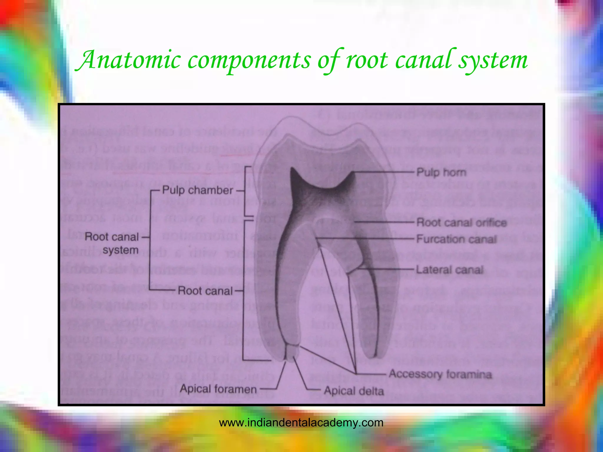
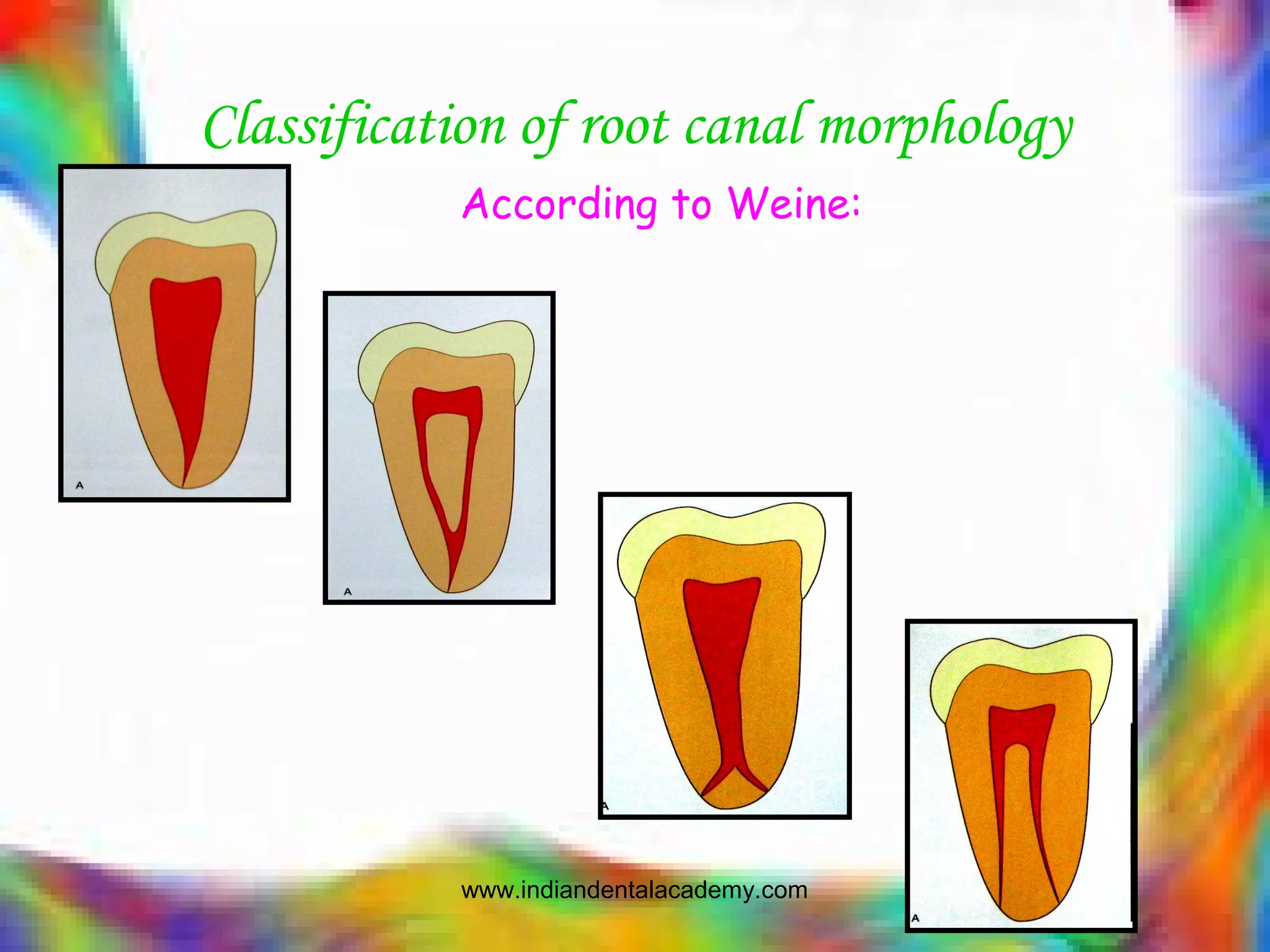
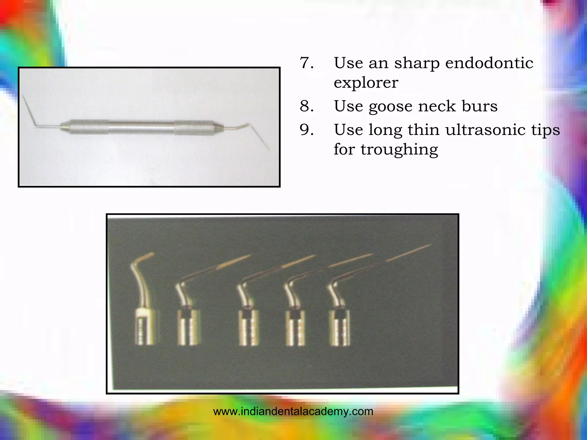
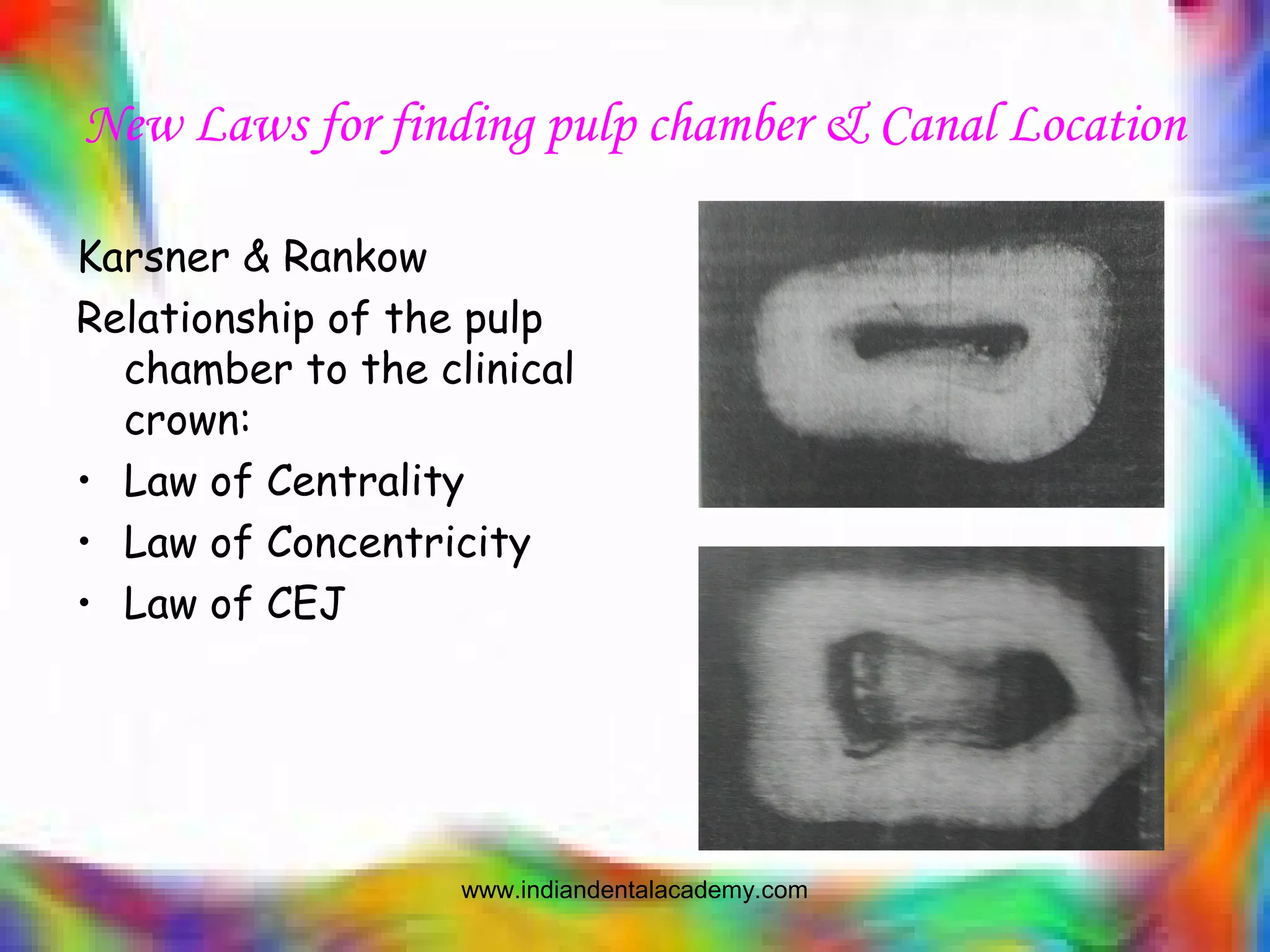
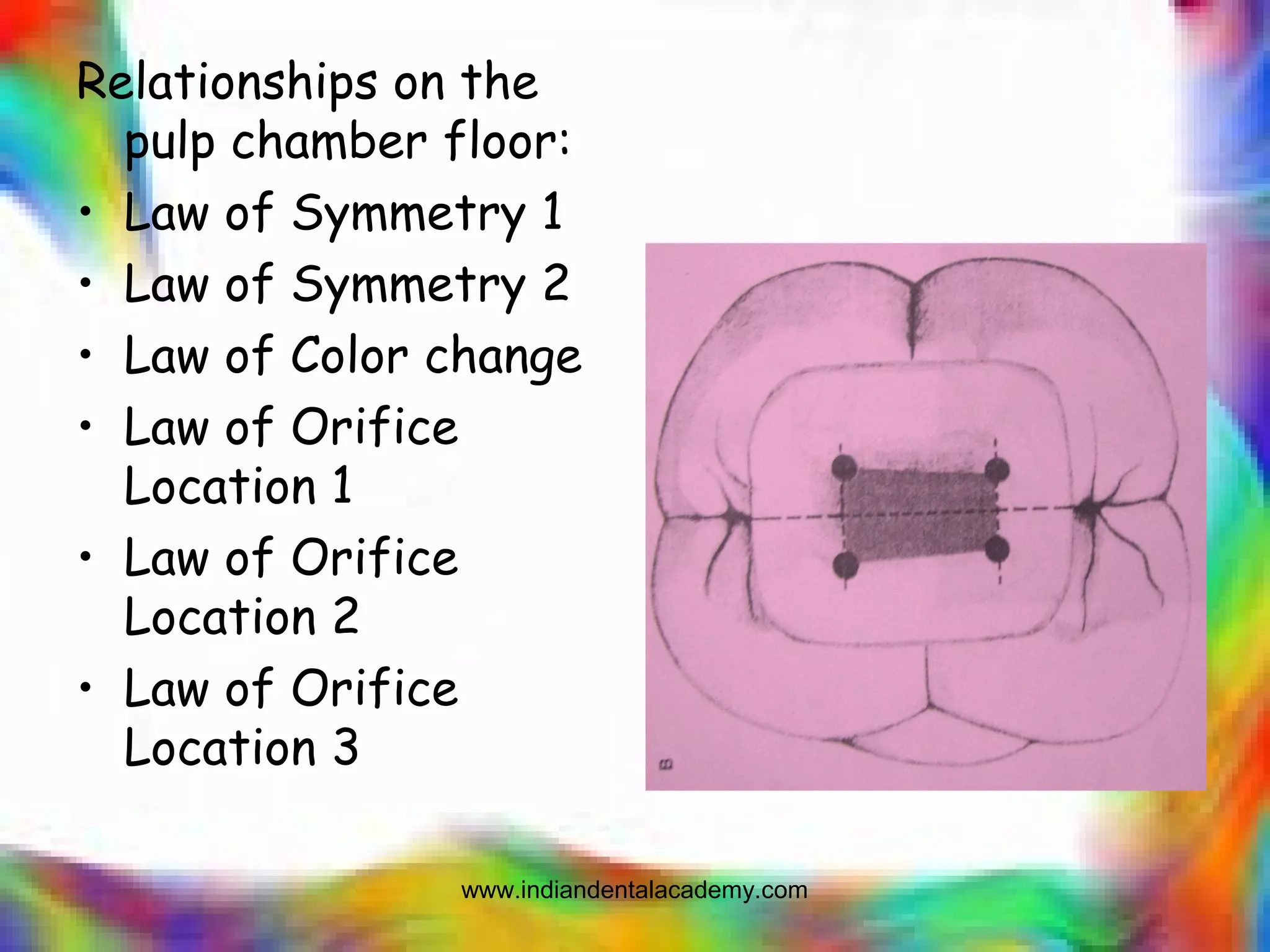
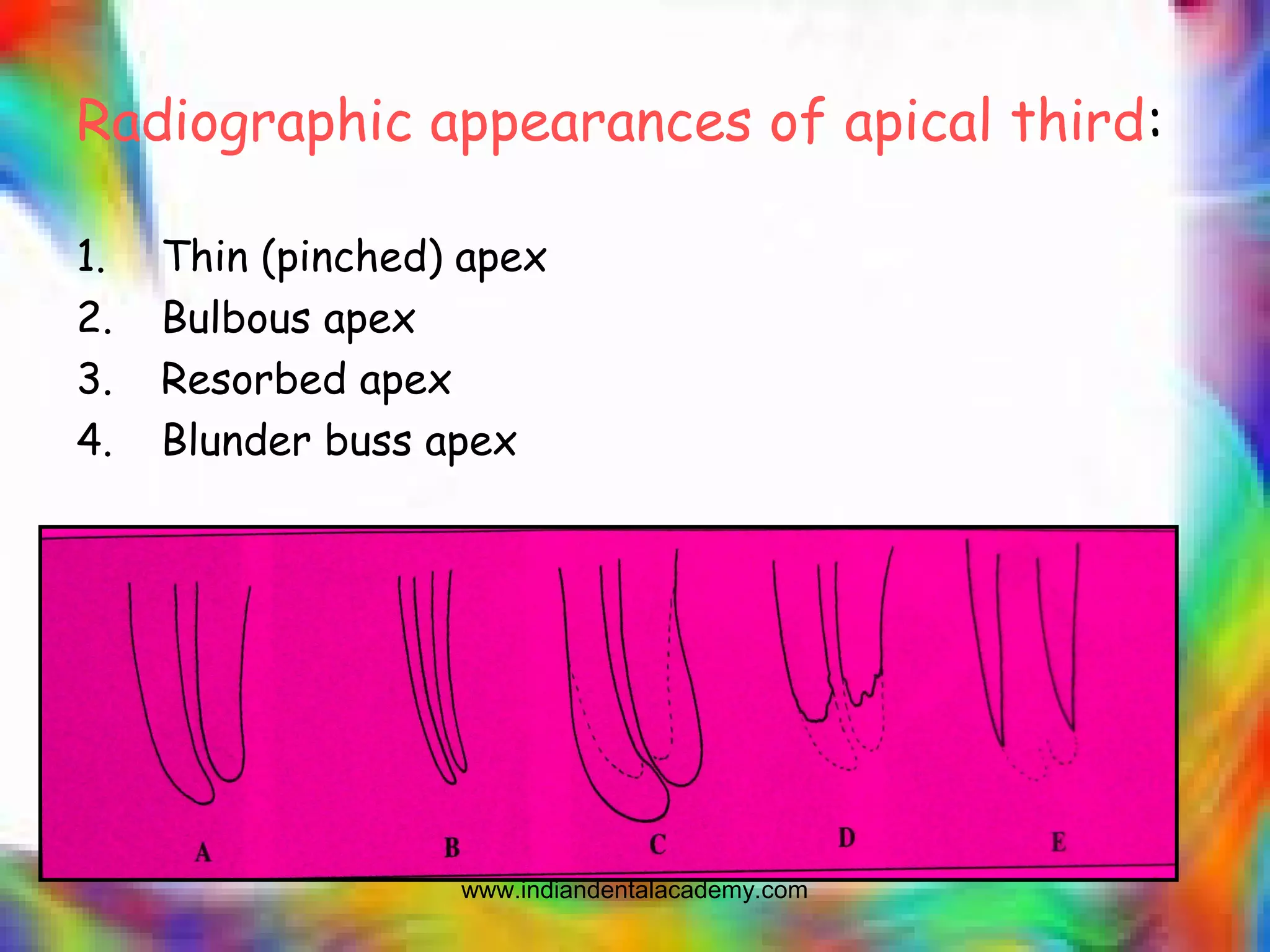
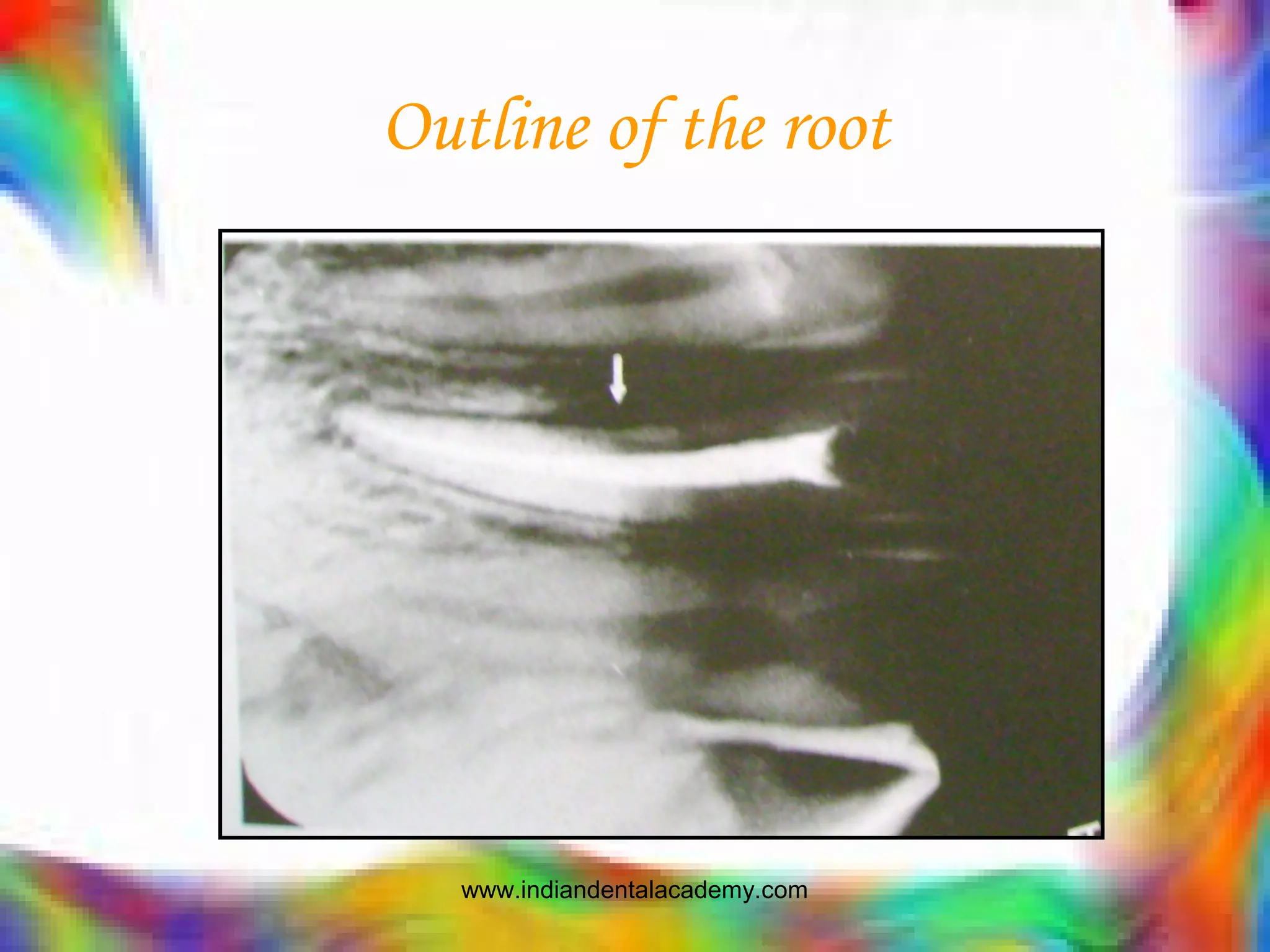
ANATOMIC CONDITIONS &
MORPHOLOGIC VARIATIONS
IN SUCCESS OF ENDODNTICS
INDIAN DENTAL ACADEMY
Leader in continuing Dental Education
www.indiandentalacademy.com
contents
• Introduction
• Anatomic components of root canal system
• Classification of root canal morphology
• General considerations
pulp chamber
orifice & coronal part of root canal
apical part of root canal
apical foramen
extra canal/s
accessory & lateral canals
buccolingual width
curved canals
age & Pathological changes
isthmus
www.indiandentalacademy.com
• Special features
C-shaped canals
developmental anomalies
blunder buss canals
• Conclusion
• References
www.indiandentalacademy.com
Introduction
• Reasons for failure of endodontic therapy
• Current advancements in the designing of
endodontic instruments and changes in the
procedural concepts are based in accordance
to the morphology of root canal system.
• Root canal anatomy being unchangeable,
judges and determines the fate of
instruments and instrumentation technique.
www.indiandentalacademy.com
Anatomic components of root canal system
www.indiandentalacademy.com
Classification of root canal morphology
According to Weine:
www.indiandentalacademy.com
According to Vertucci
www.indiandentalacademy.com
According to Gulabivala and coworkers
www.indiandentalacademy.com
GENERAL CONSIDERATIONS
PULP CHAMBER:
• Cavity situated inside the crown portion of a tooth
containing pulp tissue.
• Locating orifices
• Floor: color, contour, periphery and its nature.
• Color & contour: dark and convex.
• Periphery of the floor: distinct and appear as groove
• Nature of the floor: 50% 0f molar possess accessory
canals in the floor.
www.indiandentalacademy.com
Clinical implications:
1. Large lateral canal in the
pulp floor of a pulpally
involved molar may lead to
destruction and formation
of lesion in the furcal
bone.
2. Defective final restoration
– initiate formation of
pathological lesion in the
furca.
www.indiandentalacademy.com
Orifice & coronal part of the root canal
• Careful preparation in the early phase of
instrumentation
• Generally termed ‘flaring’ & sometimes ‘widening’
or ‘enlarging’.
www.indiandentalacademy.com
4 reasons for coronal preflaring:
1. Direct approach to apical 1/3rd.
2. Facilitates the passage of irrigation solution.
3. Debris & microorganisms found abundantly can be
eliminated which prevents inadvertent pushing of
these substance.
4. Spreaders and pluggers can be taken to adequate
depth which facilitates better obturation.
www.indiandentalacademy.com
Hints for canal orifice location
1. know the tooth
anatomy
2. Radiographs
3. Create adequate
access
4. Use adequate
illumination
5. Use magnification
6. Use the dentinal map
www.indiandentalacademy.com
7. Use an sharp endodontic
explorer
8. Use goose neck burs
9. Use long thin ultrasonic tips
for troughing
www.indiandentalacademy.com
10. Use long shank burs
11. Use microopeners and small files
12. Transillumination
www.indiandentalacademy.com
13. Use a dye
14. Sodium hypochlorite ‘champagne test’
15. Bleeding points
www.indiandentalacademy.com
New Laws for finding pulp chamber & Canal Location
Karsner & Rankow
Relationship of the pulp
chamber to the clinical
crown:
• Law of Centrality
• Law of Concentricity
• Law of CEJ
www.indiandentalacademy.com
Relationships on the
pulp chamber floor:
• Law of Symmetry 1
• Law of Symmetry 2
• Law of Color change
• Law of Orifice
Location 1
• Law of Orifice
Location 2
• Law of Orifice
Location 3
www.indiandentalacademy.com
Apical part of the root canal
• Morphologically – most
complex region
• Therapeutically –
challenging zone
• Prognostically – an
important factor
• Unfortunately – most
obscure & unclear area.
www.indiandentalacademy.com
Clinically significant features of the apical part:
1. Accessory canal:
• clinical significance:
canal 1mm long & 0.25mm
in diameter may harbor
80,000 streptococcus.
This morphologic feature
emphasizes the
importance of thorough
irrigation, cleaning,
shaping and filling of the
canal upto the apical
constriction.
www.indiandentalacademy.com
2.Divisions:
may bifurcate or
trifurcate in the apical
third and may exist as
many foramina
3.Curvature:
clinical management –
preflaring of the
coronal part
- pre curving the files
• failure to precurve or
preflare may result in
ledging, ripping,
iatrogenic canal
formation or perforation
of the apical third.
www.indiandentalacademy.com
4.Patency:
canals are almost always patent in the apical third.
clinical significance:
•knowledge of this feature is of great clinical
importance.
•Radiographic appearance of a hazy & obliterated root
canal coupled with difficulty that a clinician encounter
while tracing the orifice/s and maneuevering the
coronal part of a canal with an instrument may give a
false impression that the apical third is completely
calcified.
•This impression may not be clinically accurate; a canal
may be calcified at the orifice & coronal few mm of a
canal, but it seldom isat the apical part.
www.indiandentalacademy.com
Radiographic appearances of apical third:
1. Thin (pinched) apex
2. Bulbous apex
3. Resorbed apex
4. Blunder buss apex
www.indiandentalacademy.com
Apical foramen
Location & shape – great
endodontic significance.
Location:
Detecting the location:
1. Radiographs
2. Direction & location of
periapical lesion
3. Electronic apex locator
Clinical significance:
Overestimation: violation of
apical constriction
Underestimation: canal space
uncleaned & unfilled
www.indiandentalacademy.com
Shape:
• Usually round or oval
Current description:
1. Round
2. Oval
3. Long oval
4. flattened
www.indiandentalacademy.com
Variations in apical
constriction:
1.Traditional single
2.Tapering constriction
3.Multiconstricted
4.Parallel constriction
www.indiandentalacademy.com
Clinical significance:
• It acts as a natural stop – advantage
• Disadvantage – debris or dentinal mud get accumulated,
instrumentation become difficult and leads to mishaps
“Maintaining the original position and shape of the apical constriction are two main
objectives of root canal preparation procedure.”
Recommended steps to preserve the apical foramen:
1. Working length – measured correctly.
2. Canal patency – maintained through recapitulation.
3. Irrigation.
4. Avoid using H-file instrument before determining the working
length.
www.indiandentalacademy.com
Extra canals
• The word ‘extra canal’
• Failure to locate and treat an extra canal is one of
the common causes for failures of root canal
treatment.
• How to detect
1. Knowledge
2. Enthusiasm
3. Clinical
4. Radiograph
www.indiandentalacademy.com
SLOB rule
www.indiandentalacademy.com
Extra dark line
www.indiandentalacademy.com
Outline of the root
www.indiandentalacademy.com
Fast break
www.indiandentalacademy.com
Accessory & lateral canals
•The principal or main root canal in a root may
communicate with the surrounding periodontium at any
level through lateral & accessory canal.
•Lateral canals – perpendicular to the principal canal.
•Accessory canal – mostly found in apical third.
www.indiandentalacademy.com
Clinical significance:
1. Difficult to clean.
2. Their presence in the floor of the pulp chamber.
3. Causes post pulpectomy pain.
4. Multiple accessory canals.
Detection of lateral canals:
• Thickening of pdl.
• Development of frank lesion in the lateral waall of
the root.
• Post obturation radiograph.
www.indiandentalacademy.com
Buccolingual width
Effective endodontic treatment, the root canal of a
tooth is required to be understood in its wholeness
i.e.., 3 dimensionally
www.indiandentalacademy.com
Curved canals
How to assess the canal
curvature in the
radiograph:
1. Increased radiopacity
2. “Bull’s-eye” or “target”
appearence
www.indiandentalacademy.com
Root canal curvature measurement techniques
1. Weine angle technique
2. Long axis technique
3. Schneider angle
technique
4. Canal access angle
technique
“Failure to note this morphological
feature during endodontic
instrumentation procedure is a
common cause for failure of
root canal treatment”.
www.indiandentalacademy.com
Age & pathological changes
Secondary/tertiary dentin:
• Mostly occurs on the pulpal
floor
• Along with thickening &
elevation of pulpal floor, the
shape, size and position of the
orifice, and also the width of
cervical part of the root canal
changes.
• It is interesting & also
important to note that
dimension of the canal in the
apical third does not decrease
in size.
www.indiandentalacademy.com
Type of calcification:
1. Concentric or linear
2. Irregular
www.indiandentalacademy.com
Pulpal stones
www.indiandentalacademy.com
Apex, Apical foramen & Age
www.indiandentalacademy.com
Isthmus:
kim et al:
www.indiandentalacademy.com
SPECIAL FEATURES:
C-SHAPED CANALS:
• Thin strip of groove
• True c- shaped canal
• False c- shaped canal
• Characteristic feature
is that their propensity
to occur among Asians
and in lower second
molar.
www.indiandentalacademy.com
Clinical significance:
• Difficult to diagnose
the c-shape
radiographically
• Difficult to prepare the
canals
• Persistent hemorrhage
following initial pulp
extripation
• Interappointment pain
• No specific obturation
technique.
www.indiandentalacademy.com
DEVELOPMENTAL ANOMALIES
Dens invaginatus:
www.indiandentalacademy.com
Lingual radicular groove
• Gingivo palatal
groove
• Commonly seen in
maxillary lateral
incisors
• 2.5%
• Length & depth
www.indiandentalacademy.com
Blunder buss canal
• Canals are thin & fragile
• Proper condensation is impossible due to
absence of barrier.
• Management depends upon vitality.
www.indiandentalacademy.com
References:References:
• Pathways of the pulp; 9th
edition; Cohen
• Pathways of the pulp; 8th
edition; Cohen
• Advanced endodntics for clinicians; Jacob G.Daniel
• Endodnotic topics 2005; 10; 3-29
• Practical clinical endodontics: Churchchill
• Endodontic therapy;Weine; 6th
edition
• Endodontic practice; Grossman;11th
edition
• JOE; 2004; vol 30; N0 1; 5-16.
• JOE; 2005; No 11; Vol 31; 796-798.
• Endodontics; Stock & Gluabivala
• Ingle 5th
edition.
www.indiandentalacademy.com

Tnn /prosthodontic courses

  • 1.
    ANATOMIC CONDITIONS & MORPHOLOGICVARIATIONS IN SUCCESS OF ENDODNTICS INDIAN DENTAL ACADEMY Leader in continuing Dental Education www.indiandentalacademy.com
  • 2.
    contents • Introduction • Anatomiccomponents of root canal system • Classification of root canal morphology • General considerations pulp chamber orifice & coronal part of root canal apical part of root canal apical foramen extra canal/s accessory & lateral canals buccolingual width curved canals age & Pathological changes isthmus www.indiandentalacademy.com
  • 3.
    • Special features C-shapedcanals developmental anomalies blunder buss canals • Conclusion • References www.indiandentalacademy.com
  • 4.
    Introduction • Reasons forfailure of endodontic therapy • Current advancements in the designing of endodontic instruments and changes in the procedural concepts are based in accordance to the morphology of root canal system. • Root canal anatomy being unchangeable, judges and determines the fate of instruments and instrumentation technique. www.indiandentalacademy.com
  • 5.
    Anatomic components ofroot canal system www.indiandentalacademy.com
  • 6.
    Classification of rootcanal morphology According to Weine: www.indiandentalacademy.com
  • 7.
  • 8.
    According to Gulabivalaand coworkers www.indiandentalacademy.com
  • 9.
    GENERAL CONSIDERATIONS PULP CHAMBER: •Cavity situated inside the crown portion of a tooth containing pulp tissue. • Locating orifices • Floor: color, contour, periphery and its nature. • Color & contour: dark and convex. • Periphery of the floor: distinct and appear as groove • Nature of the floor: 50% 0f molar possess accessory canals in the floor. www.indiandentalacademy.com
  • 10.
    Clinical implications: 1. Largelateral canal in the pulp floor of a pulpally involved molar may lead to destruction and formation of lesion in the furcal bone. 2. Defective final restoration – initiate formation of pathological lesion in the furca. www.indiandentalacademy.com
  • 11.
    Orifice & coronalpart of the root canal • Careful preparation in the early phase of instrumentation • Generally termed ‘flaring’ & sometimes ‘widening’ or ‘enlarging’. www.indiandentalacademy.com
  • 12.
    4 reasons forcoronal preflaring: 1. Direct approach to apical 1/3rd. 2. Facilitates the passage of irrigation solution. 3. Debris & microorganisms found abundantly can be eliminated which prevents inadvertent pushing of these substance. 4. Spreaders and pluggers can be taken to adequate depth which facilitates better obturation. www.indiandentalacademy.com
  • 13.
    Hints for canalorifice location 1. know the tooth anatomy 2. Radiographs 3. Create adequate access 4. Use adequate illumination 5. Use magnification 6. Use the dentinal map www.indiandentalacademy.com
  • 14.
    7. Use ansharp endodontic explorer 8. Use goose neck burs 9. Use long thin ultrasonic tips for troughing www.indiandentalacademy.com
  • 15.
    10. Use longshank burs 11. Use microopeners and small files 12. Transillumination www.indiandentalacademy.com
  • 16.
    13. Use adye 14. Sodium hypochlorite ‘champagne test’ 15. Bleeding points www.indiandentalacademy.com
  • 17.
    New Laws forfinding pulp chamber & Canal Location Karsner & Rankow Relationship of the pulp chamber to the clinical crown: • Law of Centrality • Law of Concentricity • Law of CEJ www.indiandentalacademy.com
  • 18.
    Relationships on the pulpchamber floor: • Law of Symmetry 1 • Law of Symmetry 2 • Law of Color change • Law of Orifice Location 1 • Law of Orifice Location 2 • Law of Orifice Location 3 www.indiandentalacademy.com
  • 19.
    Apical part ofthe root canal • Morphologically – most complex region • Therapeutically – challenging zone • Prognostically – an important factor • Unfortunately – most obscure & unclear area. www.indiandentalacademy.com
  • 20.
    Clinically significant featuresof the apical part: 1. Accessory canal: • clinical significance: canal 1mm long & 0.25mm in diameter may harbor 80,000 streptococcus. This morphologic feature emphasizes the importance of thorough irrigation, cleaning, shaping and filling of the canal upto the apical constriction. www.indiandentalacademy.com
  • 21.
    2.Divisions: may bifurcate or trifurcatein the apical third and may exist as many foramina 3.Curvature: clinical management – preflaring of the coronal part - pre curving the files • failure to precurve or preflare may result in ledging, ripping, iatrogenic canal formation or perforation of the apical third. www.indiandentalacademy.com
  • 22.
    4.Patency: canals are almostalways patent in the apical third. clinical significance: •knowledge of this feature is of great clinical importance. •Radiographic appearance of a hazy & obliterated root canal coupled with difficulty that a clinician encounter while tracing the orifice/s and maneuevering the coronal part of a canal with an instrument may give a false impression that the apical third is completely calcified. •This impression may not be clinically accurate; a canal may be calcified at the orifice & coronal few mm of a canal, but it seldom isat the apical part. www.indiandentalacademy.com
  • 23.
    Radiographic appearances ofapical third: 1. Thin (pinched) apex 2. Bulbous apex 3. Resorbed apex 4. Blunder buss apex www.indiandentalacademy.com
  • 24.
    Apical foramen Location &shape – great endodontic significance. Location: Detecting the location: 1. Radiographs 2. Direction & location of periapical lesion 3. Electronic apex locator Clinical significance: Overestimation: violation of apical constriction Underestimation: canal space uncleaned & unfilled www.indiandentalacademy.com
  • 25.
    Shape: • Usually roundor oval Current description: 1. Round 2. Oval 3. Long oval 4. flattened www.indiandentalacademy.com
  • 26.
    Variations in apical constriction: 1.Traditionalsingle 2.Tapering constriction 3.Multiconstricted 4.Parallel constriction www.indiandentalacademy.com
  • 27.
    Clinical significance: • Itacts as a natural stop – advantage • Disadvantage – debris or dentinal mud get accumulated, instrumentation become difficult and leads to mishaps “Maintaining the original position and shape of the apical constriction are two main objectives of root canal preparation procedure.” Recommended steps to preserve the apical foramen: 1. Working length – measured correctly. 2. Canal patency – maintained through recapitulation. 3. Irrigation. 4. Avoid using H-file instrument before determining the working length. www.indiandentalacademy.com
  • 28.
    Extra canals • Theword ‘extra canal’ • Failure to locate and treat an extra canal is one of the common causes for failures of root canal treatment. • How to detect 1. Knowledge 2. Enthusiasm 3. Clinical 4. Radiograph www.indiandentalacademy.com
  • 29.
  • 30.
  • 31.
    Outline of theroot www.indiandentalacademy.com
  • 32.
  • 33.
    Accessory & lateralcanals •The principal or main root canal in a root may communicate with the surrounding periodontium at any level through lateral & accessory canal. •Lateral canals – perpendicular to the principal canal. •Accessory canal – mostly found in apical third. www.indiandentalacademy.com
  • 34.
    Clinical significance: 1. Difficultto clean. 2. Their presence in the floor of the pulp chamber. 3. Causes post pulpectomy pain. 4. Multiple accessory canals. Detection of lateral canals: • Thickening of pdl. • Development of frank lesion in the lateral waall of the root. • Post obturation radiograph. www.indiandentalacademy.com
  • 35.
    Buccolingual width Effective endodontictreatment, the root canal of a tooth is required to be understood in its wholeness i.e.., 3 dimensionally www.indiandentalacademy.com
  • 36.
    Curved canals How toassess the canal curvature in the radiograph: 1. Increased radiopacity 2. “Bull’s-eye” or “target” appearence www.indiandentalacademy.com
  • 37.
    Root canal curvaturemeasurement techniques 1. Weine angle technique 2. Long axis technique 3. Schneider angle technique 4. Canal access angle technique “Failure to note this morphological feature during endodontic instrumentation procedure is a common cause for failure of root canal treatment”. www.indiandentalacademy.com
  • 38.
    Age & pathologicalchanges Secondary/tertiary dentin: • Mostly occurs on the pulpal floor • Along with thickening & elevation of pulpal floor, the shape, size and position of the orifice, and also the width of cervical part of the root canal changes. • It is interesting & also important to note that dimension of the canal in the apical third does not decrease in size. www.indiandentalacademy.com
  • 39.
    Type of calcification: 1.Concentric or linear 2. Irregular www.indiandentalacademy.com
  • 40.
  • 41.
    Apex, Apical foramen& Age www.indiandentalacademy.com
  • 42.
  • 43.
  • 44.
    SPECIAL FEATURES: C-SHAPED CANALS: •Thin strip of groove • True c- shaped canal • False c- shaped canal • Characteristic feature is that their propensity to occur among Asians and in lower second molar. www.indiandentalacademy.com
  • 45.
    Clinical significance: • Difficultto diagnose the c-shape radiographically • Difficult to prepare the canals • Persistent hemorrhage following initial pulp extripation • Interappointment pain • No specific obturation technique. www.indiandentalacademy.com
  • 46.
  • 47.
    Lingual radicular groove •Gingivo palatal groove • Commonly seen in maxillary lateral incisors • 2.5% • Length & depth www.indiandentalacademy.com
  • 48.
    Blunder buss canal •Canals are thin & fragile • Proper condensation is impossible due to absence of barrier. • Management depends upon vitality. www.indiandentalacademy.com
  • 49.
    References:References: • Pathways ofthe pulp; 9th edition; Cohen • Pathways of the pulp; 8th edition; Cohen • Advanced endodntics for clinicians; Jacob G.Daniel • Endodnotic topics 2005; 10; 3-29 • Practical clinical endodontics: Churchchill • Endodontic therapy;Weine; 6th edition • Endodontic practice; Grossman;11th edition • JOE; 2004; vol 30; N0 1; 5-16. • JOE; 2005; No 11; Vol 31; 796-798. • Endodontics; Stock & Gluabivala • Ingle 5th edition. www.indiandentalacademy.com
  • 50.