
Recommended
More Related Content
What's hot
What's hot (20)
Similar to Pharynx
Similar to Pharynx (20)
Recently uploaded
Recently uploaded (20)
Pharynx
- 1. PHARYNX
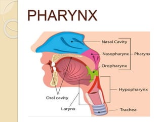
- 2. Musculomembranous tube Extent: Base of skull to C6 vertebra Interior of pharynx divided in to 3 Parts 1.NASOPHARYNX 2.OROPHARYNX 3.HYPOPHARYNX/ LARYNGOPHARYNX
- 3. NASOPHARYNX -Behind nasal cavity and above soft palate ANTERIOR WALL: deficient. Communicates with nasal cavity through posterior nasal aperture(choanae)
- 5. ROOF AND POSTERIOR WALL: Shenoid ,Atlas,Occipital bone(basilar part)
- 6. FLOOR: Soft palate and pharyngeal isthmus(space between soft palate and posterior pharyngeal wall)
- 8. FEATURES OF NASOPHARYNX 1.NASOPHARYNGEAL TONSIL: collection of lymphoid tissue under mucous membrane. When infected,enlarged to form adenoids
- 9. 2.PHARYNGEAL BURSA (Pouch of luschka):Mucous diverticulum extending in to substance of nasopharyngeal tonsil 3.PHARYNGEAL HYPOPHYSIS: Glandular tissues similar to adenohypophysis derived from remnant of Rathke’s pouch
- 10. 4.PHARYNGEAL OPENING OF AUDITORY TUBE: 1.25 cm behind inferior nasal conchae
- 11. 5. A TUBAL ELEVATION :guards the upper and posterior margin of auditory opening. Produced by collection of lymphoid tissue –TUBAL TONSIL TWO MUCOUS FOLDS:extending from tubal elevation a)salpingopharyngeal fold b)salpingo-palatine fold
- 12. 6.PHARYNGEAL RECESS(Fossa OF Rosenmuller):mucus covered deep depression behind tubal elevation
- 13. OROPHARYNX
- 14. OROPHARYNX Lies behind oral cavity supported by C2 and C3 vertebrae ANTERIOR WALL:Communicates with oralcavity through oropharyngeal isthmus
- 15. OROPHARYNGEAL ISTHMUS: Above soft palate Below posterior 1/3rd tongue On each side palatoglossal arch
- 16. Below oropharynx communicates with laryngo- pharynx at level of epiglottis ROOF: soft palate and pharyngeal isthmus FLOOR:Posterior 1/3rd of tongue
- 17. OROPHARYNX-FEATURES 1.Palatine tonsil-Lateral wall 2.lingual tonsil-lymphoid tissue in pharyngeal part of dorsum of tongue 3.upper free end of epiglottis 4.median and lateral glossoepiglottic fold 5.epiglottic valleculae – shallow fossa between median and lateral glossoepiglottic fold
- 18. LARYNGOPHARYNX behind the laryngeal inlet and posterior wall of larynx ANTERIOR: communicates with laryngeal cavity through laryngeal inlet POSTERIOR:C4 toC6 vertebra INFERIOR:Oesophagus at pharyngoesophageal junction LATERAL:Thyroid cartilage and thyrohyoid membrane
- 19. LARYNGOPHARYNX- FEATURES Anterior wall:laryngeal inlet below supported by cricoid and arytenoid cartilages Lateral wall:pyriform fossa on either side of laryngeal inlet
- 20. PYRIFORM FOSSA: (Smugglers fossa) Medially:Aryepiglottic fold laterally: mucous membrane covering the medial surface of thyroid cartilage and thyrohyoid membrane Internal laryngeal nerve and superior laryngeal vessels pierce thyrohyoid membrane Above:seperated from epiglottic vallecula by lateral glossoepiglottic fold
- 24. PHARYNGEAL WALL 4 LAYERS 1.Mucous membrane 2.pharyngobasilar fascia(pharyngeal aponeurosis) 3.muscular coat(pharyngeal muscles) 4.Buccopharyngeal
- 25. WALDEYER’S RING Collection of lymphoid tissue under epithelial lining of pharynx called tonsil Surround commencement of air and food passages These aggregations together constitute an interrupted circle-Waldeyer’s ring
- 26. Waldeyer’s ring formed by 1.pharyngeal tonsil,posterosuperiorly 2.lingual tonsil, anteriorly 3.tubal and palatine tonsils laterally
- 29. DIVERTICULUM Inferior constrictor muscle has two parts:thyropharyngeu s and cricopharyngeus Gap between these – killian’s dehiscence. Mucosa and submucosa of pharynx may bulge through this weak
- 30. LONGITUDINAL MUSCLES 1.Stylopharyngeus 2.Palatopharyngeus 3.Salpingopharyngeu s All inserted to posterior border of lamina of thyroid cartilage
- 31. PASSAVANT’S RIDGE:Fibres of palatopharyngeus and superior constrictor form a U shaped muscle loop in posterior pharyngeal wall under mucosa to form passavant’s ridge
- 32. GAPS IN PHARYNGEAL WALL
- 34. Between base of skull and superior constrictor(sinus of morgagni) 1.auditory tube 2.levator palati muscle 3.Ascending palatine artery 4.palatine branch of ascending pharyngeal artery
- 35. Between superior and middle constrictor 1.stylopharyngeus muscle 2.Glossopharyngeal nerve Between middle and inferior constrictor 1.Internal laryngeal nerve 2.Superior laryngeal vessels Below Inferior constrictor 1.Recurrent laryngeal nerve 2.Inferior laryngeal vessels
- 36. NERVE SUPPLY OF PHARYNX MOTOR:All muscles are supplied by cranial part of accessory through pharyngeal plexus EXCEPT Stylopharyngeus supplied by glossopharyngeal nerve. SENSORY: Nasopharynx-pterygopalatine ganglion Oropharynx-glossopharyngeal nerve Laryngopharynx-internal laryngeal nerve
- 37. ARTERIAL SUPPLY OF PHARYNX 1.Ascending pharyngeal artery 2.Ascending palatine and tonsillar artery 3.Greater palatine and pharyngeal artery 4.Lingual artery Venous drainage by pharyngeal venous plexus. It drains into IJV LYMPHATICS:Upper and lower deep cervical lymph nodes through retropharyngeal nodes