SlideShare a Scribd company logo
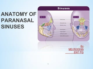
ANATOMY OF
PARANASAL
SINUSES

1
MAXILLA:

*Mxilla develops during 6-7wks
from 5 ossification centres.

*These ossification centres gives
rise to
alveolar,palatine,zygomatic&
frontal processes of maxilla &
floor of the orbit.

*Ossification centre in the medial
floor of the pyriform apperture
forms the premaxilla.

*Premaxilla gives rise to upper
incisors & lower nasal spine.

2
ETHMOID:
•Ethmoid ossifies in the
cartilagenous nasal capsule from
3 centres.
•One centre for each labyrinth
&one for perp.plate of ethmoid.
•These centres appears during 45th IUL.
•Perp.plate &crista galli
developed from the same centre
during 1st yr of life & fuses with
labyrinth during 2nd yr.

3
FRONTAL:

Develops from 2 centres
during 8th wk.

Centres are present in
superciliary ridge.

At birth frontal bone –

2halves separated by frontal
or metopic suture.

Development complete by
2yrs.

4
SPHENOID:
 Develops from presphenoidal
&postsphenoidal portions.
These portions fuse during 8th IUM.
Central portion- body & lesser wings.
Lateral portion- greater wing &pterygoid
process.
These portions fuse during 1st yr of life.
Presphenoid portion: made of 6
ossif.centres.
Lies ant. To tuberculm sella.
Continous with the lesser wings of
sphenoid.
Postsphenoidportion: 8 ossif.c
Coposed of sella tursica &dorsum sella.
Gives rise to greater wings & pterygoid
process.

5
* Frontal,maxillary and

ethmoidal sinuses arise
from evagination of
lateral nasal wall of
nasal capsule.

* Sphenoid sinus arises
from a posterior
evagination of nasal
capsule.

6
* At 25-28wks IUG 3 medially

projections formed from lateral
wall of nose from this PNS are
developed.
* Anterior projection;agger nasi.
* Inferior projection (maxilloterbinal): inf.terbinate &
max.sinus.
* Superior
projection(ethmo_terbinal):
superior terbinate, middile.t, ant.
Ethmoidal cells &corresponding
drainage channels form.
* Middle meatus invaginates
laterally to form infundibulum
&UP.
* Infundibulum grows superiorly to
form frontal recess.

7
Frontal sinus:

*It develops during 4th foetal
month as an outpouching,
medial to most superior
aspect of uncinate process.

*It is very rudimentary at

birth, it developes in
childhood by upward
continuation of embryonic
infundibulum & frontal recess.

*Embryologically it can also
devlp from ant.ethmoidal air
cells.

8
Mxillary sinus:

•It develops from primitive
ethmoidal infundibulum in
to the mass of maxilla.

•It enlarges by absorbtion
and expantion.

•At 12yrs pneumatisation

reaches under lateral
orbital wall at insertion of
zygomatic process , inferior
to the nasal floor.

9
* Ethmoid sinus:
* Develops fromlateral wall of
th

nasal capsule at 9 to10th wk.
* 6-7 folds appear,these folds
are separated by grooves.
* Folds are fuse to form 3-4
crests with anteriorly ramus
ascendus and posteriorly
ramus descendus.
* From the main fold middle,
superior ,supreme terbinates
form.
* From inferior fold inferior
terbinate form so called
maxillo-turbinal.

10
Sphenoid sinus:
It is a evagination from
sphenoethmoid recess at 3rd
month of gestation.
At 7thyr reaches floor of
sella.
Pneumatisation progress
rate 0.25mm/yr frm 4yr of
age.
In some cases internal
carotid artery and optic.n
may lie naked with in the
sinus cavity.

11
Paranasal sinuses:INTRODUCTION
These are air filled cavities in relation to nasal cavities.
*Classified in 2 groups anterior & posterior.
Four on each side:

(labyri

(labyrinth )

12
They are lined with a mucous membrane continuous with that of the
corresponding nasal fossa through their ostia.
Anterior group:

I.Frontal sinus.
II.Anterior ethmoidal cells.
III.Maxillary sinus.
Posterior group:

I.Posterior ethmoidal sinuses.
II.Sphenoid sinus.

13
Maxillary sinus:
*It is the largest of the sinuses,
with an average capacity of
about 10-20 ml in the adult.

* Is pyramidal in shape and
occupies the body of the
maxilla.

* The base lies medially,
the apex is in the zygomatic
portion of the maxilla.

* Medial wall is the wall
between the sinus and the
nasal fossa.

*

Dimensions :
height)3.3cm(
width)2-3cm(
ant.post)3-4cm(
14
* Floor :is formed by the alveolar
process and hard palate:
Œ- In children the floor lies
at, or above, the level of
the floor of the nasal
fossa.
Œ- In adults it lies about
1.25cm below the floor of
the fossa.

1,25cm

Œ- The roots of several teeth
may project into, or even
perforate, the floor.
15
The ostia of maxillary
sinus:
*Main ostium is situated
high up between the
medial wall and roof of
the cavity.
It opens into the
hiatus semilunaris.
*Accessory ostia are
sometimes present,
behind the main one.
Both main and accessory
ostia are surrounded by
a wide area of mucous
membrane unsupported
by bone.
16
Relations of maxillary sinus:
1*Orbit: is separated from
the antrum by the thin
roof of the sinus which
contains the infraorbital
nerve.

2*Teeth: may produce
elevations in the floor of
the sinus and the number
of related teeth depends
on the size of the antrum.
The second premolar and
first molar are usually
related.
17
3* Middle meatus of nose:
is related to the upper
part of the antrum.

4* Inferior meatus of nose:
is separated from the middle
part of its medial wall by bone,
which is usually thick in front
and below,but thinner above and
behind.

18
5* maxillary

artery :

is related to the posterior
wall, where it occupies the
pterygopalatine fossa.
It may be approached
through the antrum for
ligature.
6* Maxillary

division of
the Vth cranial nerve:
also traverses the pterygopalatine fossa.

19
7* Nasolacrimal

duct:

passes downwards,
medial to the antrum,
to open into the inferior
meatus.

maxillary sinus

20
ARTERIAL:

oBy facial artery branch of ECA.
oBy infra orbital & greater palatine

arteries branch of max. art which is
branch of ECA.

Infraorbital
artery

VENOUS:

oTo anterior facial vein& pterygoid
plexus.

superior dental
arteries

21
o

Maxillary division of trigeminal.n gives sensory
supply via.

o
o

Infraorbital.n ,sup.alveolar.n , greater palatine.n.

o

Also supply-pulps of canine, incisors teeth,
ant.inf.quadrant of lat.wall of nose,floor of nose,ant.
Part of nasal septum.

o
o
o
o

Middle sup. Alveolar.n-

o
o
o

Adjacent mucosa,molar teeth.

Ant superior alveolar.n- ant. Wall of antrum passing
through canalis sinosus.

Lateral wall of sinus, upper pre molar teeth.
Posterior.sup.alveolar.n
Through pterygo palatine fossa supply posterior
sinus wall.
Greater palatine.n – posterio medial wall of sinus.
Perforating branches of infra orbital.n –roof of sinus.

22
Ethmoid means sieve
like
Ethmoid is trapezoid box
narrow/taller anteriorly
wide posteriorly.
Multiple air containing
cells situated
in ethmoidal labyrinth(318)

23
Anterior group-(drains –middle
meatus)
Middle group(drains-middle
meatus)
Posterior group(drainssuperior meatus)
Haller cell..ant ethmoidal cells seen
anteriorly & below the orbit
Onadi cells… posterior ethmoidal
cells seen just in front of sphenoid

24
Type I: depth of olfactory fossa 1-3mm
(26.3%)
Type II: 4-7mm(73.3%).
TypeIII:8-16MM(0.5%).

25
Anterior ethmoidal
artery(ophthalmic artery)
Post. Ethmoidal artery
Sphenoidal
artery(maxillary artery)

Anterior ethmoid
artery

Venous drainage
Nasal veins
Ant. Ethmoidal vein
Post. Ethmoidal vein
26

posterior ethmoidal
artery
Ethmoidal aircells

recieves innervation
from anterior &posterior
ethmoidal.n & orbital
branches of
pterygopalatine
ganglion.

Postganglionic

parasympathetic fibres
for mucous secreton
from facial .n

27
Frontal sinus:
*Should be regarded as an
upward extension of an
anterior ethmoidal cell.
*It occupies a very variable
extent of the frontal bone
and may be partly loculated.
*Its average capacity is about
5-10ML in the adult.
* The right and left sinuses
are often asymmetrical.
* Dimensions…height(28-32mm)
width(24-26mm)
depth(18-20mm)

28
*They are separated by a thin
bony septum, which may be
deficient in part.
* The sinus may invade the
orbital plate of the frontal
bone and occasionally it
extends to the optic foramen.

29
Relations of frontal sinuses:
Anterior cranial fossa:
separated from the sinus by
the compact bone of its
posterior wall.
Orbit: lies below the floor of
the sinus. This is also compact
bone which may rarely be
deficient.
Skin and periosteum of forehead:
cover the anterior wall, which is
of diploic bone and is related
To supratrochlear and
supraorbital
nerves.
30
*Type1…single frontal recess

cell above the agger nasi cell,
but below the frontal sinus
*Type11…A tire of more than
one cell in frontal recess above
the agger nasi cell, but below
the frontal sinus
*Type111…large single cell
pneumatizing cephaloid into
frontal sinus
*Type1V…single isolated cell
within the frontal sinus
31
The frontonasal duct:
*It passes through the anterior
part of the ethmoidal labyrinth.
* Its length and curvature vary
considerably.
* Its lower end (ostium) usually
opens in to the infundibulum,
less often independently above
this level.

32
* Drainage into frontal recess

anterior to the
infundibulum(55%)
* Drainage above but not into the
infundibulum(30%)
* Drainage into
infundibulum(15%)
* Drainage above the bulla(1%)

33
Blood supply of frontal sinus
*Supraorbital artery
*Supratrochlear artery
Venous drainage
*Small vein that unites the Supraorbital and
Superior ophthalmic veins.
Nerve supply
*Supraorbital nerve(ophthalmic nerve)
*Supratrochlear nerve(ophthalmic nerve)
Lymphatic drainage
*Submandibular nodes

34
Sphenoidal sinus:
*Lies behind the upper part
of the nasal fossa.
* It occupies the body, and
some­times the wings and
pterygoid processes, of the
sphenoid bone.
* The average capacity is
about 7 ml in the adult.
* The right and left sinuses
are rarely symmetrical.
* They are separated by a
septum which may be
deficient in part and is
often oblique.
* Dimensions … (L..4­44mm)
W(25­34mm)h(5­33)

35
1.

conchal…(3%)

2.

Presellar(11%)
36
37
The ostium of sphenoid sinus:
*situated in the upper part of
the anterior wall of the sinus.
*It communicates with the
superior meatus indirectly
through the sphenoethmoidal
recess.

38
Relations:
*Cavernous sinus lies laterally containing the:
IIIrd,
IVth,
Vth (ophthalmic
and maxillary­
divisions) and
VIth cranial nerves,
internal carotid artery
optic nerve

39
*Above the sinus there are:
Pituitary gland,
optic chiasm,
frontal lobe of brain
olfactory tract

The pituitary gland may be approached surgically through the sinus.

40
*Posterior ethmoidal

artery(roof of sinus)
*Sphenopalatine
artery(floor of sinus)

Nerve supply

*Trigeminal (I/II div)
Lymphatics

*Retropharyngeal nodes
to upper deep cervical
nodes.

41
42
43
* Lined by mucus membrane
* Ciliated columnar epithelium
* goblet cells secretes mucus
* Cilia are more marked near
ostia.

44
*Plain sinus

radiography

*Computed

tomography

*Magnetic

resonance imaging

45
* Radiographic positions to study the paranasal

sinuses are standardised around three positions:
* 1. Two anatomical - namely coronal and sagittal
* 2. One radiographic - termed as radiographic base
line..
* The various radiographic positions used to study
paranasal sinuses are:
1. Occipito-mental view (Water's view)
2. Occipital-frontal view (Caldwell view)
3. Submento-vertical position (Hirtz position/jug
handle)
4. Lateral view
5. Oblique view 39 Degrees oblique (Rhese
position)

46
Fronta.s

orbit

ethmoids
ns

Infra orbital
margin

Max.sinus

47
Frontal.s
Ethmoid air.c
orbit

Sphenoid.b
ns

Hard palate

Max.s

Inferior.t

48
mandible
49
50
51
52
MAXILLARY.S

53
54
55
56
57
58

More Related Content

What's hot

Anatomy of parapharyngeal space and its tumours
Anatomy of parapharyngeal space and its tumoursAnatomy of parapharyngeal space and its tumours
Anatomy of parapharyngeal space and its tumours
jassicajassica
 
Mucosal folds and ventilation of middle ear
Mucosal folds and ventilation of middle ear  Mucosal folds and ventilation of middle ear
Mucosal folds and ventilation of middle ear
AlkaKapil
 
Deep Neck Spaces
Deep Neck Spaces Deep Neck Spaces
Deep Neck Spaces
sarita pandey
 
Surgical anatomy of Infratemporal fossa. by Dr. Aditya Tiwari
Surgical anatomy of Infratemporal fossa. by Dr. Aditya TiwariSurgical anatomy of Infratemporal fossa. by Dr. Aditya Tiwari
Surgical anatomy of Infratemporal fossa. by Dr. Aditya Tiwari
Aditya Tiwari
 
Surgical anatomy of nose
Surgical anatomy of noseSurgical anatomy of nose
Surgical anatomy of nose
Pratima Jaiswal
 
Anatomy of Skullbase
Anatomy of SkullbaseAnatomy of Skullbase
Anatomy of Skullbase
Jinu Iype
 
anatomy of Para nasal sinus with clinical anatomy
anatomy of Para nasal sinus with clinical anatomyanatomy of Para nasal sinus with clinical anatomy
anatomy of Para nasal sinus with clinical anatomy
Ramagopalan Surenthiran
 
Sphenoid sinus and optic nerve
Sphenoid sinus and optic nerveSphenoid sinus and optic nerve
Sphenoid sinus and optic nerve
Dr Soumya Singh
 
Middle Ear Anatomy - Arjun Antony Graison
Middle Ear Anatomy - Arjun Antony GraisonMiddle Ear Anatomy - Arjun Antony Graison
Middle Ear Anatomy - Arjun Antony Graison
Arjun Graison
 
SURGICAL ANATOMY OF DEEP NECK SPACES
SURGICAL ANATOMY OF DEEP NECK SPACESSURGICAL ANATOMY OF DEEP NECK SPACES
SURGICAL ANATOMY OF DEEP NECK SPACES
Ajay Manickam
 
Spaces of middle ear and their surgical importance
Spaces of middle ear  and their surgical importanceSpaces of middle ear  and their surgical importance
Spaces of middle ear and their surgical importance
Dr Soumya Singh
 
Nasal Cavity and Sphenoid Sinus Anatomy
Nasal Cavity and Sphenoid Sinus AnatomyNasal Cavity and Sphenoid Sinus Anatomy
Nasal Cavity and Sphenoid Sinus Anatomy
Farrukh Javeed
 
Anatomy of nose and para nasal sinuses . by DR. MD. KHURSHID PERVEJ. GMC PATIALA
Anatomy of nose and para nasal sinuses . by DR. MD. KHURSHID PERVEJ. GMC PATIALAAnatomy of nose and para nasal sinuses . by DR. MD. KHURSHID PERVEJ. GMC PATIALA
Anatomy of nose and para nasal sinuses . by DR. MD. KHURSHID PERVEJ. GMC PATIALA
kbristi12
 
Surgical anatomy of Maxillary Sinus , Lateral wall of nose and Soft Palate
Surgical anatomy of Maxillary Sinus , Lateral wall of nose and Soft Palate Surgical anatomy of Maxillary Sinus , Lateral wall of nose and Soft Palate
Surgical anatomy of Maxillary Sinus , Lateral wall of nose and Soft Palate
Dr. Hani Yousuf
 
Anatomy of deep neck spaces
Anatomy of deep neck spacesAnatomy of deep neck spaces
Anatomy of deep neck spaces
Dr. Rajendra Singh Lakhawat
 
Anatomy of larynx
Anatomy of larynxAnatomy of larynx
Anatomy of larynx
Jinu Iype
 
Sinus Anatomy Lectures: Western Residents Advanced Endsocopic Sinus Surgery
Sinus Anatomy Lectures: Western Residents Advanced Endsocopic Sinus SurgerySinus Anatomy Lectures: Western Residents Advanced Endsocopic Sinus Surgery
Sinus Anatomy Lectures: Western Residents Advanced Endsocopic Sinus Surgery
Elisabeth Ference
 
Anatomy of nose
Anatomy of noseAnatomy of nose
Anatomy of nose
Priyanka Shastri
 
Anatomy of temporal bone and it’s surgical importance
Anatomy of temporal bone and it’s surgical importanceAnatomy of temporal bone and it’s surgical importance
Anatomy of temporal bone and it’s surgical importance
sritama1988
 
Paranasal sinuses
Paranasal sinusesParanasal sinuses
Paranasal sinuses
mgmcri1234
 

What's hot (20)

Anatomy of parapharyngeal space and its tumours
Anatomy of parapharyngeal space and its tumoursAnatomy of parapharyngeal space and its tumours
Anatomy of parapharyngeal space and its tumours
 
Mucosal folds and ventilation of middle ear
Mucosal folds and ventilation of middle ear  Mucosal folds and ventilation of middle ear
Mucosal folds and ventilation of middle ear
 
Deep Neck Spaces
Deep Neck Spaces Deep Neck Spaces
Deep Neck Spaces
 
Surgical anatomy of Infratemporal fossa. by Dr. Aditya Tiwari
Surgical anatomy of Infratemporal fossa. by Dr. Aditya TiwariSurgical anatomy of Infratemporal fossa. by Dr. Aditya Tiwari
Surgical anatomy of Infratemporal fossa. by Dr. Aditya Tiwari
 
Surgical anatomy of nose
Surgical anatomy of noseSurgical anatomy of nose
Surgical anatomy of nose
 
Anatomy of Skullbase
Anatomy of SkullbaseAnatomy of Skullbase
Anatomy of Skullbase
 
anatomy of Para nasal sinus with clinical anatomy
anatomy of Para nasal sinus with clinical anatomyanatomy of Para nasal sinus with clinical anatomy
anatomy of Para nasal sinus with clinical anatomy
 
Sphenoid sinus and optic nerve
Sphenoid sinus and optic nerveSphenoid sinus and optic nerve
Sphenoid sinus and optic nerve
 
Middle Ear Anatomy - Arjun Antony Graison
Middle Ear Anatomy - Arjun Antony GraisonMiddle Ear Anatomy - Arjun Antony Graison
Middle Ear Anatomy - Arjun Antony Graison
 
SURGICAL ANATOMY OF DEEP NECK SPACES
SURGICAL ANATOMY OF DEEP NECK SPACESSURGICAL ANATOMY OF DEEP NECK SPACES
SURGICAL ANATOMY OF DEEP NECK SPACES
 
Spaces of middle ear and their surgical importance
Spaces of middle ear  and their surgical importanceSpaces of middle ear  and their surgical importance
Spaces of middle ear and their surgical importance
 
Nasal Cavity and Sphenoid Sinus Anatomy
Nasal Cavity and Sphenoid Sinus AnatomyNasal Cavity and Sphenoid Sinus Anatomy
Nasal Cavity and Sphenoid Sinus Anatomy
 
Anatomy of nose and para nasal sinuses . by DR. MD. KHURSHID PERVEJ. GMC PATIALA
Anatomy of nose and para nasal sinuses . by DR. MD. KHURSHID PERVEJ. GMC PATIALAAnatomy of nose and para nasal sinuses . by DR. MD. KHURSHID PERVEJ. GMC PATIALA
Anatomy of nose and para nasal sinuses . by DR. MD. KHURSHID PERVEJ. GMC PATIALA
 
Surgical anatomy of Maxillary Sinus , Lateral wall of nose and Soft Palate
Surgical anatomy of Maxillary Sinus , Lateral wall of nose and Soft Palate Surgical anatomy of Maxillary Sinus , Lateral wall of nose and Soft Palate
Surgical anatomy of Maxillary Sinus , Lateral wall of nose and Soft Palate
 
Anatomy of deep neck spaces
Anatomy of deep neck spacesAnatomy of deep neck spaces
Anatomy of deep neck spaces
 
Anatomy of larynx
Anatomy of larynxAnatomy of larynx
Anatomy of larynx
 
Sinus Anatomy Lectures: Western Residents Advanced Endsocopic Sinus Surgery
Sinus Anatomy Lectures: Western Residents Advanced Endsocopic Sinus SurgerySinus Anatomy Lectures: Western Residents Advanced Endsocopic Sinus Surgery
Sinus Anatomy Lectures: Western Residents Advanced Endsocopic Sinus Surgery
 
Anatomy of nose
Anatomy of noseAnatomy of nose
Anatomy of nose
 
Anatomy of temporal bone and it’s surgical importance
Anatomy of temporal bone and it’s surgical importanceAnatomy of temporal bone and it’s surgical importance
Anatomy of temporal bone and it’s surgical importance
 
Paranasal sinuses
Paranasal sinusesParanasal sinuses
Paranasal sinuses
 

Viewers also liked

Sinus anatomy and variants
Sinus anatomy and variantsSinus anatomy and variants
Sinus anatomy and variants
Dr Priyanka Vishwakarma
 
PNS
PNSPNS
CT anatomy of the paranasal sinuses
CT anatomy of the paranasal sinusesCT anatomy of the paranasal sinuses
CT anatomy of the paranasal sinuses
hazem youssef
 
Cross Sectional Anatomy of Paranasal sinus
Cross Sectional Anatomy of Paranasal sinus Cross Sectional Anatomy of Paranasal sinus
Cross Sectional Anatomy of Paranasal sinus
Sarbesh Tiwari
 
Pns New
Pns NewPns New
Pns New
seejogeorge
 
Collateral pathways in portal hypertension
Collateral pathways in portal hypertensionCollateral pathways in portal hypertension
Collateral pathways in portal hypertension
Dr Priyanka Vishwakarma
 
Paranasal sinus-2002-01-slides
Paranasal sinus-2002-01-slidesParanasal sinus-2002-01-slides
Paranasal sinus-2002-01-slides
dr esha bali
 
Orbital blood supply
Orbital blood supplyOrbital blood supply
Orbital blood supply
Othman Al-Abbadi
 
Frontal sinus procedures
Frontal sinus proceduresFrontal sinus procedures
Frontal sinus procedures
Ajay Manickam
 
External ear,tympanic membrane and auditory tube Dr.N.Mugunthan.M.S.,
External ear,tympanic membrane and auditory tube  Dr.N.Mugunthan.M.S.,External ear,tympanic membrane and auditory tube  Dr.N.Mugunthan.M.S.,
External ear,tympanic membrane and auditory tube Dr.N.Mugunthan.M.S.,
mgmcri1234
 
CT and MRI Imaging of Hepatic metastases
CT and MRI Imaging of Hepatic metastasesCT and MRI Imaging of Hepatic metastases
CT and MRI Imaging of Hepatic metastases
Dr Priyanka Vishwakarma
 
PNS (Para-nasal-sinuses) anatomy and variants
PNS (Para-nasal-sinuses) anatomy and variantsPNS (Para-nasal-sinuses) anatomy and variants
PNS (Para-nasal-sinuses) anatomy and variants
Dr. Mohit Goel
 
Maxillary sinus
Maxillary sinusMaxillary sinus
Maxillary sinus
mahmod3mmar
 
Portacaval Anastomosis
Portacaval AnastomosisPortacaval Anastomosis
Portacaval Anastomosis
Arslan Chaudhry
 
Anatomy OF ORBIT
Anatomy OF ORBITAnatomy OF ORBIT
Anatomy OF ORBIT
SSSIHMS-PG
 
Physiology of nose
Physiology of nosePhysiology of nose
Physiology of noseashesh nagar
 
Tumours of nasal cavity & paranasal sinuses
Tumours of nasal cavity & paranasal sinuses  Tumours of nasal cavity & paranasal sinuses
Tumours of nasal cavity & paranasal sinuses
Ibrahim Barakat
 
Radiology of nose and pns (by drdhiru456)
Radiology of nose and pns (by drdhiru456)Radiology of nose and pns (by drdhiru456)
Radiology of nose and pns (by drdhiru456)
Dr Dhirendra Patil
 
Nasal cavity and paranasal sinuses radiologic anatomy
Nasal cavity and paranasal sinuses radiologic anatomy Nasal cavity and paranasal sinuses radiologic anatomy
Nasal cavity and paranasal sinuses radiologic anatomy
Hamza AlGhamdi
 
Diseases of the paranasal sinuses.
Diseases of the paranasal sinuses.Diseases of the paranasal sinuses.
Diseases of the paranasal sinuses.Kapil Dhital
 

Viewers also liked (20)

Sinus anatomy and variants
Sinus anatomy and variantsSinus anatomy and variants
Sinus anatomy and variants
 
PNS
PNSPNS
PNS
 
CT anatomy of the paranasal sinuses
CT anatomy of the paranasal sinusesCT anatomy of the paranasal sinuses
CT anatomy of the paranasal sinuses
 
Cross Sectional Anatomy of Paranasal sinus
Cross Sectional Anatomy of Paranasal sinus Cross Sectional Anatomy of Paranasal sinus
Cross Sectional Anatomy of Paranasal sinus
 
Pns New
Pns NewPns New
Pns New
 
Collateral pathways in portal hypertension
Collateral pathways in portal hypertensionCollateral pathways in portal hypertension
Collateral pathways in portal hypertension
 
Paranasal sinus-2002-01-slides
Paranasal sinus-2002-01-slidesParanasal sinus-2002-01-slides
Paranasal sinus-2002-01-slides
 
Orbital blood supply
Orbital blood supplyOrbital blood supply
Orbital blood supply
 
Frontal sinus procedures
Frontal sinus proceduresFrontal sinus procedures
Frontal sinus procedures
 
External ear,tympanic membrane and auditory tube Dr.N.Mugunthan.M.S.,
External ear,tympanic membrane and auditory tube  Dr.N.Mugunthan.M.S.,External ear,tympanic membrane and auditory tube  Dr.N.Mugunthan.M.S.,
External ear,tympanic membrane and auditory tube Dr.N.Mugunthan.M.S.,
 
CT and MRI Imaging of Hepatic metastases
CT and MRI Imaging of Hepatic metastasesCT and MRI Imaging of Hepatic metastases
CT and MRI Imaging of Hepatic metastases
 
PNS (Para-nasal-sinuses) anatomy and variants
PNS (Para-nasal-sinuses) anatomy and variantsPNS (Para-nasal-sinuses) anatomy and variants
PNS (Para-nasal-sinuses) anatomy and variants
 
Maxillary sinus
Maxillary sinusMaxillary sinus
Maxillary sinus
 
Portacaval Anastomosis
Portacaval AnastomosisPortacaval Anastomosis
Portacaval Anastomosis
 
Anatomy OF ORBIT
Anatomy OF ORBITAnatomy OF ORBIT
Anatomy OF ORBIT
 
Physiology of nose
Physiology of nosePhysiology of nose
Physiology of nose
 
Tumours of nasal cavity & paranasal sinuses
Tumours of nasal cavity & paranasal sinuses  Tumours of nasal cavity & paranasal sinuses
Tumours of nasal cavity & paranasal sinuses
 
Radiology of nose and pns (by drdhiru456)
Radiology of nose and pns (by drdhiru456)Radiology of nose and pns (by drdhiru456)
Radiology of nose and pns (by drdhiru456)
 
Nasal cavity and paranasal sinuses radiologic anatomy
Nasal cavity and paranasal sinuses radiologic anatomy Nasal cavity and paranasal sinuses radiologic anatomy
Nasal cavity and paranasal sinuses radiologic anatomy
 
Diseases of the paranasal sinuses.
Diseases of the paranasal sinuses.Diseases of the paranasal sinuses.
Diseases of the paranasal sinuses.
 

Similar to ANATOMY OF PNS BY ROOHIA

E.N.T 5th year, 1st lecture (Dr. Yousif Chalabi)
E.N.T 5th year, 1st lecture (Dr. Yousif Chalabi)E.N.T 5th year, 1st lecture (Dr. Yousif Chalabi)
E.N.T 5th year, 1st lecture (Dr. Yousif Chalabi)
College of Medicine, Sulaymaniyah
 
Anatomy of para nasal sinuses, management of sinusitis.pptx
Anatomy of para nasal sinuses, management of sinusitis.pptxAnatomy of para nasal sinuses, management of sinusitis.pptx
Anatomy of para nasal sinuses, management of sinusitis.pptx
Mangusho
 
Anatomy of Nose & Paranasal sinuses.pptx
Anatomy of Nose & Paranasal sinuses.pptxAnatomy of Nose & Paranasal sinuses.pptx
Anatomy of Nose & Paranasal sinuses.pptx
Htet Ko
 
EXTERNAL ANATOMY OF NOS by dr gangaE .pptx
EXTERNAL ANATOMY OF NOS by dr gangaE .pptxEXTERNAL ANATOMY OF NOS by dr gangaE .pptx
EXTERNAL ANATOMY OF NOS by dr gangaE .pptx
abhishektengli2
 
2- Nasal cavity & Pharynx xxxxxxxxxxxx
2-  Nasal cavity  & Pharynx xxxxxxxxxxxx2-  Nasal cavity  & Pharynx xxxxxxxxxxxx
2- Nasal cavity & Pharynx xxxxxxxxxxxx
egodoc222
 
Anatomy of Nose and PNS
Anatomy of Nose and PNSAnatomy of Nose and PNS
Anatomy of Nose and PNS
Victoria2021
 
Anatomy of nose
Anatomy of noseAnatomy of nose
Anatomy of nose
NISCHAL SHRESTHA
 
Anat class nose on dec7
Anat class nose on dec7Anat class nose on dec7
Anat class nose on dec7
Giri Dharan
 
anatomyofparanasalsinuses-100511005544-phpapp01.pdf
anatomyofparanasalsinuses-100511005544-phpapp01.pdfanatomyofparanasalsinuses-100511005544-phpapp01.pdf
anatomyofparanasalsinuses-100511005544-phpapp01.pdf
UmaMaheshwariJ3
 
Paranasal sinuses
Paranasal sinuses Paranasal sinuses
Paranasal sinuses
Menna-Allah Ashraf
 
Anatomy of pns dr rk
Anatomy of pns dr rkAnatomy of pns dr rk
Anatomy of pns dr rk
raju kafle
 
Anatomy of pns
Anatomy of pnsAnatomy of pns
Anatomy of pns
raju kafle
 
Embryology and anatomy of external ear
Embryology and anatomy of external earEmbryology and anatomy of external ear
Embryology and anatomy of external ear
FalaqFaiyaz
 
Surgical anatomy of nose
Surgical anatomy of noseSurgical anatomy of nose
Surgical anatomy of nose
Augustine raj
 
Anatomy of para nasal sinuses
Anatomy of para nasal sinusesAnatomy of para nasal sinuses
Anatomy of para nasal sinuses
Balasubramanian Thiagarajan
 
Development of the Face, Tongue, Palate, Thyroid gland
Development of the Face, Tongue, Palate, Thyroid gland Development of the Face, Tongue, Palate, Thyroid gland
Development of the Face, Tongue, Palate, Thyroid gland
profgoodnewszion
 
Maxillary sinus
Maxillary sinusMaxillary sinus
Maxillary sinus
Vyshakh Mv
 
anatomy and disease of ext ear.pptx
anatomy and disease of ext ear.pptxanatomy and disease of ext ear.pptx
anatomy and disease of ext ear.pptx
sricharitha7
 
middle ear anatomy
middle ear anatomymiddle ear anatomy
middle ear anatomy
Shafiq38
 
Imaging of paranasal sinuses (including anatomy and varaints)pk1 pdf ppt
Imaging of paranasal sinuses (including anatomy and varaints)pk1 pdf  pptImaging of paranasal sinuses (including anatomy and varaints)pk1 pdf  ppt
Imaging of paranasal sinuses (including anatomy and varaints)pk1 pdf ppt
Dr pradeep Kumar
 

Similar to ANATOMY OF PNS BY ROOHIA (20)

E.N.T 5th year, 1st lecture (Dr. Yousif Chalabi)
E.N.T 5th year, 1st lecture (Dr. Yousif Chalabi)E.N.T 5th year, 1st lecture (Dr. Yousif Chalabi)
E.N.T 5th year, 1st lecture (Dr. Yousif Chalabi)
 
Anatomy of para nasal sinuses, management of sinusitis.pptx
Anatomy of para nasal sinuses, management of sinusitis.pptxAnatomy of para nasal sinuses, management of sinusitis.pptx
Anatomy of para nasal sinuses, management of sinusitis.pptx
 
Anatomy of Nose & Paranasal sinuses.pptx
Anatomy of Nose & Paranasal sinuses.pptxAnatomy of Nose & Paranasal sinuses.pptx
Anatomy of Nose & Paranasal sinuses.pptx
 
EXTERNAL ANATOMY OF NOS by dr gangaE .pptx
EXTERNAL ANATOMY OF NOS by dr gangaE .pptxEXTERNAL ANATOMY OF NOS by dr gangaE .pptx
EXTERNAL ANATOMY OF NOS by dr gangaE .pptx
 
2- Nasal cavity & Pharynx xxxxxxxxxxxx
2-  Nasal cavity  & Pharynx xxxxxxxxxxxx2-  Nasal cavity  & Pharynx xxxxxxxxxxxx
2- Nasal cavity & Pharynx xxxxxxxxxxxx
 
Anatomy of Nose and PNS
Anatomy of Nose and PNSAnatomy of Nose and PNS
Anatomy of Nose and PNS
 
Anatomy of nose
Anatomy of noseAnatomy of nose
Anatomy of nose
 
Anat class nose on dec7
Anat class nose on dec7Anat class nose on dec7
Anat class nose on dec7
 
anatomyofparanasalsinuses-100511005544-phpapp01.pdf
anatomyofparanasalsinuses-100511005544-phpapp01.pdfanatomyofparanasalsinuses-100511005544-phpapp01.pdf
anatomyofparanasalsinuses-100511005544-phpapp01.pdf
 
Paranasal sinuses
Paranasal sinuses Paranasal sinuses
Paranasal sinuses
 
Anatomy of pns dr rk
Anatomy of pns dr rkAnatomy of pns dr rk
Anatomy of pns dr rk
 
Anatomy of pns
Anatomy of pnsAnatomy of pns
Anatomy of pns
 
Embryology and anatomy of external ear
Embryology and anatomy of external earEmbryology and anatomy of external ear
Embryology and anatomy of external ear
 
Surgical anatomy of nose
Surgical anatomy of noseSurgical anatomy of nose
Surgical anatomy of nose
 
Anatomy of para nasal sinuses
Anatomy of para nasal sinusesAnatomy of para nasal sinuses
Anatomy of para nasal sinuses
 
Development of the Face, Tongue, Palate, Thyroid gland
Development of the Face, Tongue, Palate, Thyroid gland Development of the Face, Tongue, Palate, Thyroid gland
Development of the Face, Tongue, Palate, Thyroid gland
 
Maxillary sinus
Maxillary sinusMaxillary sinus
Maxillary sinus
 
anatomy and disease of ext ear.pptx
anatomy and disease of ext ear.pptxanatomy and disease of ext ear.pptx
anatomy and disease of ext ear.pptx
 
middle ear anatomy
middle ear anatomymiddle ear anatomy
middle ear anatomy
 
Imaging of paranasal sinuses (including anatomy and varaints)pk1 pdf ppt
Imaging of paranasal sinuses (including anatomy and varaints)pk1 pdf  pptImaging of paranasal sinuses (including anatomy and varaints)pk1 pdf  ppt
Imaging of paranasal sinuses (including anatomy and varaints)pk1 pdf ppt
 

More from Md Roohia

Tumours of oral cavity
Tumours of oral cavity Tumours of oral cavity
Tumours of oral cavity
Md Roohia
 
COCHLEAR IMPLANTATION over view
COCHLEAR IMPLANTATION over viewCOCHLEAR IMPLANTATION over view
COCHLEAR IMPLANTATION over view
Md Roohia
 
Case series otogenic brain abcess
Case series otogenic brain abcessCase series otogenic brain abcess
Case series otogenic brain abcess
Md Roohia
 
Case report vs with nf2
Case report vs with nf2Case report vs with nf2
Case report vs with nf2
Md Roohia
 
Surgery for paediatric sleep apnea
Surgery for paediatric sleep apneaSurgery for paediatric sleep apnea
Surgery for paediatric sleep apnea
Md Roohia
 
Rehabilitation after laryngectomy
Rehabilitation after laryngectomyRehabilitation after laryngectomy
Rehabilitation after laryngectomy
Md Roohia
 
Ossiculoplasty
OssiculoplastyOssiculoplasty
Ossiculoplasty
Md Roohia
 
Occult primary mangmnt
Occult primary mangmntOccult primary mangmnt
Occult primary mangmnt
Md Roohia
 
Oral manifestations in systemic diseases
Oral manifestations in systemic diseasesOral manifestations in systemic diseases
Oral manifestations in systemic diseases
Md Roohia
 
NASO-ORBITO-ETHMOIDAL fracture and management
NASO-ORBITO-ETHMOIDAL fracture and managementNASO-ORBITO-ETHMOIDAL fracture and management
NASO-ORBITO-ETHMOIDAL fracture and management
Md Roohia
 
management of b/l vocal cord paralysis
management of b/l vocal cord paralysismanagement of b/l vocal cord paralysis
management of b/l vocal cord paralysis
Md Roohia
 
Cochlear implantation
Cochlear implantationCochlear implantation
Cochlear implantation
Md Roohia
 
Anatomy of temporal bone and skull base
Anatomy of temporal bone and skull baseAnatomy of temporal bone and skull base
Anatomy of temporal bone and skull base
Md Roohia
 
Local flaps in head & neack reconstruction
Local flaps in head & neack reconstructionLocal flaps in head & neack reconstruction
Local flaps in head & neack reconstruction
Md Roohia
 
Neoplasms of nose and pns
Neoplasms of nose and pnsNeoplasms of nose and pns
Neoplasms of nose and pns
Md Roohia
 
Steroids in SSNHL
Steroids in SSNHLSteroids in SSNHL
Steroids in SSNHL
Md Roohia
 
Pyriform sinus tumours principles of management
Pyriform sinus tumours principles of managementPyriform sinus tumours principles of management
Pyriform sinus tumours principles of management
Md Roohia
 
Intra operative monitoring facial nerve
Intra operative monitoring facial nerveIntra operative monitoring facial nerve
Intra operative monitoring facial nerve
Md Roohia
 
Infratemporal fossa approaches
Infratemporal fossa approachesInfratemporal fossa approaches
Infratemporal fossa approaches
Md Roohia
 
craniopharyngioma
 craniopharyngioma craniopharyngioma
craniopharyngioma
Md Roohia
 

More from Md Roohia (20)

Tumours of oral cavity
Tumours of oral cavity Tumours of oral cavity
Tumours of oral cavity
 
COCHLEAR IMPLANTATION over view
COCHLEAR IMPLANTATION over viewCOCHLEAR IMPLANTATION over view
COCHLEAR IMPLANTATION over view
 
Case series otogenic brain abcess
Case series otogenic brain abcessCase series otogenic brain abcess
Case series otogenic brain abcess
 
Case report vs with nf2
Case report vs with nf2Case report vs with nf2
Case report vs with nf2
 
Surgery for paediatric sleep apnea
Surgery for paediatric sleep apneaSurgery for paediatric sleep apnea
Surgery for paediatric sleep apnea
 
Rehabilitation after laryngectomy
Rehabilitation after laryngectomyRehabilitation after laryngectomy
Rehabilitation after laryngectomy
 
Ossiculoplasty
OssiculoplastyOssiculoplasty
Ossiculoplasty
 
Occult primary mangmnt
Occult primary mangmntOccult primary mangmnt
Occult primary mangmnt
 
Oral manifestations in systemic diseases
Oral manifestations in systemic diseasesOral manifestations in systemic diseases
Oral manifestations in systemic diseases
 
NASO-ORBITO-ETHMOIDAL fracture and management
NASO-ORBITO-ETHMOIDAL fracture and managementNASO-ORBITO-ETHMOIDAL fracture and management
NASO-ORBITO-ETHMOIDAL fracture and management
 
management of b/l vocal cord paralysis
management of b/l vocal cord paralysismanagement of b/l vocal cord paralysis
management of b/l vocal cord paralysis
 
Cochlear implantation
Cochlear implantationCochlear implantation
Cochlear implantation
 
Anatomy of temporal bone and skull base
Anatomy of temporal bone and skull baseAnatomy of temporal bone and skull base
Anatomy of temporal bone and skull base
 
Local flaps in head & neack reconstruction
Local flaps in head & neack reconstructionLocal flaps in head & neack reconstruction
Local flaps in head & neack reconstruction
 
Neoplasms of nose and pns
Neoplasms of nose and pnsNeoplasms of nose and pns
Neoplasms of nose and pns
 
Steroids in SSNHL
Steroids in SSNHLSteroids in SSNHL
Steroids in SSNHL
 
Pyriform sinus tumours principles of management
Pyriform sinus tumours principles of managementPyriform sinus tumours principles of management
Pyriform sinus tumours principles of management
 
Intra operative monitoring facial nerve
Intra operative monitoring facial nerveIntra operative monitoring facial nerve
Intra operative monitoring facial nerve
 
Infratemporal fossa approaches
Infratemporal fossa approachesInfratemporal fossa approaches
Infratemporal fossa approaches
 
craniopharyngioma
 craniopharyngioma craniopharyngioma
craniopharyngioma
 

Recently uploaded

Phone Us ❤85270-49040❤ #ℂall #gIRLS In Surat By Surat @ℂall @Girls Hotel With...
Phone Us ❤85270-49040❤ #ℂall #gIRLS In Surat By Surat @ℂall @Girls Hotel With...Phone Us ❤85270-49040❤ #ℂall #gIRLS In Surat By Surat @ℂall @Girls Hotel With...
Phone Us ❤85270-49040❤ #ℂall #gIRLS In Surat By Surat @ℂall @Girls Hotel With...
Savita Shen $i11
 
For Better Surat #ℂall #Girl Service ❤85270-49040❤ Surat #ℂall #Girls
For Better Surat #ℂall #Girl Service ❤85270-49040❤ Surat #ℂall #GirlsFor Better Surat #ℂall #Girl Service ❤85270-49040❤ Surat #ℂall #Girls
For Better Surat #ℂall #Girl Service ❤85270-49040❤ Surat #ℂall #Girls
Savita Shen $i11
 
KDIGO 2024 guidelines for diabetologists
KDIGO 2024 guidelines for diabetologistsKDIGO 2024 guidelines for diabetologists
KDIGO 2024 guidelines for diabetologists
د.محمود نجيب
 
New Drug Discovery and Development .....
New Drug Discovery and Development .....New Drug Discovery and Development .....
New Drug Discovery and Development .....
NEHA GUPTA
 
Pharynx and Clinical Correlations BY Dr.Rabia Inam Gandapore.pptx
Pharynx and Clinical Correlations BY Dr.Rabia Inam Gandapore.pptxPharynx and Clinical Correlations BY Dr.Rabia Inam Gandapore.pptx
Pharynx and Clinical Correlations BY Dr.Rabia Inam Gandapore.pptx
Dr. Rabia Inam Gandapore
 
Flu Vaccine Alert in Bangalore Karnataka
Flu Vaccine Alert in Bangalore KarnatakaFlu Vaccine Alert in Bangalore Karnataka
Flu Vaccine Alert in Bangalore Karnataka
addon Scans
 
Maxilla, Mandible & Hyoid Bone & Clinical Correlations by Dr. RIG.pptx
Maxilla, Mandible & Hyoid Bone & Clinical Correlations by Dr. RIG.pptxMaxilla, Mandible & Hyoid Bone & Clinical Correlations by Dr. RIG.pptx
Maxilla, Mandible & Hyoid Bone & Clinical Correlations by Dr. RIG.pptx
Dr. Rabia Inam Gandapore
 
ARTIFICIAL INTELLIGENCE IN HEALTHCARE.pdf
ARTIFICIAL INTELLIGENCE IN  HEALTHCARE.pdfARTIFICIAL INTELLIGENCE IN  HEALTHCARE.pdf
ARTIFICIAL INTELLIGENCE IN HEALTHCARE.pdf
Anujkumaranit
 
Superficial & Deep Fascia of the NECK.pptx
Superficial & Deep Fascia of the NECK.pptxSuperficial & Deep Fascia of the NECK.pptx
Superficial & Deep Fascia of the NECK.pptx
Dr. Rabia Inam Gandapore
 
Surat @ℂall @Girls ꧁❤8527049040❤꧂@ℂall @Girls Service Vip Top Model Safe
Surat @ℂall @Girls ꧁❤8527049040❤꧂@ℂall @Girls Service Vip Top Model SafeSurat @ℂall @Girls ꧁❤8527049040❤꧂@ℂall @Girls Service Vip Top Model Safe
Surat @ℂall @Girls ꧁❤8527049040❤꧂@ℂall @Girls Service Vip Top Model Safe
Savita Shen $i11
 
POST OPERATIVE OLIGURIA and its management
POST OPERATIVE OLIGURIA and its managementPOST OPERATIVE OLIGURIA and its management
POST OPERATIVE OLIGURIA and its management
touseefaziz1
 
HOT NEW PRODUCT! BIG SALES FAST SHIPPING NOW FROM CHINA!! EU KU DB BK substit...
HOT NEW PRODUCT! BIG SALES FAST SHIPPING NOW FROM CHINA!! EU KU DB BK substit...HOT NEW PRODUCT! BIG SALES FAST SHIPPING NOW FROM CHINA!! EU KU DB BK substit...
HOT NEW PRODUCT! BIG SALES FAST SHIPPING NOW FROM CHINA!! EU KU DB BK substit...
GL Anaacs
 
Couples presenting to the infertility clinic- Do they really have infertility...
Couples presenting to the infertility clinic- Do they really have infertility...Couples presenting to the infertility clinic- Do they really have infertility...
Couples presenting to the infertility clinic- Do they really have infertility...
Sujoy Dasgupta
 
Pulmonary Thromboembolism - etilogy, types, medical- Surgical and nursing man...
Pulmonary Thromboembolism - etilogy, types, medical- Surgical and nursing man...Pulmonary Thromboembolism - etilogy, types, medical- Surgical and nursing man...
Pulmonary Thromboembolism - etilogy, types, medical- Surgical and nursing man...
VarunMahajani
 
Novas diretrizes da OMS para os cuidados perinatais de mais qualidade
Novas diretrizes da OMS para os cuidados perinatais de mais qualidadeNovas diretrizes da OMS para os cuidados perinatais de mais qualidade
Novas diretrizes da OMS para os cuidados perinatais de mais qualidade
Prof. Marcus Renato de Carvalho
 
How to Give Better Lectures: Some Tips for Doctors
How to Give Better Lectures: Some Tips for DoctorsHow to Give Better Lectures: Some Tips for Doctors
How to Give Better Lectures: Some Tips for Doctors
LanceCatedral
 
The Normal Electrocardiogram - Part I of II
The Normal Electrocardiogram - Part I of IIThe Normal Electrocardiogram - Part I of II
The Normal Electrocardiogram - Part I of II
MedicoseAcademics
 
TEST BANK for Operations Management, 14th Edition by William J. Stevenson, Ve...
TEST BANK for Operations Management, 14th Edition by William J. Stevenson, Ve...TEST BANK for Operations Management, 14th Edition by William J. Stevenson, Ve...
TEST BANK for Operations Management, 14th Edition by William J. Stevenson, Ve...
kevinkariuki227
 
Knee anatomy and clinical tests 2024.pdf
Knee anatomy and clinical tests 2024.pdfKnee anatomy and clinical tests 2024.pdf
Knee anatomy and clinical tests 2024.pdf
vimalpl1234
 
basicmodesofventilation2022-220313203758.pdf
basicmodesofventilation2022-220313203758.pdfbasicmodesofventilation2022-220313203758.pdf
basicmodesofventilation2022-220313203758.pdf
aljamhori teaching hospital
 

Recently uploaded (20)

Phone Us ❤85270-49040❤ #ℂall #gIRLS In Surat By Surat @ℂall @Girls Hotel With...
Phone Us ❤85270-49040❤ #ℂall #gIRLS In Surat By Surat @ℂall @Girls Hotel With...Phone Us ❤85270-49040❤ #ℂall #gIRLS In Surat By Surat @ℂall @Girls Hotel With...
Phone Us ❤85270-49040❤ #ℂall #gIRLS In Surat By Surat @ℂall @Girls Hotel With...
 
For Better Surat #ℂall #Girl Service ❤85270-49040❤ Surat #ℂall #Girls
For Better Surat #ℂall #Girl Service ❤85270-49040❤ Surat #ℂall #GirlsFor Better Surat #ℂall #Girl Service ❤85270-49040❤ Surat #ℂall #Girls
For Better Surat #ℂall #Girl Service ❤85270-49040❤ Surat #ℂall #Girls
 
KDIGO 2024 guidelines for diabetologists
KDIGO 2024 guidelines for diabetologistsKDIGO 2024 guidelines for diabetologists
KDIGO 2024 guidelines for diabetologists
 
New Drug Discovery and Development .....
New Drug Discovery and Development .....New Drug Discovery and Development .....
New Drug Discovery and Development .....
 
Pharynx and Clinical Correlations BY Dr.Rabia Inam Gandapore.pptx
Pharynx and Clinical Correlations BY Dr.Rabia Inam Gandapore.pptxPharynx and Clinical Correlations BY Dr.Rabia Inam Gandapore.pptx
Pharynx and Clinical Correlations BY Dr.Rabia Inam Gandapore.pptx
 
Flu Vaccine Alert in Bangalore Karnataka
Flu Vaccine Alert in Bangalore KarnatakaFlu Vaccine Alert in Bangalore Karnataka
Flu Vaccine Alert in Bangalore Karnataka
 
Maxilla, Mandible & Hyoid Bone & Clinical Correlations by Dr. RIG.pptx
Maxilla, Mandible & Hyoid Bone & Clinical Correlations by Dr. RIG.pptxMaxilla, Mandible & Hyoid Bone & Clinical Correlations by Dr. RIG.pptx
Maxilla, Mandible & Hyoid Bone & Clinical Correlations by Dr. RIG.pptx
 
ARTIFICIAL INTELLIGENCE IN HEALTHCARE.pdf
ARTIFICIAL INTELLIGENCE IN  HEALTHCARE.pdfARTIFICIAL INTELLIGENCE IN  HEALTHCARE.pdf
ARTIFICIAL INTELLIGENCE IN HEALTHCARE.pdf
 
Superficial & Deep Fascia of the NECK.pptx
Superficial & Deep Fascia of the NECK.pptxSuperficial & Deep Fascia of the NECK.pptx
Superficial & Deep Fascia of the NECK.pptx
 
Surat @ℂall @Girls ꧁❤8527049040❤꧂@ℂall @Girls Service Vip Top Model Safe
Surat @ℂall @Girls ꧁❤8527049040❤꧂@ℂall @Girls Service Vip Top Model SafeSurat @ℂall @Girls ꧁❤8527049040❤꧂@ℂall @Girls Service Vip Top Model Safe
Surat @ℂall @Girls ꧁❤8527049040❤꧂@ℂall @Girls Service Vip Top Model Safe
 
POST OPERATIVE OLIGURIA and its management
POST OPERATIVE OLIGURIA and its managementPOST OPERATIVE OLIGURIA and its management
POST OPERATIVE OLIGURIA and its management
 
HOT NEW PRODUCT! BIG SALES FAST SHIPPING NOW FROM CHINA!! EU KU DB BK substit...
HOT NEW PRODUCT! BIG SALES FAST SHIPPING NOW FROM CHINA!! EU KU DB BK substit...HOT NEW PRODUCT! BIG SALES FAST SHIPPING NOW FROM CHINA!! EU KU DB BK substit...
HOT NEW PRODUCT! BIG SALES FAST SHIPPING NOW FROM CHINA!! EU KU DB BK substit...
 
Couples presenting to the infertility clinic- Do they really have infertility...
Couples presenting to the infertility clinic- Do they really have infertility...Couples presenting to the infertility clinic- Do they really have infertility...
Couples presenting to the infertility clinic- Do they really have infertility...
 
Pulmonary Thromboembolism - etilogy, types, medical- Surgical and nursing man...
Pulmonary Thromboembolism - etilogy, types, medical- Surgical and nursing man...Pulmonary Thromboembolism - etilogy, types, medical- Surgical and nursing man...
Pulmonary Thromboembolism - etilogy, types, medical- Surgical and nursing man...
 
Novas diretrizes da OMS para os cuidados perinatais de mais qualidade
Novas diretrizes da OMS para os cuidados perinatais de mais qualidadeNovas diretrizes da OMS para os cuidados perinatais de mais qualidade
Novas diretrizes da OMS para os cuidados perinatais de mais qualidade
 
How to Give Better Lectures: Some Tips for Doctors
How to Give Better Lectures: Some Tips for DoctorsHow to Give Better Lectures: Some Tips for Doctors
How to Give Better Lectures: Some Tips for Doctors
 
The Normal Electrocardiogram - Part I of II
The Normal Electrocardiogram - Part I of IIThe Normal Electrocardiogram - Part I of II
The Normal Electrocardiogram - Part I of II
 
TEST BANK for Operations Management, 14th Edition by William J. Stevenson, Ve...
TEST BANK for Operations Management, 14th Edition by William J. Stevenson, Ve...TEST BANK for Operations Management, 14th Edition by William J. Stevenson, Ve...
TEST BANK for Operations Management, 14th Edition by William J. Stevenson, Ve...
 
Knee anatomy and clinical tests 2024.pdf
Knee anatomy and clinical tests 2024.pdfKnee anatomy and clinical tests 2024.pdf
Knee anatomy and clinical tests 2024.pdf
 
basicmodesofventilation2022-220313203758.pdf
basicmodesofventilation2022-220313203758.pdfbasicmodesofventilation2022-220313203758.pdf
basicmodesofventilation2022-220313203758.pdf
 

ANATOMY OF PNS BY ROOHIA

  • 2. MAXILLA: *Mxilla develops during 6-7wks from 5 ossification centres. *These ossification centres gives rise to alveolar,palatine,zygomatic& frontal processes of maxilla & floor of the orbit. *Ossification centre in the medial floor of the pyriform apperture forms the premaxilla. *Premaxilla gives rise to upper incisors & lower nasal spine. 2
  • 3. ETHMOID: •Ethmoid ossifies in the cartilagenous nasal capsule from 3 centres. •One centre for each labyrinth &one for perp.plate of ethmoid. •These centres appears during 45th IUL. •Perp.plate &crista galli developed from the same centre during 1st yr of life & fuses with labyrinth during 2nd yr. 3
  • 4. FRONTAL: Develops from 2 centres during 8th wk. Centres are present in superciliary ridge. At birth frontal bone – 2halves separated by frontal or metopic suture. Development complete by 2yrs. 4
  • 5. SPHENOID:  Develops from presphenoidal &postsphenoidal portions. These portions fuse during 8th IUM. Central portion- body & lesser wings. Lateral portion- greater wing &pterygoid process. These portions fuse during 1st yr of life. Presphenoid portion: made of 6 ossif.centres. Lies ant. To tuberculm sella. Continous with the lesser wings of sphenoid. Postsphenoidportion: 8 ossif.c Coposed of sella tursica &dorsum sella. Gives rise to greater wings & pterygoid process. 5
  • 6. * Frontal,maxillary and ethmoidal sinuses arise from evagination of lateral nasal wall of nasal capsule. * Sphenoid sinus arises from a posterior evagination of nasal capsule. 6
  • 7. * At 25-28wks IUG 3 medially projections formed from lateral wall of nose from this PNS are developed. * Anterior projection;agger nasi. * Inferior projection (maxilloterbinal): inf.terbinate & max.sinus. * Superior projection(ethmo_terbinal): superior terbinate, middile.t, ant. Ethmoidal cells &corresponding drainage channels form. * Middle meatus invaginates laterally to form infundibulum &UP. * Infundibulum grows superiorly to form frontal recess. 7
  • 8. Frontal sinus: *It develops during 4th foetal month as an outpouching, medial to most superior aspect of uncinate process. *It is very rudimentary at birth, it developes in childhood by upward continuation of embryonic infundibulum & frontal recess. *Embryologically it can also devlp from ant.ethmoidal air cells. 8
  • 9. Mxillary sinus: •It develops from primitive ethmoidal infundibulum in to the mass of maxilla. •It enlarges by absorbtion and expantion. •At 12yrs pneumatisation reaches under lateral orbital wall at insertion of zygomatic process , inferior to the nasal floor. 9
  • 10. * Ethmoid sinus: * Develops fromlateral wall of th nasal capsule at 9 to10th wk. * 6-7 folds appear,these folds are separated by grooves. * Folds are fuse to form 3-4 crests with anteriorly ramus ascendus and posteriorly ramus descendus. * From the main fold middle, superior ,supreme terbinates form. * From inferior fold inferior terbinate form so called maxillo-turbinal. 10
  • 11. Sphenoid sinus: It is a evagination from sphenoethmoid recess at 3rd month of gestation. At 7thyr reaches floor of sella. Pneumatisation progress rate 0.25mm/yr frm 4yr of age. In some cases internal carotid artery and optic.n may lie naked with in the sinus cavity. 11
  • 12. Paranasal sinuses:INTRODUCTION These are air filled cavities in relation to nasal cavities. *Classified in 2 groups anterior & posterior. Four on each side: (labyri (labyrinth ) 12 They are lined with a mucous membrane continuous with that of the corresponding nasal fossa through their ostia.
  • 13. Anterior group: I.Frontal sinus. II.Anterior ethmoidal cells. III.Maxillary sinus. Posterior group: I.Posterior ethmoidal sinuses. II.Sphenoid sinus. 13
  • 14. Maxillary sinus: *It is the largest of the sinuses, with an average capacity of about 10-20 ml in the adult. * Is pyramidal in shape and occupies the body of the maxilla. * The base lies medially, the apex is in the zygomatic portion of the maxilla. * Medial wall is the wall between the sinus and the nasal fossa. * Dimensions : height)3.3cm( width)2-3cm( ant.post)3-4cm( 14
  • 15. * Floor :is formed by the alveolar process and hard palate: Œ- In children the floor lies at, or above, the level of the floor of the nasal fossa. Œ- In adults it lies about 1.25cm below the floor of the fossa. 1,25cm Œ- The roots of several teeth may project into, or even perforate, the floor. 15
  • 16. The ostia of maxillary sinus: *Main ostium is situated high up between the medial wall and roof of the cavity. It opens into the hiatus semilunaris. *Accessory ostia are sometimes present, behind the main one. Both main and accessory ostia are surrounded by a wide area of mucous membrane unsupported by bone. 16
  • 17. Relations of maxillary sinus: 1*Orbit: is separated from the antrum by the thin roof of the sinus which contains the infraorbital nerve. 2*Teeth: may produce elevations in the floor of the sinus and the number of related teeth depends on the size of the antrum. The second premolar and first molar are usually related. 17
  • 18. 3* Middle meatus of nose: is related to the upper part of the antrum. 4* Inferior meatus of nose: is separated from the middle part of its medial wall by bone, which is usually thick in front and below,but thinner above and behind. 18
  • 19. 5* maxillary artery : is related to the posterior wall, where it occupies the pterygopalatine fossa. It may be approached through the antrum for ligature. 6* Maxillary division of the Vth cranial nerve: also traverses the pterygopalatine fossa. 19
  • 20. 7* Nasolacrimal duct: passes downwards, medial to the antrum, to open into the inferior meatus. maxillary sinus 20
  • 21. ARTERIAL: oBy facial artery branch of ECA. oBy infra orbital & greater palatine arteries branch of max. art which is branch of ECA. Infraorbital artery VENOUS: oTo anterior facial vein& pterygoid plexus. superior dental arteries 21
  • 22. o Maxillary division of trigeminal.n gives sensory supply via. o o Infraorbital.n ,sup.alveolar.n , greater palatine.n. o Also supply-pulps of canine, incisors teeth, ant.inf.quadrant of lat.wall of nose,floor of nose,ant. Part of nasal septum. o o o o Middle sup. Alveolar.n- o o o Adjacent mucosa,molar teeth. Ant superior alveolar.n- ant. Wall of antrum passing through canalis sinosus. Lateral wall of sinus, upper pre molar teeth. Posterior.sup.alveolar.n Through pterygo palatine fossa supply posterior sinus wall. Greater palatine.n – posterio medial wall of sinus. Perforating branches of infra orbital.n –roof of sinus. 22
  • 23. Ethmoid means sieve like Ethmoid is trapezoid box narrow/taller anteriorly wide posteriorly. Multiple air containing cells situated in ethmoidal labyrinth(318) 23
  • 24. Anterior group-(drains –middle meatus) Middle group(drains-middle meatus) Posterior group(drainssuperior meatus) Haller cell..ant ethmoidal cells seen anteriorly & below the orbit Onadi cells… posterior ethmoidal cells seen just in front of sphenoid 24
  • 25. Type I: depth of olfactory fossa 1-3mm (26.3%) Type II: 4-7mm(73.3%). TypeIII:8-16MM(0.5%). 25
  • 26. Anterior ethmoidal artery(ophthalmic artery) Post. Ethmoidal artery Sphenoidal artery(maxillary artery) Anterior ethmoid artery Venous drainage Nasal veins Ant. Ethmoidal vein Post. Ethmoidal vein 26 posterior ethmoidal artery
  • 27. Ethmoidal aircells recieves innervation from anterior &posterior ethmoidal.n & orbital branches of pterygopalatine ganglion. Postganglionic parasympathetic fibres for mucous secreton from facial .n 27
  • 28. Frontal sinus: *Should be regarded as an upward extension of an anterior ethmoidal cell. *It occupies a very variable extent of the frontal bone and may be partly loculated. *Its average capacity is about 5-10ML in the adult. * The right and left sinuses are often asymmetrical. * Dimensions…height(28-32mm) width(24-26mm) depth(18-20mm) 28
  • 29. *They are separated by a thin bony septum, which may be deficient in part. * The sinus may invade the orbital plate of the frontal bone and occasionally it extends to the optic foramen. 29
  • 30. Relations of frontal sinuses: Anterior cranial fossa: separated from the sinus by the compact bone of its posterior wall. Orbit: lies below the floor of the sinus. This is also compact bone which may rarely be deficient. Skin and periosteum of forehead: cover the anterior wall, which is of diploic bone and is related To supratrochlear and supraorbital nerves. 30
  • 31. *Type1…single frontal recess cell above the agger nasi cell, but below the frontal sinus *Type11…A tire of more than one cell in frontal recess above the agger nasi cell, but below the frontal sinus *Type111…large single cell pneumatizing cephaloid into frontal sinus *Type1V…single isolated cell within the frontal sinus 31
  • 32. The frontonasal duct: *It passes through the anterior part of the ethmoidal labyrinth. * Its length and curvature vary considerably. * Its lower end (ostium) usually opens in to the infundibulum, less often independently above this level. 32
  • 33. * Drainage into frontal recess anterior to the infundibulum(55%) * Drainage above but not into the infundibulum(30%) * Drainage into infundibulum(15%) * Drainage above the bulla(1%) 33
  • 34. Blood supply of frontal sinus *Supraorbital artery *Supratrochlear artery Venous drainage *Small vein that unites the Supraorbital and Superior ophthalmic veins. Nerve supply *Supraorbital nerve(ophthalmic nerve) *Supratrochlear nerve(ophthalmic nerve) Lymphatic drainage *Submandibular nodes 34
  • 35. Sphenoidal sinus: *Lies behind the upper part of the nasal fossa. * It occupies the body, and some­times the wings and pterygoid processes, of the sphenoid bone. * The average capacity is about 7 ml in the adult. * The right and left sinuses are rarely symmetrical. * They are separated by a septum which may be deficient in part and is often oblique. * Dimensions … (L..4­44mm) W(25­34mm)h(5­33) 35
  • 37. 37
  • 38. The ostium of sphenoid sinus: *situated in the upper part of the anterior wall of the sinus. *It communicates with the superior meatus indirectly through the sphenoethmoidal recess. 38
  • 39. Relations: *Cavernous sinus lies laterally containing the: IIIrd, IVth, Vth (ophthalmic and maxillary­ divisions) and VIth cranial nerves, internal carotid artery optic nerve 39
  • 40. *Above the sinus there are: Pituitary gland, optic chiasm, frontal lobe of brain olfactory tract The pituitary gland may be approached surgically through the sinus. 40
  • 41. *Posterior ethmoidal artery(roof of sinus) *Sphenopalatine artery(floor of sinus) Nerve supply *Trigeminal (I/II div) Lymphatics *Retropharyngeal nodes to upper deep cervical nodes. 41
  • 42. 42
  • 43. 43
  • 44. * Lined by mucus membrane * Ciliated columnar epithelium * goblet cells secretes mucus * Cilia are more marked near ostia. 44
  • 46. * Radiographic positions to study the paranasal sinuses are standardised around three positions: * 1. Two anatomical - namely coronal and sagittal * 2. One radiographic - termed as radiographic base line.. * The various radiographic positions used to study paranasal sinuses are: 1. Occipito-mental view (Water's view) 2. Occipital-frontal view (Caldwell view) 3. Submento-vertical position (Hirtz position/jug handle) 4. Lateral view 5. Oblique view 39 Degrees oblique (Rhese position) 46
  • 49. 49
  • 50. 50
  • 51. 51
  • 52. 52
  • 54. 54
  • 55. 55
  • 56. 56
  • 57. 57
  • 58. 58