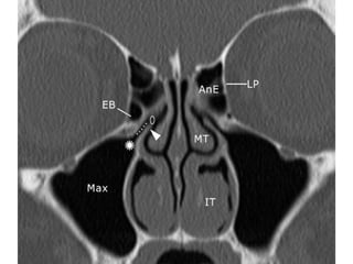
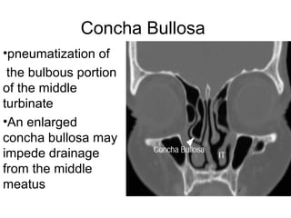
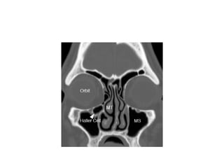
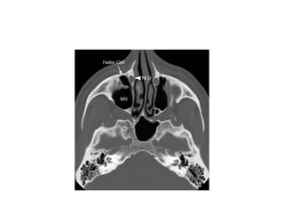
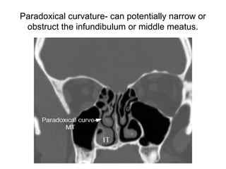
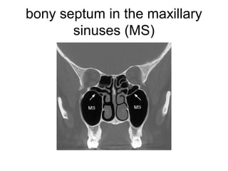
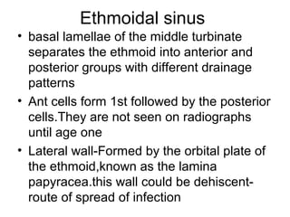
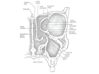
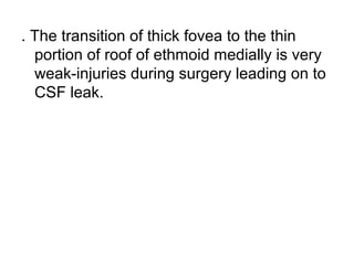
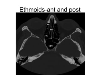
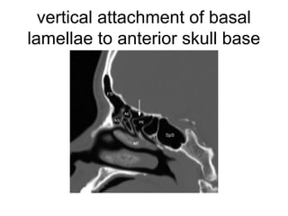
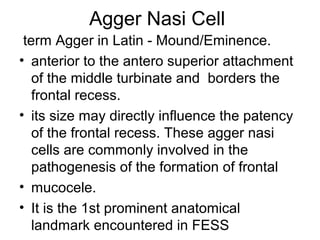
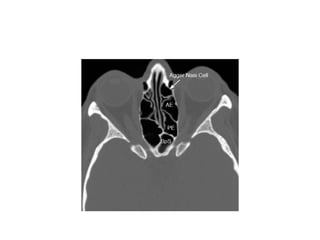
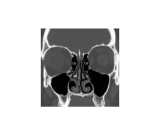
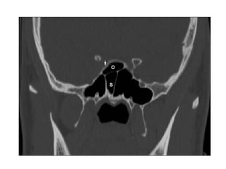
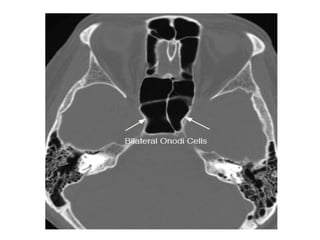
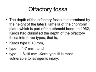
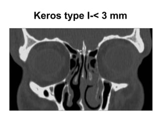
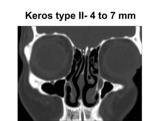
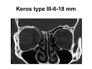
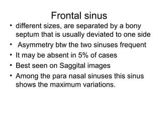
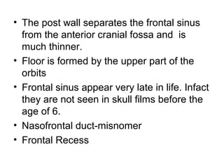
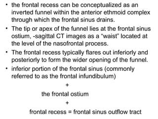
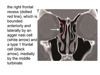
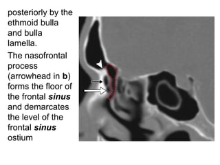
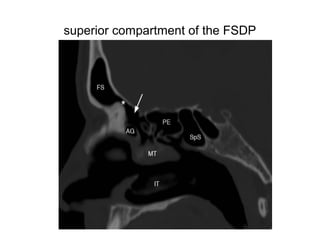
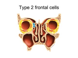
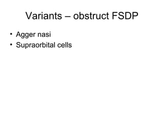
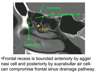
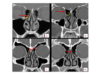
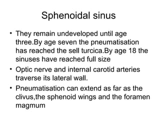
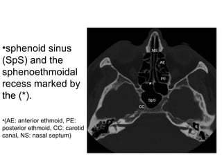
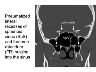
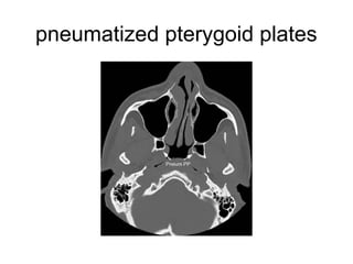
The document discusses the anatomy and variants of the paranasal sinuses relevant to functional endoscopic sinus surgery (FESS). It describes the four paired sinuses and their drainage pathways. Key anatomical structures for drainage include the osteomeatal complex and frontal recess. Common anatomic variants are described such as concha bullosa, Haller cells, and Onodi cells which can impact sinus drainage. Radiologists should evaluate pre-operative scans for variants that may obstruct drainage or pose surgical hazards. A systematic checklist is recommended to identify issues important for surgical planning.
Lateral skull base anatomy and applied science by Dr, bomkar bamBomkar Bam
the lateral skull base is complex anatomy that is usually students finds difficult to understand. here concise literature is made to understand the skull base more easily.
Lateral skull base anatomy and applied science by Dr, bomkar bamBomkar Bam
the lateral skull base is complex anatomy that is usually students finds difficult to understand. here concise literature is made to understand the skull base more easily.
Imaging of paranasal sinuses (including anatomy and varaints)pk1 pdf pptDr pradeep Kumar
This is very good powerpoint presentation of imaging anatomy and variants of paranasal sinuses and imaging pathology as well as multiple pathological imaging findings and images.it will helps for radiologist and radiology resident and even ent resident. our references is CT and mri whole body by Haaga and various internet sources. THANKS.
Detailed discussion on tumors and other pathologies of paranasal sinus and their management. Surgical anatomy and approaches are also discussed. Complications of PNS surgeries are discussed briefly
The framework of the nose consists of bone and cartilage. Two small nasal bones and extensions of the maxillae form the bridge of the nose, which is the bony portion. The remainder of the framework is cartilage and is the flexible portion. Connective tissue and skin cover the framework.
Air enters the nasal cavity from the outside through two openings: the nostrils or external nares. The openings from the nasal cavity into the pharynx are the internal nares. Nose hairs at the entrance to the nose trap large inhaled particles.
Paranasal sinuses are air-filled cavities in the frontal, maxilae, ethmoid, and sphenoid bones. These sinuses, which have the same names as the bones in which they are located, surround the nasal cavity and open into it. They function to reduce the weight of the skull, to produce mucus, and to influence voice quality by acting as resonating chambers.
Lung Cancer: Artificial Intelligence, Synergetics, Complex System Analysis, S...Oleg Kshivets
RESULTS: Overall life span (LS) was 2252.1±1742.5 days and cumulative 5-year survival (5YS) reached 73.2%, 10 years – 64.8%, 20 years – 42.5%. 513 LCP lived more than 5 years (LS=3124.6±1525.6 days), 148 LCP – more than 10 years (LS=5054.4±1504.1 days).199 LCP died because of LC (LS=562.7±374.5 days). 5YS of LCP after bi/lobectomies was significantly superior in comparison with LCP after pneumonectomies (78.1% vs.63.7%, P=0.00001 by log-rank test). AT significantly improved 5YS (66.3% vs. 34.8%) (P=0.00000 by log-rank test) only for LCP with N1-2. Cox modeling displayed that 5YS of LCP significantly depended on: phase transition (PT) early-invasive LC in terms of synergetics, PT N0—N12, cell ratio factors (ratio between cancer cells- CC and blood cells subpopulations), G1-3, histology, glucose, AT, blood cell circuit, prothrombin index, heparin tolerance, recalcification time (P=0.000-0.038). Neural networks, genetic algorithm selection and bootstrap simulation revealed relationships between 5YS and PT early-invasive LC (rank=1), PT N0—N12 (rank=2), thrombocytes/CC (3), erythrocytes/CC (4), eosinophils/CC (5), healthy cells/CC (6), lymphocytes/CC (7), segmented neutrophils/CC (8), stick neutrophils/CC (9), monocytes/CC (10); leucocytes/CC (11). Correct prediction of 5YS was 100% by neural networks computing (area under ROC curve=1.0; error=0.0).
CONCLUSIONS: 5YS of LCP after radical procedures significantly depended on: 1) PT early-invasive cancer; 2) PT N0--N12; 3) cell ratio factors; 4) blood cell circuit; 5) biochemical factors; 6) hemostasis system; 7) AT; 8) LC characteristics; 9) LC cell dynamics; 10) surgery type: lobectomy/pneumonectomy; 11) anthropometric data. Optimal diagnosis and treatment strategies for LC are: 1) screening and early detection of LC; 2) availability of experienced thoracic surgeons because of complexity of radical procedures; 3) aggressive en block surgery and adequate lymph node dissection for completeness; 4) precise prediction; 5) adjuvant chemoimmunoradiotherapy for LCP with unfavorable prognosis.
New Drug Discovery and Development .....NEHA GUPTA
The "New Drug Discovery and Development" process involves the identification, design, testing, and manufacturing of novel pharmaceutical compounds with the aim of introducing new and improved treatments for various medical conditions. This comprehensive endeavor encompasses various stages, including target identification, preclinical studies, clinical trials, regulatory approval, and post-market surveillance. It involves multidisciplinary collaboration among scientists, researchers, clinicians, regulatory experts, and pharmaceutical companies to bring innovative therapies to market and address unmet medical needs.
Ethanol (CH3CH2OH), or beverage alcohol, is a two-carbon alcohol
that is rapidly distributed in the body and brain. Ethanol alters many
neurochemical systems and has rewarding and addictive properties. It
is the oldest recreational drug and likely contributes to more morbidity,
mortality, and public health costs than all illicit drugs combined. The
5th edition of the Diagnostic and Statistical Manual of Mental Disorders
(DSM-5) integrates alcohol abuse and alcohol dependence into a single
disorder called alcohol use disorder (AUD), with mild, moderate,
and severe subclassifications (American Psychiatric Association, 2013).
In the DSM-5, all types of substance abuse and dependence have been
combined into a single substance use disorder (SUD) on a continuum
from mild to severe. A diagnosis of AUD requires that at least two of
the 11 DSM-5 behaviors be present within a 12-month period (mild
AUD: 2–3 criteria; moderate AUD: 4–5 criteria; severe AUD: 6–11 criteria).
The four main behavioral effects of AUD are impaired control over
drinking, negative social consequences, risky use, and altered physiological
effects (tolerance, withdrawal). This chapter presents an overview
of the prevalence and harmful consequences of AUD in the U.S.,
the systemic nature of the disease, neurocircuitry and stages of AUD,
comorbidities, fetal alcohol spectrum disorders, genetic risk factors, and
pharmacotherapies for AUD.
HOT NEW PRODUCT! BIG SALES FAST SHIPPING NOW FROM CHINA!! EU KU DB BK substit...GL Anaacs
Contact us if you are interested:
Email / Skype : kefaya1771@gmail.com
Threema: PXHY5PDH
New BATCH Ku !!! MUCH IN DEMAND FAST SALE EVERY BATCH HAPPY GOOD EFFECT BIG BATCH !
Contact me on Threema or skype to start big business!!
Hot-sale products:
NEW HOT EUTYLONE WHITE CRYSTAL!!
5cl-adba precursor (semi finished )
5cl-adba raw materials
ADBB precursor (semi finished )
ADBB raw materials
APVP powder
5fadb/4f-adb
Jwh018 / Jwh210
Eutylone crystal
Protonitazene (hydrochloride) CAS: 119276-01-6
Flubrotizolam CAS: 57801-95-3
Metonitazene CAS: 14680-51-4
Payment terms: Western Union,MoneyGram,Bitcoin or USDT.
Deliver Time: Usually 7-15days
Shipping method: FedEx, TNT, DHL,UPS etc.Our deliveries are 100% safe, fast, reliable and discreet.
Samples will be sent for your evaluation!If you are interested in, please contact me, let's talk details.
We specializes in exporting high quality Research chemical, medical intermediate, Pharmaceutical chemicals and so on. Products are exported to USA, Canada, France, Korea, Japan,Russia, Southeast Asia and other countries.
micro teaching on communication m.sc nursing.pdfAnurag Sharma
Microteaching is a unique model of practice teaching. It is a viable instrument for the. desired change in the teaching behavior or the behavior potential which, in specified types of real. classroom situations, tends to facilitate the achievement of specified types of objectives.
Prix Galien International 2024 Forum ProgramLevi Shapiro
June 20, 2024, Prix Galien International and Jerusalem Ethics Forum in ROME. Detailed agenda including panels:
- ADVANCES IN CARDIOLOGY: A NEW PARADIGM IS COMING
- WOMEN’S HEALTH: FERTILITY PRESERVATION
- WHAT’S NEW IN THE TREATMENT OF INFECTIOUS,
ONCOLOGICAL AND INFLAMMATORY SKIN DISEASES?
- ARTIFICIAL INTELLIGENCE AND ETHICS
- GENE THERAPY
- BEYOND BORDERS: GLOBAL INITIATIVES FOR DEMOCRATIZING LIFE SCIENCE TECHNOLOGIES AND PROMOTING ACCESS TO HEALTHCARE
- ETHICAL CHALLENGES IN LIFE SCIENCES
- Prix Galien International Awards Ceremony
NVBDCP.pptx Nation vector borne disease control programSapna Thakur
NVBDCP was launched in 2003-2004 . Vector-Borne Disease: Disease that results from an infection transmitted to humans and other animals by blood-feeding arthropods, such as mosquitoes, ticks, and fleas. Examples of vector-borne diseases include Dengue fever, West Nile Virus, Lyme disease, and malaria.
Couples presenting to the infertility clinic- Do they really have infertility...Sujoy Dasgupta
Dr Sujoy Dasgupta presented the study on "Couples presenting to the infertility clinic- Do they really have infertility? – The unexplored stories of non-consummation" in the 13th Congress of the Asia Pacific Initiative on Reproduction (ASPIRE 2024) at Manila on 24 May, 2024.
Tom Selleck Health: A Comprehensive Look at the Iconic Actor’s Wellness Journeygreendigital
Tom Selleck, an enduring figure in Hollywood. has captivated audiences for decades with his rugged charm, iconic moustache. and memorable roles in television and film. From his breakout role as Thomas Magnum in Magnum P.I. to his current portrayal of Frank Reagan in Blue Bloods. Selleck's career has spanned over 50 years. But beyond his professional achievements. fans have often been curious about Tom Selleck Health. especially as he has aged in the public eye.
Follow us on: Pinterest
Introduction
Many have been interested in Tom Selleck health. not only because of his enduring presence on screen but also because of the challenges. and lifestyle choices he has faced and made over the years. This article delves into the various aspects of Tom Selleck health. exploring his fitness regimen, diet, mental health. and the challenges he has encountered as he ages. We'll look at how he maintains his well-being. the health issues he has faced, and his approach to ageing .
Early Life and Career
Childhood and Athletic Beginnings
Tom Selleck was born on January 29, 1945, in Detroit, Michigan, and grew up in Sherman Oaks, California. From an early age, he was involved in sports, particularly basketball. which played a significant role in his physical development. His athletic pursuits continued into college. where he attended the University of Southern California (USC) on a basketball scholarship. This early involvement in sports laid a strong foundation for his physical health and disciplined lifestyle.
Transition to Acting
Selleck's transition from an athlete to an actor came with its physical demands. His first significant role in "Magnum P.I." required him to perform various stunts and maintain a fit appearance. This role, which he played from 1980 to 1988. necessitated a rigorous fitness routine to meet the show's demands. setting the stage for his long-term commitment to health and wellness.
Fitness Regimen
Workout Routine
Tom Selleck health and fitness regimen has evolved. adapting to his changing roles and age. During his "Magnum, P.I." days. Selleck's workouts were intense and focused on building and maintaining muscle mass. His routine included weightlifting, cardiovascular exercises. and specific training for the stunts he performed on the show.
Selleck adjusted his fitness routine as he aged to suit his body's needs. Today, his workouts focus on maintaining flexibility, strength, and cardiovascular health. He incorporates low-impact exercises such as swimming, walking, and light weightlifting. This balanced approach helps him stay fit without putting undue strain on his joints and muscles.
Importance of Flexibility and Mobility
In recent years, Selleck has emphasized the importance of flexibility and mobility in his fitness regimen. Understanding the natural decline in muscle mass and joint flexibility with age. he includes stretching and yoga in his routine. These practices help prevent injuries, improve posture, and maintain mobilit
Acute scrotum is a general term referring to an emergency condition affecting the contents or the wall of the scrotum.
There are a number of conditions that present acutely, predominantly with pain and/or swelling
A careful and detailed history and examination, and in some cases, investigations allow differentiation between these diagnoses. A prompt diagnosis is essential as the patient may require urgent surgical intervention
Testicular torsion refers to twisting of the spermatic cord, causing ischaemia of the testicle.
Testicular torsion results from inadequate fixation of the testis to the tunica vaginalis producing ischemia from reduced arterial inflow and venous outflow obstruction.
The prevalence of testicular torsion in adult patients hospitalized with acute scrotal pain is approximately 25 to 50 percent
Recomendações da OMS sobre cuidados maternos e neonatais para uma experiência pós-natal positiva.
Em consonância com os ODS – Objetivos do Desenvolvimento Sustentável e a Estratégia Global para a Saúde das Mulheres, Crianças e Adolescentes, e aplicando uma abordagem baseada nos direitos humanos, os esforços de cuidados pós-natais devem expandir-se para além da cobertura e da simples sobrevivência, de modo a incluir cuidados de qualidade.
Estas diretrizes visam melhorar a qualidade dos cuidados pós-natais essenciais e de rotina prestados às mulheres e aos recém-nascidos, com o objetivo final de melhorar a saúde e o bem-estar materno e neonatal.
Uma “experiência pós-natal positiva” é um resultado importante para todas as mulheres que dão à luz e para os seus recém-nascidos, estabelecendo as bases para a melhoria da saúde e do bem-estar a curto e longo prazo. Uma experiência pós-natal positiva é definida como aquela em que as mulheres, pessoas que gestam, os recém-nascidos, os casais, os pais, os cuidadores e as famílias recebem informação consistente, garantia e apoio de profissionais de saúde motivados; e onde um sistema de saúde flexível e com recursos reconheça as necessidades das mulheres e dos bebês e respeite o seu contexto cultural.
Estas diretrizes consolidadas apresentam algumas recomendações novas e já bem fundamentadas sobre cuidados pós-natais de rotina para mulheres e neonatos que recebem cuidados no pós-parto em unidades de saúde ou na comunidade, independentemente dos recursos disponíveis.
É fornecido um conjunto abrangente de recomendações para cuidados durante o período puerperal, com ênfase nos cuidados essenciais que todas as mulheres e recém-nascidos devem receber, e com a devida atenção à qualidade dos cuidados; isto é, a entrega e a experiência do cuidado recebido. Estas diretrizes atualizam e ampliam as recomendações da OMS de 2014 sobre cuidados pós-natais da mãe e do recém-nascido e complementam as atuais diretrizes da OMS sobre a gestão de complicações pós-natais.
O estabelecimento da amamentação e o manejo das principais intercorrências é contemplada.
Recomendamos muito.
Vamos discutir essas recomendações no nosso curso de pós-graduação em Aleitamento no Instituto Ciclos.
Esta publicação só está disponível em inglês até o momento.
Prof. Marcus Renato de Carvalho
www.agostodourado.com
ARTIFICIAL INTELLIGENCE IN HEALTHCARE.pdfAnujkumaranit
Artificial intelligence (AI) refers to the simulation of human intelligence processes by machines, especially computer systems. It encompasses tasks such as learning, reasoning, problem-solving, perception, and language understanding. AI technologies are revolutionizing various fields, from healthcare to finance, by enabling machines to perform tasks that typically require human intelligence.
1. Paranasal Sinuses Anatomy &
Variants-
A Systematic Approach To
Imaging Before FESS
Dr Priyanka Vishwakarma
2. Four Paired Sinuses –
• Ethmoid
• Maxillary
• Frontal
• Sphenoid
• The sinuses develop as outgrowths from
the nasal cavity; hence they all drain
directly or indirectly into the nose
3. Meati
• superior meatus drains the posterior ethmoid
air cells and the sphenoid sinus via the
sphenoethmoidal recess
• middle meatus drains the frontal sinus via the
nasofrontal duct/frontal recess, the maxillary
sinus via the maxillary ostium, and the anterior
ethmoid air cells via the ethmoid cell ostia.
• The nasolacrimal duct drains into the inferior
meatus
• spheno-ethmoidal recess, above and posterior
to the superior concha, receives the opening of
the sphenoidal sinus
4. Osteomeatal unit
Common Drainage Pathway Of The Ant.
Group of Sinuses.-Coronal scan
The osteomeatal unit (OMU) includes the
• uncinate process
• Ethmoid infundibulum
• Ethmoid Bulla
• Middle Meatus
• Hiatus Semilunaris
Most common site of inflammatory disease
7. Maxillary sinus
• Largest and most constant pns.
• Pyramidal in shape- base is usually
medial, with its apex in the zygomatic
process of the maxilla
• Base -lat nasal wall-ostium
• Posterior wall/Temporal- pterygomaxillary
fossa
• Roof -Formed by roof of the orbit- infra
orbital foramen containing the infra orbital
vessels and nerves
• Ant-maxilla facial surface
8. Variants Related To the maxillary Sinus
Concha bullosa
Paradoxical curvature of MT
Haller Cell
Septae
Dehiscent floor-1st
,2nd
Molar Infn
10. Haller cells
• Ethmoidal air cells belonging to the
anterior ethmoidal group.
• Also known as the infra orbital cells
• Adhere to roof of maxillary sinus forming
the lat wall of infundibulum
• Enlargement of these cells can impede
the maxillary sinus drainage
15. Ethmoidal sinus
• basal lamellae of the middle turbinate
separates the ethmoid into anterior and
posterior groups with different drainage
patterns
• Ant cells form 1st followed by the posterior
cells.They are not seen on radiographs
until age one
• Lateral wall-Formed by the orbital plate of
the ethmoid,known as the lamina
papyracea.this wall could be dehiscent-
route of spread of infection
16.
17. . The transition of thick fovea to the thin
portion of roof of ethmoid medially is very
weak-injuries during surgery leading on to
CSF leak.
20. Related Variants
• A cell above the orbit is called a
supraorbital cell.found in 15% of pt
• Invasion of an ethmoid cell into the floor of
the frontal sinus is called a frontal cell(type
1-4)
21. Agger Nasi Cell
term Agger in Latin - Mound/Eminence.
• anterior to the antero superior attachment
of the middle turbinate and borders the
frontal recess.
• its size may directly influence the patency
of the frontal recess. These agger nasi
cells are commonly involved in the
pathogenesis of the formation of frontal
• mucocele.
• It is the 1st prominent anatomical
landmark encountered in FESS
22.
23. ethmoid bulla
• superior to uncinate processes.
• Ethmoid bulla air cells are part of the
anterior ethmoid sinuses and make up the
superior border of the hiatus semilunaris.
• variable pneumatization.
24.
25. Onodi Cells
• posterior ethmoidal cells extending supero
lateral to the sphenoid sinus & can either
abut to or impinging upon the optic nerve.
• When these Onodi cells abut or surround
the optic nerve, the nerve is at risk when
surgical excision of these cells is
performed.
• It is also a potential cause of incomplete
sphenoidectomy.
26.
27.
28. Olfactory fossa
• The depth of the olfactory fossa is determined by
the height of the lateral lamella of the cribriform
plate, which is part of the ethmoid bone. In 1962,
Keros had classified the depth of the olfactory
fossa into three types, that is,
• Keros type I: <3 mm,
• type II: 4-7 mm , and
• type III: 8-16 mm.-Kero type III is most
vulnerable to iatrogenic injury.
32. Frontal sinus
• different sizes, are separated by a bony
septum that is usually deviated to one side
• Asymmetry btw the two sinuses frequent
• It may be absent in 5% of cases
• Best seen on Saggital images
• Among the para nasal sinuses this sinus
shows the maximum variations.
33. • The post wall separates the frontal sinus
from the anterior cranial fossa and is
much thinner.
• Floor is formed by the upper part of the
orbits
• Frontal sinus appear very late in life. Infact
they are not seen in skull films before the
age of 6.
• Nasofrontal duct-misnomer
• Frontal Recess
34. • the frontal recess can be conceptualized as an
inverted funnel within the anterior ethmoid complex
through which the frontal sinus drains.
• The tip or apex of the funnel lies at the frontal sinus
ostium, -sagittal CT images as a “waist” located at
the level of the nasofrontal process.
• The frontal recess typically flares out inferiorly and
posteriorly to form the wider opening of the funnel.
• inferior portion of the frontal sinus (commonly
referred to as the frontal infundibulum)
+
the frontal ostium
+
frontal recess = frontal sinus outflow tract
35. the right frontal
recess (dotted
red line), which is
bounded
anteriorly and
laterally by an
agger nasi cell
(white arrow) and
a type 1 frontal
cell (black
arrow), medially
by the middle
turbinate
36.
posteriorly by the
ethmoid bulla
and bulla
lamella.
The nasofrontal
process
(arrowhead in b)
forms the floor of
the frontal sinus
and demarcates
the level of the
frontal sinus
ostium
38. Frontal outflow tract shows conglomeratization of
air cells.
Types of frontal sinus air cells include:
• I – Type I frontal cell (a single air cell above
agger nasi)
• II – Type II frontal cell (a series of air cells above
agger nasi but below the orbital roof)
• III – Type III frontal cell (this cell extends into the
frontal sinus but is contiguous with agger nasi
• cell)
• IV – Type IV frontal cell lies completely within
the frontal sinus
41. Type 4 frontal cell
situated entirely
within the right
frontal sinus &
bordered by the
anterior frontal
sinus wall. The
type 4 cell does
not abut the
agger nasi cell.
48. • The uncinate process may be attached to:
• Lamina papyracea or agger nasi (lamina
terminalis). The frontal recess opens
directly into middle meatus,medial to UP
The lamina terminalis is the blind pouch
between the UP and lamina papyracea
• Skull base or middle turbinate. The frontal
recess drains into the ethmoid
infundibulum lateral to UP
• Orbital floor or inferior aspect of the lamina
papyracea (silent sinus syndrome,
atelectatic uncinate process). This variant
is associated with hypoplastic, ipsilateral
49.
50. Sphenoidal sinus
• They remain undeveloped until age
three.By age seven the pneumatisation
has reached the sell turcica.By age 18 the
sinuses have reached full size
• Optic nerve and internal carotid arteries
traverse its lateral wall.
• Pneumatisation can extend as far as the
clivus,the sphenoid wings and the foramen
magmum
51. •sphenoid sinus
(SpS) and the
sphenoethmoidal
recess marked by
the (*).
•(AE: anterior ethmoid, PE:
posterior ethmoid, CC: carotid
canal, NS: nasal septum)
54. variations of intersinus septum
• 1.A single midline intersinus septum
extending on to the anterior wall of sella.
• 2. Multiple incomplete septae may be
seen
• 3. Accessory septa may be present.
These could be seen terminating on to the
carotid canal or optic
58. FESS-a roadmap to the otorhinolaryngologist prior to
surgery.
There are two main questions that the radiologist should
address:
1. Are there anatomic features on the computed
tomography (CT) scan that predispose the patient to
impaired mucociliary clearance?
2. Are there anatomic features that pose a surgical
hazard?
59. Checklist-Systematic
• the extent of sinus opacification,
• patency of sinus drainage pathways,
• anatomic variants(obstruct drainage
pathways &limit Surgical access),
• critical variants, (CP,LP,SphS
dehiscence)and
• condition of soft tissues of the brain, neck,
and orbits.-extrasinus extent of the
disease
The maxillary sinus, the largest of the sinuses, is within the body of the maxilla. It is shaped like a pyramid; its base is usually medial, with its apex in the zygomatic process of the maxilla. Its roof is the floor of the orbit, and its floor is the alveolar process of the maxilla. The maxillary sinus drains into the middle meatus by means of the semilunar hiatus. The floor of the maxillary sinus is slightly below the level of the nasal cavity, and it is related to the upper teeth
If present, a Haller cell can cause narrowing of the infundibulum and maxillary sinus ostuim potentially causing obstruction
Roof of ethmoid
olfactory fossae are deeper and the lateral lamellae are longer
olfactory fossae are very deep
Normal frontal recess anatomy. Coronal (a) and sagittal (b) CT images show the right frontal recess (dotted red line), which is bounded anteriorly and laterally by an agger nasi cell (white arrow) and a type 1 frontal cell (black arrow), medially by the middle turbinate, and posteriorly by the ethmoid bulla and bulla lamella. The nasofrontal process (arrowhead in b) forms the floor of the frontal sinus and demarcates the level of the frontal sinus ostium.
Sagittal image shows frontal sinus ostium (*) and arrow pointing to the superior compartment of the FSDP. (FS: frontal sinus, AG: agger nasi, PE: posterior ethmoid, SpS: sphenoid sinus, MT: middle turbinate, IT: inferior turbinate)
Type 2 frontal cells. (a, b) Coronal (a) and parasagittal (b) drawings show a tier of type 2 frontal cells (blue areas) sitting atop an agger nasi cell. (c, d) Coronal (c) and sagittal (d) CT images show a tier of two type 2 frontal cells (arrows) sitting directly atop an agger nasi cell (*).
Type 3 frontal cell. Coronal (a) and parasagittal (b) drawings show a type 3 frontal cell (blue area) sitting atop an agger nasi cell. The type 3 cell extends superiorly from the frontal recess through the frontal ostium and into the frontal sinus.
Type 4 frontal cell. (a, b) Coronal (a) and parasagittal (b) drawings show a type 4 frontal cell (blue area) situated entirely within the right frontal sinus and bordered by the anterior frontal sinus wall. The type 4 cell does not abut the agger nasi cell. (c, d) Coronal (c) and sagittal (d) CT images show an opacified type 4 frontal cell (arrow) in the frontal sinus.
Axial CT image shows the supraorbital ethmoid cell (arrow), which is clearly differentiated from the frontal sinus (*) by a discrete bony septum.
pneumatized crista galli. Pneumatized crista galli may communicate with the frontal recess and can potentially obstruct the frontal sinus ostium
which arises from the frontal sinus septum
Axial image shows sphenoid sinus (SpS) and the sphenoethmoidal recess marked by the (*). (AE: anterior ethmoid, PE: posterior ethmoid, CC: carotid canal, NS: nasal septum)
Coronal image of the sphenoid sinus (SpS) and neighboring structures. (FR: foramen rotundum, VC: vidian canal, OC: optic canal, AC: anterior clinoid, PtP: pterygoid plate)