The document provides details on the anatomy of the middle ear, including its walls, structures, and measurements. Key points:
- The middle ear is an air-filled cavity in the temporal bone between the external ear canal and inner ear. It is divided into three regions and contains the ossicles, muscles, and facial nerve.
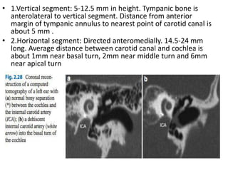
- The walls of the middle ear cavity include the lateral, inferior, posterior, superior, anterior, and medial walls. Important structures on the walls include the tympanic membrane, ossicles, facial nerve canal, oval and round windows.
- The middle ear cavity measures approximately 2mm wide at its center, widening to 6mm superiorly and 4mm inferiorly.