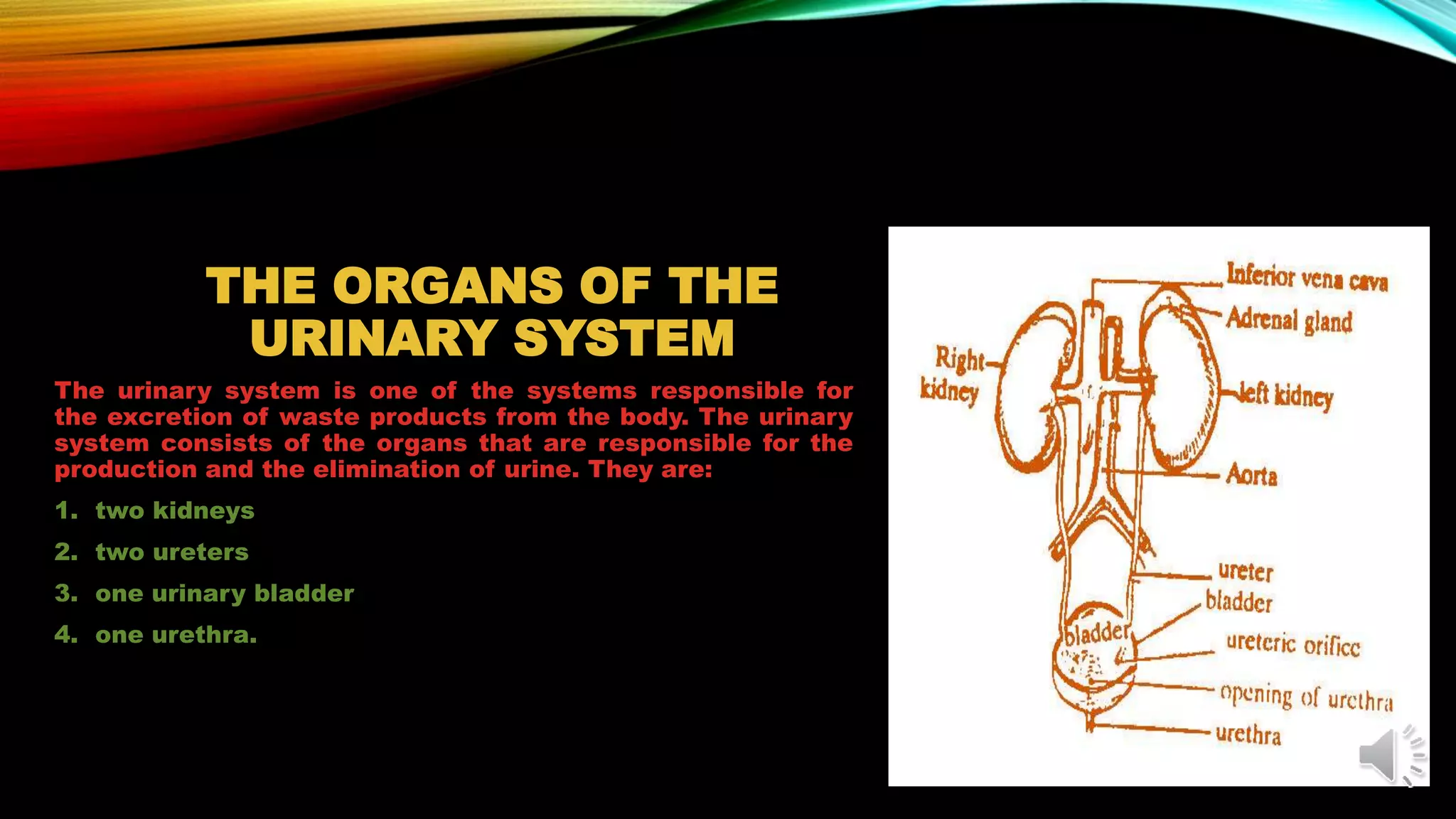
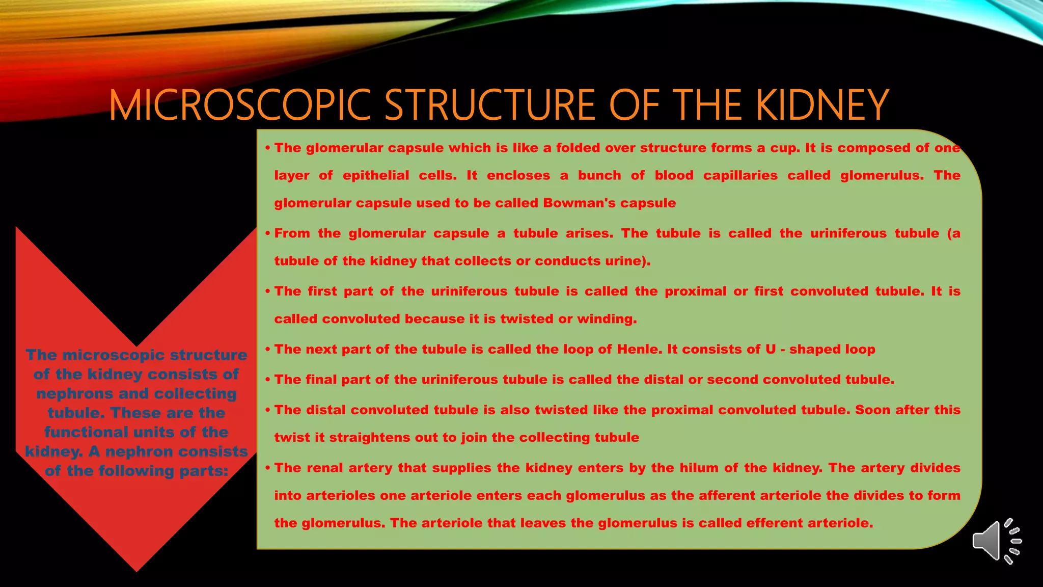
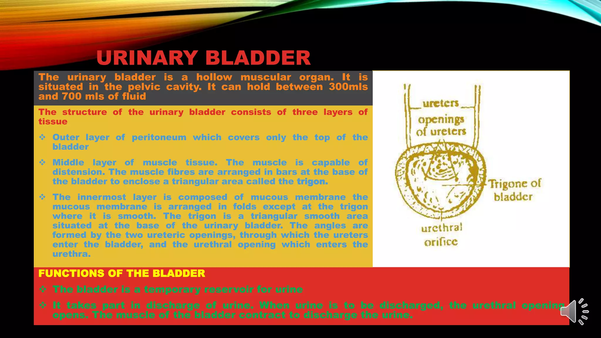
ANATOMY AND PHYSIOLOGY
THE URINARY OR EXCRETORY SYSTEM
Presentation by:
Bamigboye G.S
LEARNING OBJECTIVES
• By this end of this unit, you should be able to:
i. component organs of the urinary system;
ii. describe the structure and functions of the kidney;
iii. describe the structure of the ureter;
iv. describe the structure of the bladder;
v. describe the structure of the urethra and
vi. explain how urine is formed and its composition.
WORD STUDY
• The following words that are used in this unit that may be unfamiliar to you are:
Microscopic: something tiny or small that the naked eyes cannot see
Macroscopic: something that the ordinary eyes can see
Organs : are the structure in the system or the entire body
THE ORGANS OF THE
URINARY SYSTEM
The urinary system is one of the systems responsible for
the excretion of waste products from the body. The urinary
system consists of the organs that are responsible for the
production and the elimination of urine. They are:
1. two kidneys
2. two ureters
3. one urinary bladder
4. one urethra.
THE KIDNEY
The kidney is a bean - shaped organ that is located
at the back of the abdominal cavity, at the level of
the last thoracic and first 3 lumbar vertebrae i.e
above the waistline. They are usually two in number.
One on each side of the vertebral column. They are
covered by a pad of fat that serves as a protective
mechanism for the kidney against cold and injury.
Toward the midline border of the kidney is a concave
notch known as the hilium. This is the point that the
renal arteries enter the kidney while the renal veins,
ureters, nerves and lymphatic vessels leave the
kidney.
The kidneys are dark red in colour, Each is 11 cm (1
over in length, 5cm (2 in) in width and about 3cm 1
over in) thickness and weighs about 130 - 150 grams.
STRUCTURE OF THE KIDNEY
• The structure of the kidney is conveniently described in two parts,
namely macroscopic structure and microscopic structure.
• The macroscopic structure refers to parts of the kidney that can be
seen with the naked eye.
• The microscopic structure refers to parts of the kidney that can be
seen with the aid of a microscope.
MACROSCOPIC STRUCTURE OF THE KIDNEY
• The kidney has an outer capsule composed of fibrous tissue.
• The cortex is the superficial layer of the substance of the kidney,
situated immediately under the capsule
• The medulla is the deeper layer of the substance of the kidney
situated under the cortex. It consists of pyramids formed by
collecting tubules. The pyramids enter structures shaped like cups
called calyces (singular = calyx)
• The hilum which is the concave medial border through which
structures enter and leave.
MICROSCOPIC STRUCTURE OF THE KIDNEY
The microscopic structure
of the kidney consists of
nephrons and collecting
tubule. These are the
functional units of the
kidney. A nephron consists
of the following parts:
• The glomerular capsule which is like a folded over structure forms a cup. It is composed of one
layer of epithelial cells. It encloses a bunch of blood capillaries called glomerulus. The
glomerular capsule used to be called Bowman's capsule
• From the glomerular capsule a tubule arises. The tubule is called the uriniferous tubule (a
tubule of the kidney that collects or conducts urine).
• The first part of the uriniferous tubule is called the proximal or first convoluted tubule. It is
called convoluted because it is twisted or winding.
• The next part of the tubule is called the loop of Henle. It consists of U - shaped loop
• The final part of the uriniferous tubule is called the distal or second convoluted tubule.
• The distal convoluted tubule is also twisted like the proximal convoluted tubule. Soon after this
twist it straightens out to join the collecting tubule
• The renal artery that supplies the kidney enters by the hilum of the kidney. The artery divides
into arterioles one arteriole enters each glomerulus as the afferent arteriole the divides to form
the glomerulus. The arteriole that leaves the glomerulus is called efferent arteriole.
FORMATION OF URINE
• Waste is excreted from the human body, mainly in the form of urine.
Our kidneys play a major role in the process of excretion.
Constituents of normal human urine include 95 percent water and 5
percent solid wastes. It is produced in the nephron, which is the
structural and functional unit of the kidney. Urine formation in our
body is mainly carried out in three phases namely
1. Filtration
2. Reabsorption
3. Secretion
• Filtration occurs in the glomerulus where blood is filtered. This process occurs across the three layers-
epithelium of Bowman’s capsule, endothelium of glomerular blood vessels, and a membrane between these two
layers.
• Blood is filtered in such a way that all the constituents of the plasma reach the Bowman’s capsule, except
proteins. Therefore, this process is known as ultrafiltration.
•Around 99 percent of the filtrate obtained is reabsorbed by the renal tubules. This is known as
reabsorption. This is achieved by active and passive transport.
• The next step in urine formation is the tubular secretion. Here, tubular cells secrete substances like
hydrogen ion, potassium ion, etc into the filtrate. By this process, the ionic, acid-base and the balance
of other body fluids are maintained. The secreted ions combine with the filtrate and form urine. The
urine passes out of the nephron tubule into a collecting duct.
Urine
•The urine produced is 95% water and 5% nitrogenous wastes. Wastes such as urea, ammonia,
creatinine are excreted in the urine. Apart from these, the potassium, sodium and calcium ions
are also excreted.
FUNCTIONS OF THE KIDNEY
The main function of
the kidney is
formation of urine.
Other functions are
also accomplished
through the
formation of urine
Maintenance of
water balance. When
water intake is
increased,
especially when the
body is not
sweating, urinary
output is increased
so as to keep the
body fluid in
balance.
Maintenance of
alkalinity of blood.
When much of the
substances, such as
protein, that can
make blood acid are
eaten, the kidney
will allow the excess
to leave the body
through the urine.
Such substances are
as a result secreted
into the tubule to be
removed along with
urine.
Maintenance of
osmotic pressure of
blood. When the
osmotic pressure of
blood is high , the
tubules will reabsorb
more water into the
blood so as to dilute
the blood and
reduce the osmotic
pressure
Electrolyte balance
in the body. Sodium
and potassium is the
most common
electrolytes involved
in body processes.
The body will
remove which ever
is in excess through
the urine.
The ureters consist of two narrow tubes about 25cm
long and 3mm in diameter. Each one extends from the
pelvis of one kidney to the base of the urinary bladder.
URETERS
The function of the ureters is to convey urine from the
kidney to the bladder by means of peristaltic action.
The structure consists of three coats
1. Outer layer composed of fibrous tissue
2. Middle layer composed of muscle tissue and
3. Innermost layer composed of mucous membrane
URINARY BLADDER
The urinary bladder is a hollow muscular organ. It is
situated in the pelvic cavity. It can hold between 300mls
and 700 mls of fluid
FUNCTIONS OF THE BLADDER
 The bladder is a temporary reservoir for urine
 It takes part in discharge of urine. When urine is to be discharged, the urethral opening
opens. The muscle of the bladder contract to discharge the urine.
The structure of the urinary bladder consists of three layers of
tissue
 Outer layer of peritoneum which covers only the top of the
bladder
 Middle layer of muscle tissue. The muscle is capable of
distension. The muscle fibres are arranged in bars at the base of
the bladder to enclose a triangular area called the trigon.
 The innermost layer is composed of mucous membrane the
mucous membrane is arranged in folds except at the trigon
where it is smooth. The trigon is a triangular smooth area
situated at the base of the urinary bladder. The angles are
formed by the two ureteric openings, through which the ureters
enter the bladder, and the urethral opening which enters the
urethra.
The urethra is a narrow canal which extends from the base of the bladder to the
exterior or outside.
The female urethra is only about 4cm long but the male urethra is about 15cm long. In
addition, the male urethra is a common path way for urine and semen from the
reproductive system.
URETHRA
The function of the urethra is to convey urine
from the bladder to the exterior or outside.
The structure consists of three layers as follows:
1. Outer layer composed of muscle
2. Middle layer composed of vascular connective tissue and
3. Inner layer of mucous membrane
SUMMARY
Urinary system consists of two kidneys, two ureters, one urinary bladder,
and one urethra.
The structure of the kidney consists of macroscopic and microscopic
structure.
The macroscopic structure consists of renal capsule, cortex and medulla
The microscopic structures consist of nephrons.
Formation of urine occurs in three phases simple: filtration, selective
reabsorption, and secretion.
In addition to urine formation, the kidneys maintain water balance
alkalinity of blood, osmotic pressure of blood and electrolyte balance
The ureters convey urine from the kidneys to the bladder.
The urinary bladder is a temporary reservoir for urine, and takes part in
discharge of urine.
The urethra discharge urine from the bladder to the exterior.
The male urethra conveys both urine and semen from reproductive
system.
Anatomy and physiology (urinary system)

Anatomy and physiology (urinary system)

  • 1.
    ANATOMY AND PHYSIOLOGY THEURINARY OR EXCRETORY SYSTEM Presentation by: Bamigboye G.S
  • 2.
    LEARNING OBJECTIVES • Bythis end of this unit, you should be able to: i. component organs of the urinary system; ii. describe the structure and functions of the kidney; iii. describe the structure of the ureter; iv. describe the structure of the bladder; v. describe the structure of the urethra and vi. explain how urine is formed and its composition.
  • 3.
    WORD STUDY • Thefollowing words that are used in this unit that may be unfamiliar to you are: Microscopic: something tiny or small that the naked eyes cannot see Macroscopic: something that the ordinary eyes can see Organs : are the structure in the system or the entire body
  • 4.
    THE ORGANS OFTHE URINARY SYSTEM The urinary system is one of the systems responsible for the excretion of waste products from the body. The urinary system consists of the organs that are responsible for the production and the elimination of urine. They are: 1. two kidneys 2. two ureters 3. one urinary bladder 4. one urethra.
  • 5.
    THE KIDNEY The kidneyis a bean - shaped organ that is located at the back of the abdominal cavity, at the level of the last thoracic and first 3 lumbar vertebrae i.e above the waistline. They are usually two in number. One on each side of the vertebral column. They are covered by a pad of fat that serves as a protective mechanism for the kidney against cold and injury. Toward the midline border of the kidney is a concave notch known as the hilium. This is the point that the renal arteries enter the kidney while the renal veins, ureters, nerves and lymphatic vessels leave the kidney. The kidneys are dark red in colour, Each is 11 cm (1 over in length, 5cm (2 in) in width and about 3cm 1 over in) thickness and weighs about 130 - 150 grams.
  • 6.
    STRUCTURE OF THEKIDNEY • The structure of the kidney is conveniently described in two parts, namely macroscopic structure and microscopic structure. • The macroscopic structure refers to parts of the kidney that can be seen with the naked eye. • The microscopic structure refers to parts of the kidney that can be seen with the aid of a microscope.
  • 7.
    MACROSCOPIC STRUCTURE OFTHE KIDNEY • The kidney has an outer capsule composed of fibrous tissue. • The cortex is the superficial layer of the substance of the kidney, situated immediately under the capsule • The medulla is the deeper layer of the substance of the kidney situated under the cortex. It consists of pyramids formed by collecting tubules. The pyramids enter structures shaped like cups called calyces (singular = calyx) • The hilum which is the concave medial border through which structures enter and leave.
  • 8.
    MICROSCOPIC STRUCTURE OFTHE KIDNEY The microscopic structure of the kidney consists of nephrons and collecting tubule. These are the functional units of the kidney. A nephron consists of the following parts: • The glomerular capsule which is like a folded over structure forms a cup. It is composed of one layer of epithelial cells. It encloses a bunch of blood capillaries called glomerulus. The glomerular capsule used to be called Bowman's capsule • From the glomerular capsule a tubule arises. The tubule is called the uriniferous tubule (a tubule of the kidney that collects or conducts urine). • The first part of the uriniferous tubule is called the proximal or first convoluted tubule. It is called convoluted because it is twisted or winding. • The next part of the tubule is called the loop of Henle. It consists of U - shaped loop • The final part of the uriniferous tubule is called the distal or second convoluted tubule. • The distal convoluted tubule is also twisted like the proximal convoluted tubule. Soon after this twist it straightens out to join the collecting tubule • The renal artery that supplies the kidney enters by the hilum of the kidney. The artery divides into arterioles one arteriole enters each glomerulus as the afferent arteriole the divides to form the glomerulus. The arteriole that leaves the glomerulus is called efferent arteriole.
  • 9.
    FORMATION OF URINE •Waste is excreted from the human body, mainly in the form of urine. Our kidneys play a major role in the process of excretion. Constituents of normal human urine include 95 percent water and 5 percent solid wastes. It is produced in the nephron, which is the structural and functional unit of the kidney. Urine formation in our body is mainly carried out in three phases namely 1. Filtration 2. Reabsorption 3. Secretion
  • 10.
    • Filtration occursin the glomerulus where blood is filtered. This process occurs across the three layers- epithelium of Bowman’s capsule, endothelium of glomerular blood vessels, and a membrane between these two layers. • Blood is filtered in such a way that all the constituents of the plasma reach the Bowman’s capsule, except proteins. Therefore, this process is known as ultrafiltration. •Around 99 percent of the filtrate obtained is reabsorbed by the renal tubules. This is known as reabsorption. This is achieved by active and passive transport. • The next step in urine formation is the tubular secretion. Here, tubular cells secrete substances like hydrogen ion, potassium ion, etc into the filtrate. By this process, the ionic, acid-base and the balance of other body fluids are maintained. The secreted ions combine with the filtrate and form urine. The urine passes out of the nephron tubule into a collecting duct. Urine •The urine produced is 95% water and 5% nitrogenous wastes. Wastes such as urea, ammonia, creatinine are excreted in the urine. Apart from these, the potassium, sodium and calcium ions are also excreted.
  • 11.
    FUNCTIONS OF THEKIDNEY The main function of the kidney is formation of urine. Other functions are also accomplished through the formation of urine Maintenance of water balance. When water intake is increased, especially when the body is not sweating, urinary output is increased so as to keep the body fluid in balance. Maintenance of alkalinity of blood. When much of the substances, such as protein, that can make blood acid are eaten, the kidney will allow the excess to leave the body through the urine. Such substances are as a result secreted into the tubule to be removed along with urine. Maintenance of osmotic pressure of blood. When the osmotic pressure of blood is high , the tubules will reabsorb more water into the blood so as to dilute the blood and reduce the osmotic pressure Electrolyte balance in the body. Sodium and potassium is the most common electrolytes involved in body processes. The body will remove which ever is in excess through the urine.
  • 12.
    The ureters consistof two narrow tubes about 25cm long and 3mm in diameter. Each one extends from the pelvis of one kidney to the base of the urinary bladder. URETERS The function of the ureters is to convey urine from the kidney to the bladder by means of peristaltic action. The structure consists of three coats 1. Outer layer composed of fibrous tissue 2. Middle layer composed of muscle tissue and 3. Innermost layer composed of mucous membrane
  • 13.
    URINARY BLADDER The urinarybladder is a hollow muscular organ. It is situated in the pelvic cavity. It can hold between 300mls and 700 mls of fluid FUNCTIONS OF THE BLADDER  The bladder is a temporary reservoir for urine  It takes part in discharge of urine. When urine is to be discharged, the urethral opening opens. The muscle of the bladder contract to discharge the urine. The structure of the urinary bladder consists of three layers of tissue  Outer layer of peritoneum which covers only the top of the bladder  Middle layer of muscle tissue. The muscle is capable of distension. The muscle fibres are arranged in bars at the base of the bladder to enclose a triangular area called the trigon.  The innermost layer is composed of mucous membrane the mucous membrane is arranged in folds except at the trigon where it is smooth. The trigon is a triangular smooth area situated at the base of the urinary bladder. The angles are formed by the two ureteric openings, through which the ureters enter the bladder, and the urethral opening which enters the urethra.
  • 14.
    The urethra isa narrow canal which extends from the base of the bladder to the exterior or outside. The female urethra is only about 4cm long but the male urethra is about 15cm long. In addition, the male urethra is a common path way for urine and semen from the reproductive system. URETHRA The function of the urethra is to convey urine from the bladder to the exterior or outside. The structure consists of three layers as follows: 1. Outer layer composed of muscle 2. Middle layer composed of vascular connective tissue and 3. Inner layer of mucous membrane
  • 15.
    SUMMARY Urinary system consistsof two kidneys, two ureters, one urinary bladder, and one urethra. The structure of the kidney consists of macroscopic and microscopic structure. The macroscopic structure consists of renal capsule, cortex and medulla The microscopic structures consist of nephrons. Formation of urine occurs in three phases simple: filtration, selective reabsorption, and secretion. In addition to urine formation, the kidneys maintain water balance alkalinity of blood, osmotic pressure of blood and electrolyte balance The ureters convey urine from the kidneys to the bladder. The urinary bladder is a temporary reservoir for urine, and takes part in discharge of urine. The urethra discharge urine from the bladder to the exterior. The male urethra conveys both urine and semen from reproductive system.