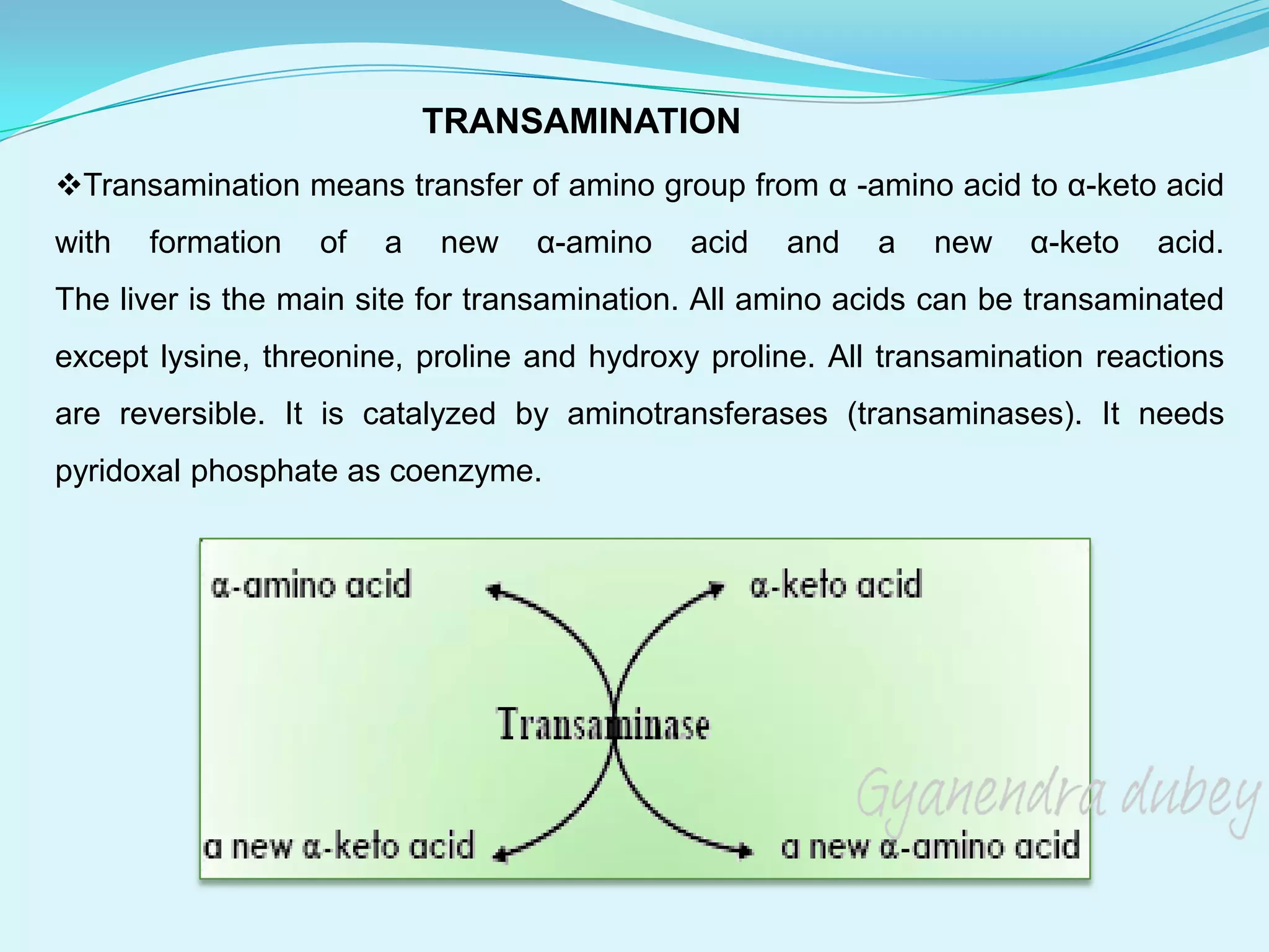
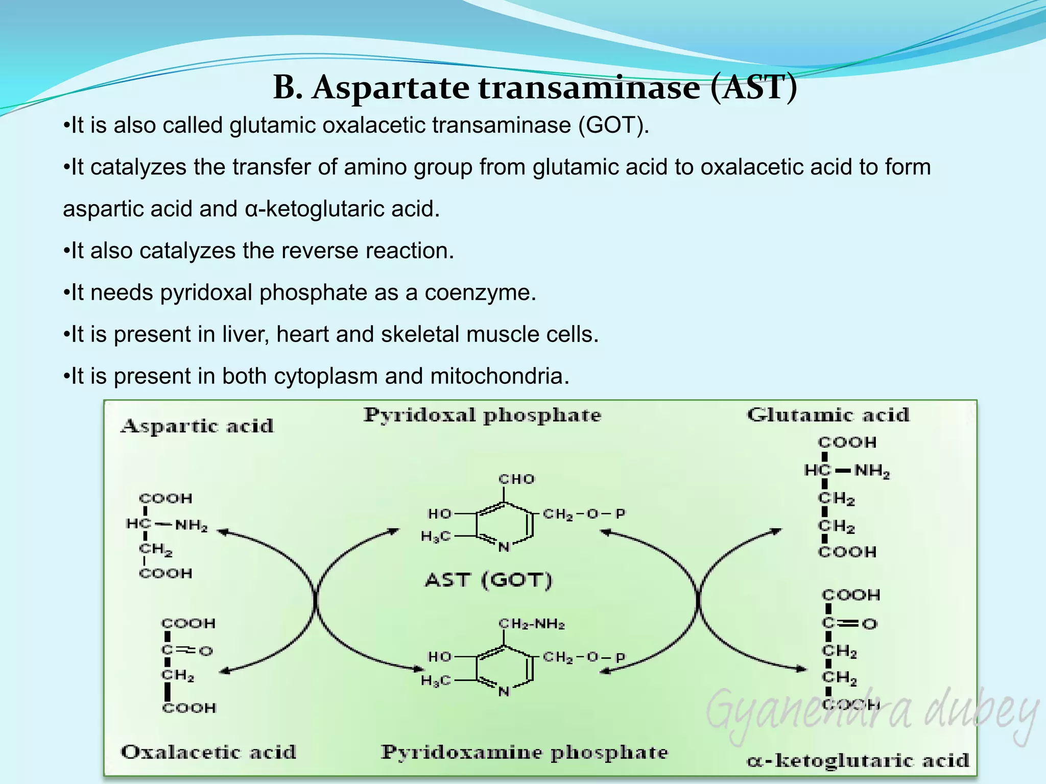
Metabolism of Amino Acids
The proteins on degradation (proteolysis) release individual amino acids. Amino acids are not
just the structural components of proteins. Each of the 20 naturally occurring amino acids
undergoes its own metabolism and performs specific functions.
Protein metabolism is more appropriately learnt as metabolism of amino acids.
The amino acids undergo certain common reactions like transamination followed by
deamination for the liberation of ammonia. The amino group of the amino acids is utilized for
the formation of urea which is an excretory end product of protein metabolism.
The carbon skeleton of the amino acids is first converted to keto acids (by
transamination) which meet one or more of the following fates:
1. Utilized to generate energy.
2. Used for the synthesis of glucose.
3. Diverted for the formation of fat or ketone bodies.
4. Involved in the production of non-essential amino acids.
A general picture of amino acid metabolism is depicted in Fig.
TRANSAMINATION
Transamination means transfer of amino group from α -amino acid to α-keto acid
with formation of a new α-amino acid and a new α-keto acid.
The liver is the main site for transamination. All amino acids can be transaminated
except lysine, threonine, proline and hydroxy proline. All transamination reactions
are reversible. It is catalyzed by aminotransferases (transaminases). It needs
pyridoxal phosphate as coenzyme.
Role of pyridoxal phosphate in Transamination
Pyridoxal phosphate acts as an intermediate carrier for amino group. Pyridoxal
phosphate accepts the amino group from amino acid to form pyridoxamine
phosphate, which in turn gives the amino group to α-keto acid.
Examples of transaminases
A. Alanine transaminase
B. Aspartate transaminase
C. Glutamate transaminase
A. Alanine transaminase
•It is also called glutamic pyruvic transaminase (GPT).
•It catalyzes the transfer of amino group from glutamic acid to pyruvic acid to form alanine and
α-ketoglutaric acid.
•It also catalyzes the reverse reaction.
•It needs pyridoxal phosphate as a coenzyme.
•It is present in the cytoplasm of liver cells.
B. Aspartate transaminase (AST)
•It is also called glutamic oxalacetic transaminase (GOT).
•It catalyzes the transfer of amino group from glutamic acid to oxalacetic acid to form
aspartic acid and α-ketoglutaric acid.
•It also catalyzes the reverse reaction.
•It needs pyridoxal phosphate as a coenzyme.
•It is present in liver, heart and skeletal muscle cells.
•It is present in both cytoplasm and mitochondria.
C. Glutamate transaminase
•It catalyzes the transfer of amino group from any amino acid (except lysine, threonine,
proline and hydroxy proline) to α-ketoglutaric acid to form glutamic acid and the
corresponding α-keto acid.
•It also catalyzes the reverse reaction.
•It needs pyridoxal phosphate as a coenzyme.
•It is widely distributed in all tissues.
Clinical significance of serum transaminases
Transaminases are intracellular enzyme.
Their levels in blood plasma are low under normal conditions.
ALT (GPT) is present mainly in the cytoplasm of liver cells.
AST (GOT) is present in both cytoplasm and mitochondria in liver, heart and skeletal
muscle

Any damage to these organs will increase the level of transaminases in blood.
In liver diseases, there is an increase in both serum ALT (SGPT) and AST (SGOT) levels.
In acute liver diseases, e.g. acute viral hepatitis, the increase is more in SGPT.
In chronic liver diseases, e.g. liver cirrhosis the increase is more in SGOT.
In heart diseases, e.g. myocardial infarction, there is an increase in SGOT only.
In skeletal muscle diseases, e.g. myasthenia gravis, there is an increase in SGOT only.
DEAMINATION
Deamination means the removal of amino group from α-amino acid in the form of
ammonia with formation of α-keto acid
The liver and kidney are the main sites for deamination .
Deamination may be oxidative or non-oxidative
A. Oxidative deamination
It is catalyzed by one of the following enzymes:
1.L-amino acid oxidases
2.D-amino acid oxidases
3.Glutamate dehydrogenase
B. Non-oxidative deamination
It is catalyzed by one of the following enzymes:
1.Dehydratases
2.Desulfhydrases
A. Oxidative deamination
1- L amino acid oxidase
•This enzyme is present in the liver and kidney. Its activity is low.
•It is an aerobic dehydrogenase that needs FMN as a coenzyme.
•It deaminates most of the naturally occurring L-amino acids
2- D amino acid oxidase
•D- amino acids are present in plants and bacterial cell wall.
•They are not used in protein biosynthesis in humans and animals.
•D-amino acids are deaminated by D-amino acid oxidase resulting in ammonia and α-
keto acids.
•D-amino acid oxidase is present in the liver.
•It is an aerobic dehydrogenase.
•It needs FAD as a coenzyme.
3- Glutamate dehydrogenase
•This enzyme is present in most tissues.
•It is present both in cytoplasm and mitochondria.
•Its activity is high.
•It is an anaerobic dehydrogenase.
•It needs NAD or NADP as a coenzyme.
•It deaminates glutamic acid resulting in α-ketoglutaric acid and ammonia.
B. Non-oxidative deamination
1. Dehydratase
•This enzyme deaminates amino acids containing hydroxyl group e.g.
serine, homoserine and threonine.
•It needs pyridoxal phosphate as coenzyme.
2-Desulfhydrase
•This enzyme deaminates sulpher containing amino aids e.g. cysteine and
cystine, methionine.
•It needs pyridoxal phosphate as a coenzyme.
Note- Most of the naturally occurring α-amino acids are catabolized by
transamination with α-ketoglutaric acid followed by deamination of the produced
glutamic acid, a condition called transdeamination
DECARBOXYLATION
•Decarboxylation means removal of CO2 from amino acid with formation of
corresponding amines.
•It is catalyzed by decarboxylase enzyme.
•It needs pyridoxal phosphate as a coenzyme.
Examples of decarboxylation reaction include:
1. Decarboxylation of histidine to form histamine
2. Decarboxylation of tyrosine to form tyramine
UREA (ORNITHINE) CYCLE
•Ammonia (which is produced as a result of deamination of amino acid) is a polar and
very toxic substance freely passing through physical barriers, as well as the blood-brain
barrier. When its concentration increases in the body result in ammonia toxicity and may
lead to the coma.
•It is removed from the tissues, mainly as urea, which is formed in the liver by the
process called UREA (ORNITHINE) CYCLE.
• It is also removed as glutamine from the brain.
•Urea cycle is the process by which ammonia a high toxic water insoluble substances is
converted into less toxic and water soluble as excretory product.
Reactions in urea cycle:
•Urea, a non-toxic compound, is transported via the bloodstream to the kidneys where it
is excreted with the urine. Urea cycle is located in the matrix of mitochondria and cytosol
of liver cells. This pathway is an energy-consuming process in which the three
substrates enter – ammonia, carbon dioxide (bicarbonate) and aspartate (its amino
group). Mitochondrial carbamoyl phosphate synthetase is the regulatory enzyme.
Ornithine cycle communicates with the Krebs cycle via oxaloacetate and fumarate.
Urea formation involves five reactions:
1) Carbamoyl phosphate formation is catalyzed by mitochondrial carbamoyl
phosphate synthetase.
NH4+ + HCO3– + ATP → carbamoyl phosphate + 2 ADP + Pi
2) Citrulline formation is catalyzed by ornithine transcarbamoylase:
Ornithine + carbamoyl phosphate → citrulline + Pi
Citrulline is passed into the cytosol.
3) Argininosuccinate formation is catalyzed by argininosuccinate synthetase:
Citrulline + Asp + ATP → argininosuccinate + AMP + PPi
4) Argininosuccinate break down is catalyzed by argininosuccinate lyase:
Argininosuccinate → arginine + fumarate
5) Hydrolysis of arginine is catalyzed by arginase:
Arginine + H2O → ornithine + urea.
Ornithine returns into the mitochondrial matrix.
Regulation of Ornithine cycle:
Carbamoyl phosphate synthetase, the main regulatory enzyme of ornithine cycle, is
activated by N-acetylglutamate. Enzyme N-acetylglutamate synthetase catalyzes the
reaction between AcCoA and glutamate which produces N-acetylglutamate. The amino
acid arginine increases the enzyme activity. Transcription of urea cycle enzymes is
increased in high-protein diet or by increasing protein catabolism (e.g. starvation),
therefore in the increased supply of amino acids. The urea cycle belongs among proton-
producing reactions, its activity is reduced at lower pH – acidosis.
Metabolic disorders of urea cycle
Various disorders due to the deficiencies of the enzymes of the urea cycle have been
reported.
Gyanendra Dubey

Amino acid metabolism 1.pdf

  • 2.
    Metabolism of AminoAcids The proteins on degradation (proteolysis) release individual amino acids. Amino acids are not just the structural components of proteins. Each of the 20 naturally occurring amino acids undergoes its own metabolism and performs specific functions. Protein metabolism is more appropriately learnt as metabolism of amino acids. The amino acids undergo certain common reactions like transamination followed by deamination for the liberation of ammonia. The amino group of the amino acids is utilized for the formation of urea which is an excretory end product of protein metabolism. The carbon skeleton of the amino acids is first converted to keto acids (by transamination) which meet one or more of the following fates: 1. Utilized to generate energy. 2. Used for the synthesis of glucose. 3. Diverted for the formation of fat or ketone bodies. 4. Involved in the production of non-essential amino acids.
  • 3.
    A general pictureof amino acid metabolism is depicted in Fig.
  • 4.
    TRANSAMINATION Transamination means transferof amino group from α -amino acid to α-keto acid with formation of a new α-amino acid and a new α-keto acid. The liver is the main site for transamination. All amino acids can be transaminated except lysine, threonine, proline and hydroxy proline. All transamination reactions are reversible. It is catalyzed by aminotransferases (transaminases). It needs pyridoxal phosphate as coenzyme.
  • 5.
    Role of pyridoxalphosphate in Transamination Pyridoxal phosphate acts as an intermediate carrier for amino group. Pyridoxal phosphate accepts the amino group from amino acid to form pyridoxamine phosphate, which in turn gives the amino group to α-keto acid.
  • 6.
    Examples of transaminases A.Alanine transaminase B. Aspartate transaminase C. Glutamate transaminase A. Alanine transaminase •It is also called glutamic pyruvic transaminase (GPT). •It catalyzes the transfer of amino group from glutamic acid to pyruvic acid to form alanine and α-ketoglutaric acid. •It also catalyzes the reverse reaction. •It needs pyridoxal phosphate as a coenzyme. •It is present in the cytoplasm of liver cells.
  • 7.
    B. Aspartate transaminase(AST) •It is also called glutamic oxalacetic transaminase (GOT). •It catalyzes the transfer of amino group from glutamic acid to oxalacetic acid to form aspartic acid and α-ketoglutaric acid. •It also catalyzes the reverse reaction. •It needs pyridoxal phosphate as a coenzyme. •It is present in liver, heart and skeletal muscle cells. •It is present in both cytoplasm and mitochondria.
  • 8.
    C. Glutamate transaminase •Itcatalyzes the transfer of amino group from any amino acid (except lysine, threonine, proline and hydroxy proline) to α-ketoglutaric acid to form glutamic acid and the corresponding α-keto acid. •It also catalyzes the reverse reaction. •It needs pyridoxal phosphate as a coenzyme. •It is widely distributed in all tissues.
  • 9.
    Clinical significance ofserum transaminases Transaminases are intracellular enzyme. Their levels in blood plasma are low under normal conditions. ALT (GPT) is present mainly in the cytoplasm of liver cells. AST (GOT) is present in both cytoplasm and mitochondria in liver, heart and skeletal muscle  Any damage to these organs will increase the level of transaminases in blood. In liver diseases, there is an increase in both serum ALT (SGPT) and AST (SGOT) levels. In acute liver diseases, e.g. acute viral hepatitis, the increase is more in SGPT. In chronic liver diseases, e.g. liver cirrhosis the increase is more in SGOT. In heart diseases, e.g. myocardial infarction, there is an increase in SGOT only. In skeletal muscle diseases, e.g. myasthenia gravis, there is an increase in SGOT only.
  • 10.
    DEAMINATION Deamination means theremoval of amino group from α-amino acid in the form of ammonia with formation of α-keto acid The liver and kidney are the main sites for deamination . Deamination may be oxidative or non-oxidative A. Oxidative deamination It is catalyzed by one of the following enzymes: 1.L-amino acid oxidases 2.D-amino acid oxidases 3.Glutamate dehydrogenase B. Non-oxidative deamination It is catalyzed by one of the following enzymes: 1.Dehydratases 2.Desulfhydrases
  • 11.
    A. Oxidative deamination 1-L amino acid oxidase •This enzyme is present in the liver and kidney. Its activity is low. •It is an aerobic dehydrogenase that needs FMN as a coenzyme. •It deaminates most of the naturally occurring L-amino acids 2- D amino acid oxidase •D- amino acids are present in plants and bacterial cell wall. •They are not used in protein biosynthesis in humans and animals. •D-amino acids are deaminated by D-amino acid oxidase resulting in ammonia and α- keto acids. •D-amino acid oxidase is present in the liver. •It is an aerobic dehydrogenase. •It needs FAD as a coenzyme.
  • 12.
    3- Glutamate dehydrogenase •Thisenzyme is present in most tissues. •It is present both in cytoplasm and mitochondria. •Its activity is high. •It is an anaerobic dehydrogenase. •It needs NAD or NADP as a coenzyme. •It deaminates glutamic acid resulting in α-ketoglutaric acid and ammonia.
  • 13.
    B. Non-oxidative deamination 1.Dehydratase •This enzyme deaminates amino acids containing hydroxyl group e.g. serine, homoserine and threonine. •It needs pyridoxal phosphate as coenzyme. 2-Desulfhydrase •This enzyme deaminates sulpher containing amino aids e.g. cysteine and cystine, methionine. •It needs pyridoxal phosphate as a coenzyme.
  • 14.
    Note- Most ofthe naturally occurring α-amino acids are catabolized by transamination with α-ketoglutaric acid followed by deamination of the produced glutamic acid, a condition called transdeamination
  • 15.
    DECARBOXYLATION •Decarboxylation means removalof CO2 from amino acid with formation of corresponding amines. •It is catalyzed by decarboxylase enzyme. •It needs pyridoxal phosphate as a coenzyme. Examples of decarboxylation reaction include: 1. Decarboxylation of histidine to form histamine 2. Decarboxylation of tyrosine to form tyramine
  • 16.
    UREA (ORNITHINE) CYCLE •Ammonia(which is produced as a result of deamination of amino acid) is a polar and very toxic substance freely passing through physical barriers, as well as the blood-brain barrier. When its concentration increases in the body result in ammonia toxicity and may lead to the coma. •It is removed from the tissues, mainly as urea, which is formed in the liver by the process called UREA (ORNITHINE) CYCLE. • It is also removed as glutamine from the brain. •Urea cycle is the process by which ammonia a high toxic water insoluble substances is converted into less toxic and water soluble as excretory product. Reactions in urea cycle: •Urea, a non-toxic compound, is transported via the bloodstream to the kidneys where it is excreted with the urine. Urea cycle is located in the matrix of mitochondria and cytosol of liver cells. This pathway is an energy-consuming process in which the three substrates enter – ammonia, carbon dioxide (bicarbonate) and aspartate (its amino group). Mitochondrial carbamoyl phosphate synthetase is the regulatory enzyme. Ornithine cycle communicates with the Krebs cycle via oxaloacetate and fumarate.
  • 17.
    Urea formation involvesfive reactions: 1) Carbamoyl phosphate formation is catalyzed by mitochondrial carbamoyl phosphate synthetase. NH4+ + HCO3– + ATP → carbamoyl phosphate + 2 ADP + Pi 2) Citrulline formation is catalyzed by ornithine transcarbamoylase: Ornithine + carbamoyl phosphate → citrulline + Pi Citrulline is passed into the cytosol. 3) Argininosuccinate formation is catalyzed by argininosuccinate synthetase: Citrulline + Asp + ATP → argininosuccinate + AMP + PPi 4) Argininosuccinate break down is catalyzed by argininosuccinate lyase: Argininosuccinate → arginine + fumarate 5) Hydrolysis of arginine is catalyzed by arginase: Arginine + H2O → ornithine + urea. Ornithine returns into the mitochondrial matrix.
  • 19.
    Regulation of Ornithinecycle: Carbamoyl phosphate synthetase, the main regulatory enzyme of ornithine cycle, is activated by N-acetylglutamate. Enzyme N-acetylglutamate synthetase catalyzes the reaction between AcCoA and glutamate which produces N-acetylglutamate. The amino acid arginine increases the enzyme activity. Transcription of urea cycle enzymes is increased in high-protein diet or by increasing protein catabolism (e.g. starvation), therefore in the increased supply of amino acids. The urea cycle belongs among proton- producing reactions, its activity is reduced at lower pH – acidosis. Metabolic disorders of urea cycle Various disorders due to the deficiencies of the enzymes of the urea cycle have been reported.
  • 20.