478047882-Disorders-of-the-Eye.pdf full explanation
1. Nursing Management
Diagnostics
Signs and Symptoms
Nursing Diagnosis
Medical Management
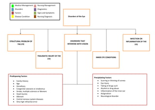
Disorders
Factors
Disease Condition
DISORDERS THAT
INTERFERE WITH VISION
STRUCTURAL PROBLEM OF
THE EYE
INFECTION OR
INFLAMMATION OF THE
EYE
INNER EYE CONDITIONS
TRAUMATIC INJURY OF THE
EYE
Predisposing Factors
Family History
Age
Hereditary
Congenital cataracts or strabismus
Stroke, multiple sclerosis or Meniere
Head injuries
Albinism
Central nervous system diseases
Very high refractive error
Precipitating Factors
Scarring or thinning of cornea
Eye injury
Taking of drugs such
Alcohol or drug abuse
Inflammation of the inner ear
Astigmatism
Neurological disorder
Disorders of the Eye
2. ASTIGMATISM
Congenital or acquired unevenness of the
curvature of the cornea. This causes light rays
coming to the retina to not be all refracted in the
same way
Irregular curvature of the
lens
Uneven quality of vision
Refracting power is not
uniform in all meridians of
both lens or cornea
Diagnostic Tests
Snellen Chart – Can be used to
measure visual acuity
Phoroptor/Refractor – Contains
lenses of different strengths that
can be moved into your view
Keratometry – Measures the
curvature of the cornea
Irregular curvature on the
cornea
Signs/Symptoms
Uneven quality of vision
May have difficulty reading or
following instructions
May report headache and vertigo
after doing close work
Eyestrain
Headache
Treatment
Corrective lenses – Helps to relieve the
symptoms and restore functional vision
Contact lenses – Helps to actually smooth
out the curvature of the cornea
Orthokeratology – Treatment that uses
rigid contact lenses to temporarily correct
the irregular curvature of the cornea
LASIK Surgery, Photorefractive
Keratectomy (PRK), and Radial
Keratotomy (RK)
Nursing Diagnosis
Disturbed sensory perception (visual) related
to visual problems
Risk for fall related to decreased vision
Fear and anxiety related to visual impairment
and loss of autonomy
Knowledge deficit related to impaired vision
3. REFRACTIVE ERROR
Light Refraction refers to the matter in which
light is bent as it passes through the lens. This
causes a ray of light to fall directly on the retina.
Genetic Factors
DECREASED VISION
Changes in choroid and
retina
Elongation and Stretching of
Sclera
Increase of axial length of
the eyeball
Degenerative changes in
Retina
Diagnostic Tests
Snellen Chart – Can be used to
measure visual acuity
Phoroptor/Refractor – Contains
lenses of different strengths that
can be moved into your view
Keratometry – Measures the
curvature of the cornea
Myopia (Nearsightedness)
Hyperopia (Farsightedness)
Signs/Symptoms
Hyperopia
o Blurry vision at a close
range and clear at a far
range
Headaches or dizziness after
completing schoolwork
Myopia
o Blurry vision at a far range
and clear at a close range
o Tries to focus on objects by
squinting eyes
o After LASIK Surgery:
Disturbed tear function for
1 or more months and need
to be managed by artificial
tears
Treatment
Hyperopia
o Normal hyperopia of a
preschooler needs no correction.
For some, this diminishes at about
5 years old as a result of
developmental changes.
o Glasses with convex lens
Myopia
o Corrective (Concave) lenses
o Laser surgery [Laser in Situ
Keratomileusis] (LASIK)
LASIK Surgery, Photorefractive
Keratectomy (PRK), and Radial
Keratotomy (RK)
Signs/Symptoms
Myopia
o Blurry vision at a far range and clear at
a close range
o Tries to focus on objects by squinting
eyes
o After LASIK Surgery: Disturbed tear
function for 1 or more months and
need to be managed by artificial tears
4. NYSTAGMUS
Nystagmus is rapid, irregular eye movement,
either vertically or horizontally; a symptom of an
underlying disease condition
Infantile: most
often develops by 2-
3 mo of age; The
eyes tend to move
in a horizontal
swinging fashion. It
is often associated
with other
conditions, such as
albinism, congenital
absence of the iris
(the colored part of
the eye),
underdeveloped
optic nerves and
congenital cataract
Acquired: Develops
later in childhood or
adulthood. The
cause is often
unknown, but it
may be due to
central nervous
system and
metabolic disorders
or alcohol and drug
toxicity
Diagnostic Tests
Ear exam
Neurological exam
Brain MRI
Brain CT scan
Recording of the eye movement
Pt history
Visual acuity measurements
Refraction
Spasmus nutans:
usually occurs at 6
mo - 3 yo but can
improve at 2-8 yo;
Children with this
form of nystagmus
often nod and tilt
their heads
Signs and Symptoms
Uncontrollable eye
movement
Inability to focus
Light sensitivity
Trouble balancing
Dizziness
Treatment
Corrective lenses
Eye muscle surgery
Use large-printed books
Use of magnifying
devices
Increased lighting
Nursing Diagnosis
Risk for injury related to
impaired sensory
function.
Disturbed sensory
perception related to
structural damage.
Knowledge deficit
related to impaired
vision.
5. AMBYLOPIA
“lazy eye,” or subnormal vision in one
eye; the child may be using only one eye
for vision while “resting” the other eye
Deprivation amblyopia
Visual loss (amblyopia) in
eye with higher refractive
error
Refractive (anisometropic)
amblyopia
Diagnostic Tests
Vision test
Strabismic amblyopia
Signs and Symptoms
20/50 vision
A child may cry when
one eye is covered
(covering the good
eye)
Treatment
Occlusion therapy:
The good eye is
covered by a patch
held firmly in place
which forces the
child to use the
poor eye, thus
developing vision
in that eye
Atropine: causes
pupil dilation and
blurred vision
Strabismus surgery
Vision therapy
Prosthetic contact
lenses
Atropine eye
drops
Computer
programs that
stimulate neural
changes
(RevitalVision)
Nursing Diagnosis
Disturbed sensory perception (visual) related to visual
problems
Risk for fall related to decreased vision
Fear and anxiety related to visual impairment and loss
of autonomy
Knowledge deficit related to impaired vision
Clearer image favored
Difference in refractive
error between the two eyes
Misalignment of the eyes
results in abnormal
binocular interaction.
Eventual unconscious
suppression of visual
stimulation to an affected
eye creates amblyopia
Eyes fail to receive clearly
formed images on the retina
Due to a cataract, other
opacity, or obstruction
(hemangioma of lid)
6. STRUCTURAL PROBLEM OF
THE EYE
Predisposing Factors
Congenital, age(common in elderly),
development of myasthenia gravis
History of strabismus in family,
congenital
Predisposing Factors
Injury to levator muscle, injury to
third cranial nerve.
PTOSIS
Movement of the eyelids'
muscle (levator muscle) is
affected, thus the
appearance of a droopy
eyelid occurs
amblyopia (from lack of use
of the closed eye).
Obstructs vision
Ptosis can be unilateral or
lateral
Diagnostic Tests
Observation of eyelid movements
Assessment of the functions of third
cranial nerve
Signs and Symptoms
Children tend to wrinkle their
forehead and raise their eyebrows
more than usual in attempt to lift
eyelid further
Cock head back to see under lower
lid
Dilated pupil
Inability to rotate the eye globe
upward, medially or downward
Weakness of accommodation
(looking at near objects)
Treatment
Surgical correction
Inability to raise the upper eyelid the usual
distance, so the eyelid always remains slightly
closed.
Nursing Intervention
Provide support and
guidance to child
when walking around
Teach parents the
importance of
correcting ptosis at a
young age to prevent
irreversible effects of
amblyopia.
Provide eye drops to
help moisten eyeballs
Help child in eye
exercises
Nursing Diagnosis
Impaired comfort related to lack of use of closed eye
that causes eyes to dry.
Risk for chronic low esteem related to appearance of
drooping eyelids
7. STRABISMUS
Unequally aligned eyes (cross
eyes)
Deviations can be
Exotropia
Esotropia
hypertropia
The resting position of the
one eye, is not aligned to be
forwards.
Diagnostic Tests
Eye Test
Cover test
Hirschberg
test
Caused by imbalance of the
extraocular muscles that
control the movement of
the eyeglobes
Signs and Symptoms
Abnormal
appearance of the
resting level of eyes
Eyestrain
Headache
Tired, irritated eyes
Nausea
Vomiting
Nursing Intervention
Aid children in
ambulating to
avoid injury
Aromatherapy to
help ease nausea
and vomiting
Eye exercises
(orthoptics)
Nursing Diagnosis
Risk for injury related to impaired sensory function.
Disturbed sensory perception related to structural
damage.
Knowledge deficit related to impaired vision.
Resting position can be:
divergent (turned
out)
convergent (turned
in)
vertical strabismus
(pupil may be
higher than the
other)
Strabismus can be:
Monocular or same
eye deviates
constantly
Alternating
strabismus where
one eye and then
the other deviates
Treatment
Glasses or lenses
to basic correct
visual defect
Surgery to align
extraocular
muscles
Types: concomitant and
nonconcomitant
8. INFECTION OR
INFLAMMATION OF THE
EYE
Predisposing Factors
Congenital, age
Have diabetes
Predisposing Factors
Have dry skin
Are experiencing hormonal changes
Have blepharitis
Have certain skin conditions
Lack of Handwashing
STY
An internal sty results from
inflammation of
a meibomian gland, one of
the modified sebaceous
glands that lie close to the
eyeball along the margin of
the eyelids. It may be
caused by an infectious (i.e.,
staphylococcal) or
noninfectious process.
a painless, chronic swelling
of the meibomian gland will
then occur
Infected part of the eye
begins with a reddened
color on the infected part of
the eye
Diagnostic Tests
Your doctor will usually diagnose
a sty just by looking at your eyelid.
Symptoms
Burning sensation in the eye
Crusting of the eyelid margins
Droopiness of the eyelids
Itchiness on the eyeball
Sensitivity to light
Tearing
A feeling that something is stuck in
the eye
Discomfort when blinking
Treatment
Most styes go away on their
own without any treatment,
as soon as the stye ruptures
symptoms tend to improve
rapidly
Inflammation of the eyelid associated with a small
collection of pus and is caused mostly by the
staphylococcus bacteria
Nursing Intervention
DO NOT burst stye by
yourself
Apply warm
compress
o held against
the eye 5 to
10 minutes, 3
to 4 times
each day
o to also
encourage
the pus to
drain away
Pain relievers
o ibuprofen or
acetaminoph
en
9. CONJUNCTIVITIS
Pink eye (conjunctivitis) is an inflammation or
infection of the transparent membrane
(conjunctiva) that lines your eyelid and covers the
white part of your eyeball.
Bacterial conjunctivitis
Bacterial conjunctivitis is
caused by bacteria such as
staphylococci, streptococci
or haemophilus
Small blood vessels in the
conjunctiva become
inflamed causing the whites
of your eyes to appear
reddish or pink
Viral conjunctivitis
Viral conjunctivitis is usually
occurs after a cold or a sore
throat and it is highly
contagious.
Diagnostic Tests
Slit-lamp
exam
Cultures
Allergic conjunctivitis
Allergic conjunctivitis is
often caused by dust mites,
pollen and cosmetics and is
common in people who
have hay fever, asthma and
eczema.
Signs/Symptoms
The pink eye effect is
one of the first signs
of conjunctivitis. In
severe cases, the eyes
may be glued shut on
waking.
Dryness and itchiness
of the infected eye
occurs
Redness present at,
discharge may also be
present
Treatment
Topical antibiotic
treatment will
relieve symptoms
and shorten the
length of the
illness. Treatment
with antibiotic
drops/ointment
will also prevent
the risk of more
widespread
extraocular
disease.
(Chloramphenicol
or Fusidic acid)
Nursing Diagnosis
1. Acute Pain related to inflammation of the conjunctiva
2. Anxiety related to lack of knowledge about the
disease process
3. Self-concept disturbance related to a change in the
eyelid (swelling / edema).
Inflammation or infection of
the transparent membrane
10. CHALAZION
a small, usually painless, lump or
swelling that appears on the eyelid.
Granulomatous
inflammatory response
happens
A chalazion forms and may
enlarge and break through
the tarsal plate to the
external portion of the lid
Edema due to blockage of
Meibomian glands occur
Lipid breakdown products
from bacterial enzymes or
sebaceous secretions leak
into surrounding tissue
Signs and symptoms:
redness in the
eyelid
tenderness
swelling
Nursing Management
Do patient teaching about gentle but firm
massages to promote drainage of
obstructed gland
Apply warm compresses to help melt
viscous lipids
Encourage nutritional supplementation
with essential fatty acids
Nursing Diagnosis
Risk for injury r/t frequent touching of lump in
eyelid
Risk for infection r/t improper care of eyelid
secretions
Medical management:
Antibiotic eye ointment or antibiotic pills
Injection of steroid medicine if it gets
worse
11. TRAUMATIC INJURY OF THE
EYE
Predisposing Factors
Congenital, age
Have diabetes
Predisposing Factors
Have dry skin
Are experiencing hormonal changes
Have blepharitis
Have certain skin conditions
Lack of Handwashing
CONTUSION
Blunt trauma occurs to eye
Hemorrhage gradually
reabsorbed over time
Hemorrhage around eye
occurs
Nsg Dx: Altered Body Image related to eye
contusion,
Signs and Symptoms
Hemorrhage around eye
Pain around eye
Limited motion of the eye
Loss of vision
Maxillary fracture
Treatment:
Ice pack
Pain reliever if pain
exists
Surgery if maxillary
fracture exists
Blunt trauma caused by being hit in the eye by a blunt
object I.e baseball, fist, soccer ball, or car dashboard
resulting to a “black eye”
Assessment:
Inspect eye globe
Ask children how
many fingers they
can count from a
distance of 6 feet or
letting them read a
page from a distance
Ask children if they
have difficulty seeing
Evaluate extraocular
eye movements for
adequate function
12. EYELID INJURY
Foreign object strikes
eye
If laceration is in the
inner canthus, disrupts
lacrimal drainage
system (dacryostenosis)
Eyelid becomes
damaged or lacerated
Nsg Dx: Pain r/t injury
Signs and Symptoms
Pain around eye
Limited motion of the eye
Loss of vision
Injury that may accompany eye globe injury or may be
present after a foreign body has struck the eye
Assessment:
Check for lacerations
Treatment
Go to doctor
(ophthalmologist)
If laceration is deep,
may cause permanent
ptosis
13. INNER EYE CONDITIONS
Predisposing Factors
- Age group of children at 1 year of
age
- Gender, often in girls
- Increasing age
- Genetic predisposition
Precipitating Factors
- Scarring at the canal of Schlemm
- Galactosemia
- Steroid use
- Radiation exposure
- Retinoblastoma
- Retinopathy
- Smoking
- Obesity
- Diabetes
CONGENITAL GLAUCOMA
A developmental anomaly
in the angle of the anterior
chamber prevents proper
drainage into the canal.
After it has increased in size
to the extent that it can, the
pressure in the eye globe
continues to rise,
compressing and ultimately
destroying the optic nerve.
The increased fluid content
that accumulates causes the
globe of the eye to increase
in size.
Diagnostics
Examination of the front part of the
eye
Examination of the fundus
Tonometry
Signs and Symptoms
Seems painfully sensitive to light
Tears up a lot
Depending on how far the disease has
worsened, other eye symptoms can include:
A cloudy cornea (the front layer of
your eye that’s normally clear)
One or both eyes larger than
normal
Redness
Treatment
Goniotomy or
trabeoculotomy
acetazolamide
(Diamox)
Laser Therapy
A developmental anomaly in the angle of the
anterior chamber prevents proper drainage into
the canal. Later in life, glaucoma occurs when the
canal becomes blocked.
Nursing Diagnosis
Disturbed sensory
perception related to
congenital glaucoma
Glaucoma occurs when the
canal becomes blocked
14. CATARACT
A cataract is the gradually
developing opacity of the lens
or lens capsule of the eye.
If the lens becomes less
transparent and obstructs
the passage of light to the
retina, the patient suffers
from a progressive loss of
vision.
Rays of light pass through
this lens to reach the retina,
where images are formed.
Diagnostics
Visual acuity test.
Slit-lamp
examination.
Retinal exam.
A cataract is the loss of
transparency of the
crystalline lens, the eye’s
natural lens located behind
the pupil.
Signs and Symptoms
- White pupil opening
- Blurred vision in children
- Lack of response to a smile or
inability to reach and grasp a
nearby object for infants
- Nystagmus
- Increasing difficulty with vision at
night
- Sensitivity to light and glare
- Need for brighter light for reading
and other activities
- Seeing "halos" around lights
- Frequent changes in eyeglass or
contact lens prescription
- Fading or yellowing of colors
- Double vision in a single eye
Treatment
Surgical removal of the cloudy lens
Phacoemulsification: emulsifying the
cataract and aspirating it
Insertion of an internal intraocular lens
Myriatic agent
Nursing Diagnosis
- Risk for injury related to loss of vision as
evidenced by cataract