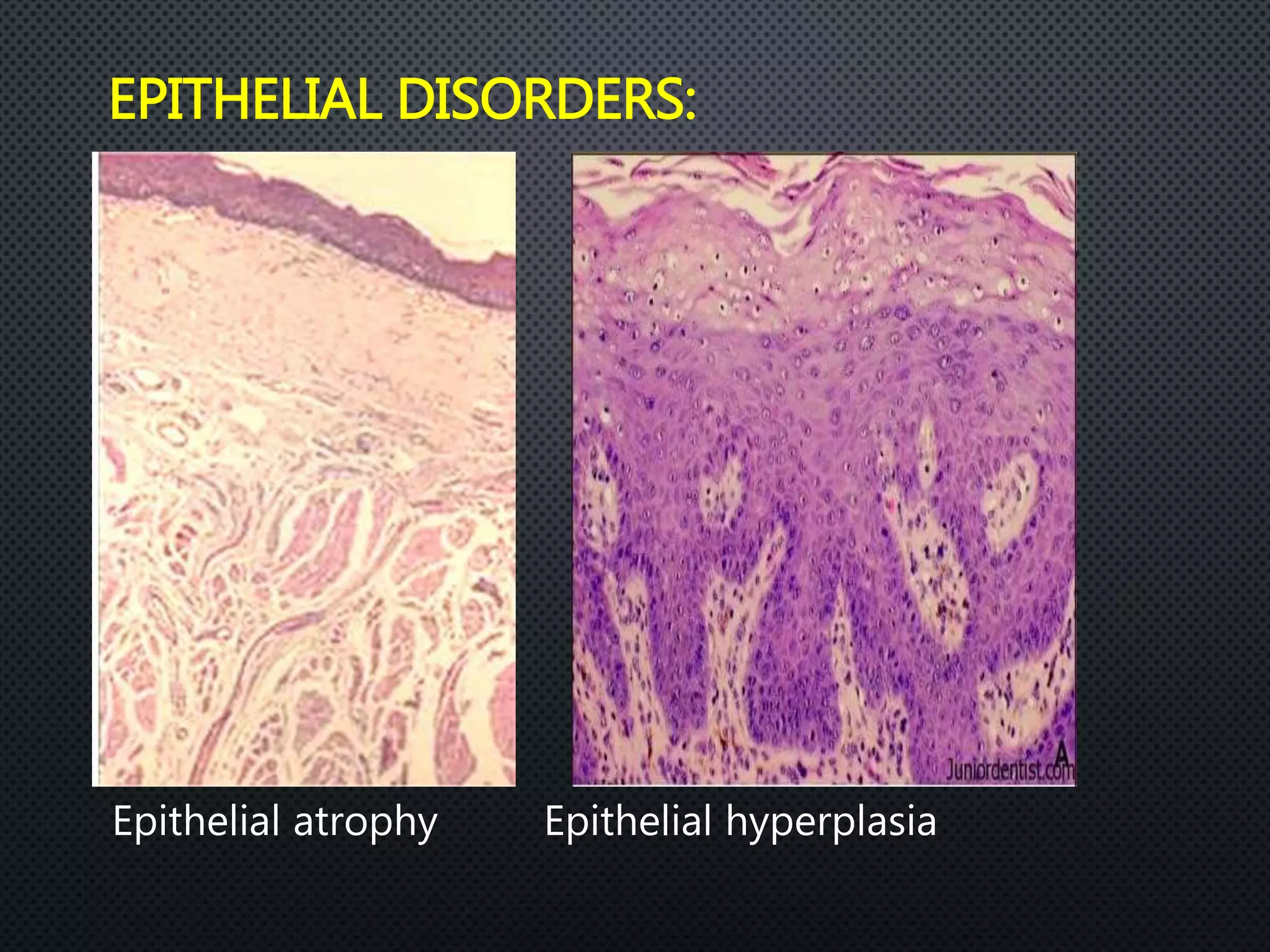
The oral epithelium acts as a protective barrier for the oral mucosa. It is composed of keratinocytes that undergo proliferation and maturation as they move from the basal layer to the surface. Keratinocytes express different cytokeratins as they mature and differentiate. The oral epithelium can be keratinized or non-keratinized depending on its location. A basement membrane anchors the epithelium. Various cellular disorders can affect the oral epithelium. Non-keratinocytes including melanocytes, Langerhans cells, and lymphocytes are also present in small numbers.
PHYSICAL PROPERTIES
CHEMICAL PROPERTIES
STRUCTURE OF ENAMEL
DEVELOPMENT OF ENAMEL
EPITHELIAL ENAMEL ORGAN
AMELOGENESIS
LIFE CYCLE OF AMELOBLASTS
AGE CHANGES IN ENAMEL
DEFECTS OF AMELOGENESIS
CLINICAL IMPLICATIONS
PHYSICAL PROPERTIES
CHEMICAL PROPERTIES
STRUCTURE OF ENAMEL
DEVELOPMENT OF ENAMEL
EPITHELIAL ENAMEL ORGAN
AMELOGENESIS
LIFE CYCLE OF AMELOBLASTS
AGE CHANGES IN ENAMEL
DEFECTS OF AMELOGENESIS
CLINICAL IMPLICATIONS
JUNCTIONAL EPITHELIUM
It is a highly specialized epithelial tissue which divides faster than any other normal epithelium.
The mean turnover time of junctional epithelium is 5–6 days.
The junctional epithelium is basically a stratified, squamous, non-keratinizing epithelium comprising two layers: basal & suprabasal layers.
The junctional epithelium differs from the gingival oral epithelium & sulcular epithelium in origin & structure.
This specialized epithelium ranges in thickness from few cells at its most apical portion to between 15 & 30 cells at its most coronal portion adjacent to the sulcular epithelium, & the cells align themselves in a plane parallel to the tooth surface.
The length of this epithelium is approximately 0.25–1.35 mm.
The presentation discusses about tooth enamel in detail including its formation, characteristics, structure and histological features along with its clinical considerations. It is well supported with diagrams for better understanding of the text.
Suggestions and feedback will be well appreciated.
Coronal and radicular pulp
Apical foramen
Accessory canal
Functions of dental pulp
Components of dental pulp
Functions of pulpal extracellular matrix
Organization of cells in the pulp
The principle cells of the pulp
The pathways of collagen synthesis
Matrix and ground substances
Vasculature and lymphatic supply
Innervation of Dentin- pulp complex
Disorders of the dental pulp
Advances in pulp vitality testing
The Indian Dental Academy is the Leader in continuing dental education , training dentists in all aspects of dentistry and
offering a wide range of dental certified courses in different formats.for more details please visit
www.indiandentalacademy.com
oral mucous membranes-2 /certified fixed orthodontic courses by Indian dental...Indian dental academy
The Indian Dental Academy is the Leader in continuing dental education , training dentists in all aspects of dentistry and
offering a wide range of dental certified courses in different formats.for more details please visit
www.indiandentalacademy.com
epithelium covers body surfaces, lines body cavities and constitute glands.so it is important to know about epithelium in detail to deal with tissue of different type and origin.
A presentation on the topic of microscopic section of gingiva. This topic is mostly looked on by periodontists. A very important chapter in the speciality in dentistry of periodontology and implantology department. Basic understanding of microscopic features and clinical features of gingiva is an important topic for post graduate as well as undergraduate students in the dental field.
Keratinized tissue, also known as keratinized mucosa, refers to the band of tissue surrounding your teeth at the point where they meet the gums. The word "keratinized" is used to describe cells that produce large amounts of a protein called keratin, making them strong and better at forming barriers. Local irritation interferes with keratinization, and healthy gingiva is more keratinized than diseased, irritated gingiva. Nonepithelial cells are also present in the oral gingival epithelium. These include melanocytes, and Langerhans cells in the stratum spinosum. In the oral cavity, keratinized mucosa is found in the gingiva and palate mucosa, whereas the non-keratinized mucosa is found in the buccal mucosa.
https://userupload.net/3ppacneii1wj
Toxicologic Pathology (Second Edition), 2010
INTRODUCTION
The oral mucosa is, in many ways, similar to the skin in its architecture, function, and reaction patterns. This section only emphasizes those characteristics of the oral mucosa that influence or result in a distinct group of pathologic entities.
Because of its location at the entrance of the digestive and respiratory tracts and its proximity to the teeth, the oral mucosa is subjected to numerous natural and man-made xenobiotics. The peculiar architecture and absorption characteristics of the oral mucosa, especially in areas of extreme thinness, coupled with the rich microorganism flora of the mouth, makes the oral mucosa a peculiar site deserving separate discussion.
PDL is complex vascular structure which surround tooth root and connect it to the alveolar bone. In this we discuss development, macroscopic and microscopic features, functions and it’s supply and clinical consideration.
DISSERTATION on NEW DRUG DISCOVERY AND DEVELOPMENT STAGES OF DRUG DISCOVERYNEHA GUPTA
The process of drug discovery and development is a complex and multi-step endeavor aimed at bringing new pharmaceutical drugs to market. It begins with identifying and validating a biological target, such as a protein, gene, or RNA, that is associated with a disease. This step involves understanding the target's role in the disease and confirming that modulating it can have therapeutic effects. The next stage, hit identification, employs high-throughput screening (HTS) and other methods to find compounds that interact with the target. Computational techniques may also be used to identify potential hits from large compound libraries.
Following hit identification, the hits are optimized to improve their efficacy, selectivity, and pharmacokinetic properties, resulting in lead compounds. These leads undergo further refinement to enhance their potency, reduce toxicity, and improve drug-like characteristics, creating drug candidates suitable for preclinical testing. In the preclinical development phase, drug candidates are tested in vitro (in cell cultures) and in vivo (in animal models) to evaluate their safety, efficacy, pharmacokinetics, and pharmacodynamics. Toxicology studies are conducted to assess potential risks.
Before clinical trials can begin, an Investigational New Drug (IND) application must be submitted to regulatory authorities. This application includes data from preclinical studies and plans for clinical trials. Clinical development involves human trials in three phases: Phase I tests the drug's safety and dosage in a small group of healthy volunteers, Phase II assesses the drug's efficacy and side effects in a larger group of patients with the target disease, and Phase III confirms the drug's efficacy and monitors adverse reactions in a large population, often compared to existing treatments.
After successful clinical trials, a New Drug Application (NDA) is submitted to regulatory authorities for approval, including all data from preclinical and clinical studies, as well as proposed labeling and manufacturing information. Regulatory authorities then review the NDA to ensure the drug is safe, effective, and of high quality, potentially requiring additional studies. Finally, after a drug is approved and marketed, it undergoes post-marketing surveillance, which includes continuous monitoring for long-term safety and effectiveness, pharmacovigilance, and reporting of any adverse effects.
- Video recording of this lecture in English language: https://youtu.be/kqbnxVAZs-0
- Video recording of this lecture in Arabic language: https://youtu.be/SINlygW1Mpc
- Link to download the book free: https://nephrotube.blogspot.com/p/nephrotube-nephrology-books.html
- Link to NephroTube website: www.NephroTube.com
- Link to NephroTube social media accounts: https://nephrotube.blogspot.com/p/join-nephrotube-on-social-media.html
Ozempic: Preoperative Management of Patients on GLP-1 Receptor Agonists Saeid Safari
Preoperative Management of Patients on GLP-1 Receptor Agonists like Ozempic and Semiglutide
ASA GUIDELINE
NYSORA Guideline
2 Case Reports of Gastric Ultrasound
Title: Sense of Taste
Presenter: Dr. Faiza, Assistant Professor of Physiology
Qualifications:
MBBS (Best Graduate, AIMC Lahore)
FCPS Physiology
ICMT, CHPE, DHPE (STMU)
MPH (GC University, Faisalabad)
MBA (Virtual University of Pakistan)
Learning Objectives:
Describe the structure and function of taste buds.
Describe the relationship between the taste threshold and taste index of common substances.
Explain the chemical basis and signal transduction of taste perception for each type of primary taste sensation.
Recognize different abnormalities of taste perception and their causes.
Key Topics:
Significance of Taste Sensation:
Differentiation between pleasant and harmful food
Influence on behavior
Selection of food based on metabolic needs
Receptors of Taste:
Taste buds on the tongue
Influence of sense of smell, texture of food, and pain stimulation (e.g., by pepper)
Primary and Secondary Taste Sensations:
Primary taste sensations: Sweet, Sour, Salty, Bitter, Umami
Chemical basis and signal transduction mechanisms for each taste
Taste Threshold and Index:
Taste threshold values for Sweet (sucrose), Salty (NaCl), Sour (HCl), and Bitter (Quinine)
Taste index relationship: Inversely proportional to taste threshold
Taste Blindness:
Inability to taste certain substances, particularly thiourea compounds
Example: Phenylthiocarbamide
Structure and Function of Taste Buds:
Composition: Epithelial cells, Sustentacular/Supporting cells, Taste cells, Basal cells
Features: Taste pores, Taste hairs/microvilli, and Taste nerve fibers
Location of Taste Buds:
Found in papillae of the tongue (Fungiform, Circumvallate, Foliate)
Also present on the palate, tonsillar pillars, epiglottis, and proximal esophagus
Mechanism of Taste Stimulation:
Interaction of taste substances with receptors on microvilli
Signal transduction pathways for Umami, Sweet, Bitter, Sour, and Salty tastes
Taste Sensitivity and Adaptation:
Decrease in sensitivity with age
Rapid adaptation of taste sensation
Role of Saliva in Taste:
Dissolution of tastants to reach receptors
Washing away the stimulus
Taste Preferences and Aversions:
Mechanisms behind taste preference and aversion
Influence of receptors and neural pathways
Impact of Sensory Nerve Damage:
Degeneration of taste buds if the sensory nerve fiber is cut
Abnormalities of Taste Detection:
Conditions: Ageusia, Hypogeusia, Dysgeusia (parageusia)
Causes: Nerve damage, neurological disorders, infections, poor oral hygiene, adverse drug effects, deficiencies, aging, tobacco use, altered neurotransmitter levels
Neurotransmitters and Taste Threshold:
Effects of serotonin (5-HT) and norepinephrine (NE) on taste sensitivity
Supertasters:
25% of the population with heightened sensitivity to taste, especially bitterness
Increased number of fungiform papillae
Basavarajeeyam is a Sreshta Sangraha grantha (Compiled book ), written by Neelkanta kotturu Basavaraja Virachita. It contains 25 Prakaranas, First 24 Chapters related to Rogas& 25th to Rasadravyas.
Here is the updated list of Top Best Ayurvedic medicine for Gas and Indigestion and those are Gas-O-Go Syp for Dyspepsia | Lavizyme Syrup for Acidity | Yumzyme Hepatoprotective Capsules etc
These simplified slides by Dr. Sidra Arshad present an overview of the non-respiratory functions of the respiratory tract.
Learning objectives:
1. Enlist the non-respiratory functions of the respiratory tract
2. Briefly explain how these functions are carried out
3. Discuss the significance of dead space
4. Differentiate between minute ventilation and alveolar ventilation
5. Describe the cough and sneeze reflexes
Study Resources:
1. Chapter 39, Guyton and Hall Textbook of Medical Physiology, 14th edition
2. Chapter 34, Ganong’s Review of Medical Physiology, 26th edition
3. Chapter 17, Human Physiology by Lauralee Sherwood, 9th edition
4. Non-respiratory functions of the lungs https://academic.oup.com/bjaed/article/13/3/98/278874
4. INTRODUCTION
THE TISSUE THAT FORMS THE SURFACE OF THE ORAL MUCOSA
ACTS AS A BARRIER
IT IS OF STRATIFIED SQUAMOUS TYPE
MAINTAIN STRUCTURAL INTEGRITY
DERIVED FROM
5. TYPES OF ORAL EPITHELIUM
ORTHOKERATINISED PARAKERATINISED NON KERATINISED
7. KERATINOCYTES
KERATINOCYTES CONSISTS OF 2 FUNCTIONAL POPULATIONS
• PROGENITOR POPULATION – PERFORMING
EPITHELIAL PROLIFERATION
• MATURING POPULATION – PERFORMING EPITHELIAL
MATURATION
8. Keratins (previously also called cytokeratins) are
filament forming proteins of epithelial cells and are
essential for normal tissue structure and function
Forms the cytoskeleton of all the epithelial cells,
along with microfilaments & microtubules.
Provide mechanical linkage & distribute force over
wide area
9. Based on distribution
Soft keratin
Hard keratin
Based on X-ray diffraction pattern
Alpha
Beta
Feather keratins
Amorphous keratins
10. Based on amino acid sequence and charge
o Type I: Acidic proteins
: Keratins 9-20
o Type II: Basic or neutral proteins
: Keratins 1-8
Based on molecular weight
o Low molecular weight keratins(40kDa)
o Intermediate molecular weight keratins
o High molecular weight keratins(67kDa)
11. Known as intermediate filament associated proteins
These include
Filaggrin
Trichohyalin
Desmosomal proteins
Proteins of cornified cell envelope
12. FILAGGRIN
• CATIONIC PROTEIN; AIDS IN
DENSE PACKING OF KERATIN
• SYNTHESIZED IN THE
GRANULAR CELL LAYER
• FACILITATES DISULFIDE BOND
FORMATION
• MARKER FOR KERATINIZED
TYPE OF EPITHELIUM
13. TRICHOHYALIN
• EXPRESSED IN THE KERATINIZING FILIFORM PAPILLA OF
TONGUE
• SINGLE STRANDED ALPHA-HELICAL ROD THAT BIND
KERATIN
• FUNCTION AS INTRACELLULAR CEMENT
• ALSO FUNCTIONS AS CROSS BRIDGING PROTEINS.
14. DESMOSOMAL PROTEINS
• LINKS EPITHELIAL CELLS TO EACH OTHER; ATTACHES KERATIN
CYTOSKELETON TO CELL SURFACE
• INTEGRAL PROTEINS: DESMOGLEIN & DESMOCOLLIN
• CYTOPLASMIC PLAQUE PROTEIN: DESMOPLAKIN & PLAKOGLOBIN
• PLAQUE ASSOCIATED PROTEINS: PLAKOPHILIN, ENVOPLAKIN &
PERIPLAKIN
15. PROTEINS OF CORNIFIED CELL ENVELOPE
• DEPOSITED ON THE INNER FACE OF PLASMA MEMBRANE OF
KERATINOCYTES.
• BARRIER FUNCTION OF STRATIFIED KERATINIZED EPITHELIA
• EXPRESSED IN
• MOST ABUNDANT CE PROTEINS
oLORICRIN
oINVOLUCRIN
oSMALL PROLINE RICH PROTEINS (SPR’S)
16. They together makes up 10% of cell population in
the oral epithelium
In light microscope –
No tonofilaments
No maturation
17. Different non- keratinocytes in oral epithelium are
Melanocytes
Langerhans cells
Merkel cells
Inflammatory cells
22. Progenitor cells present in the basal layer
Dividing cells tend to occur on clusters
Progenitor compartment consists of 2 functional sub
population of cells
◦ Small stem cells-
◦ Large amplifying cells-
23.
24.
25. Turnover time of the epithelium is the time it takes
for a cell to divide and pass through the entire
epithelium
Turnover time
Skin – 52 – 75 days
Gut – 4 – 14 days
Gingiva – 41 – 57 days
Cheek – 25 days
26. Proliferation is controlled by biologically active
substances called cytokines
31. Stratum spinosum
Otherwise
Situated
Contacts only at points known as intercellular bridges or desmosomes
Basal and prickle cell constitutes
Stratum germinivatum
32. LAMELLAR BODIES, LAMELLAR GRANULES,
MEMBRANE COATING GRANULES OR
KERATINOSOMES.
THESE GRANULES ARE
• SMALL
• MEMBRANE BOUND
• SIZE – 250 NM
• CONTAIN GLYCOLIPID
• ORIGINATE FROM GOLGI SYSTEM
ODLAND BODIES
33. Stratum granulosm
Next to spinous layer
Consists of large flattened cells
Cells contain small granules that stain immensely with
hematoxylin
34. KERATOHYALIN GRANULES
• CHARACTERISTIC FEATURE
• UNDER LM,EM
• IRREGULAR IN SHAPE
• SIZE 0.5 – 1 MICROMETER
• SYNTHESIZED BY RIBOSOMES
• KERATOHYALIN GRANULES ASSOCIATED WITH
TONOFIBRILS ,
• FILAGGRIN, LORICRIN
35. Stratum corneaum
Surface layer
Composed of very flat cells
Eosinophilic, do not contain any nuclei
This pattern of maturation is called orthokeratinization
Some times in some mucosa, retain the shrunken nuclei
called as parakeratinisation
36. Non keratinization
◦ Usually the lining mucosa
◦ Basal & prickle layers resemble that of keratinized
except the prickle cells of non- keratinized epithelium
are slightly larger and intercellular bridges are less
conspisious
37. Above the prickle layer, divided into 2 zones
Stratum intermedium
Stratum superficiale
# No granular layer
# Superficial layer contain plump nucleus
# Not stain intensely with eosin
38. spinous cell layer (s. intermedium)
In prickle cell layer, increase in size is more than that of
keratinized epithelium
Tonofilaments remain dispersed
Contain membrane bound granules
They are circular in shape, with an amorphous coat
39. Stratum superficiale
The cells of the superficial layer,
Are more flattened
Contain dispersed tonofilaments and nuclei
Number of cell organelles are diminished
Not dehydrated
41. Depends on the
Thickness of the epithelium
Pattern of maturation
Thinnest epithelium allow better penetration
Permeability barrier is due to the lipids derived
from the membrane coating granules
43. • FORMED BY
• LM – AMORPHOUS, DENSE LAYER OF VARIABLE THICKNESS
• PAS STAIN - WELL DEFINED MAGENTA LAYER
• IN ELECTRON MICROSCOPE BM CONSISTS OF 3 LAYER
• LAMINA LUCIDA
• LAMINA DENSA
• LAMINA FIBRO-RETICULARIS
BASEMENT MEMBRANE
44. • MAIN CONSTITUENTS OF BM ARE
• THE GLYCOSAMINOGLYCAN, HEPARIN SULPHATE
• FIBROUS PROTEIN –COLLAGEN TYPE-IV , VII
• STRUCTURAL GLYCOPROTEINS – FIBRONECTIN , LAMININ &
ENACTIN
45. Functional role
Compartmentalize tissues
Anchor cell sheets
Play major role in control of cell migration
Act as an stimulus
Serve as a barrier
As epithelium devoid of blood vessels
Fenestrations in BM
51. ORAL EPITHELIUM
• SERRATED AND NON-SERRATED BASAL CELLS
• CYTOKERATINS EXPRESSION
• SPECIAL STAINS FOR NON-KERATINOCYTES
52.
53. SERRATED HEAVILY PACKED WITH TONOFILAMENTS WHICH
ARE ADAPTATIONS FOR ATTATCHMENT
NON-SERRATED - SLOWLY DIVIDING CELLS WHICH SERVE TO
PROTECT GENETIC INFORMATION OF TISSUE
• PERIMETER OF BOTH NUCLEUS & CYTOPLASM
• CYTOPLASM IS PRIMITIVE & CONTAIN
55. CYTOKERATINS EXPRESSION IN EPITHELIAL
TUMOR CELLS:
• best IHC marker for Merkel-cell
carcinomas
Ck 20
• markers for poorly differentiated
squamous cell carcinoma
Ck 13
• a marker for odontogenic epithelial
origin
Ck 14 & 19
56. Keratins can be used as differentiation markers in
normal oral epithelia:
• markers for simple epithelial differentiation
K8/18
• markers for keratinized epithelium
K1/10
• markers for non-keratinized epithelium.
K4/13
• considered as hyperproliferative markers
• expressed in sites of high epidermal keratinocyte turnover and in pathological
hyperproliferative conditions affecting the skin
K6/16