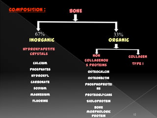
ALVEOLAR
  BONE

           1
Definition
Osteogenesis
Composition
Gross structure of
bone
microscopic structure of
bone
Osseous topography
Fenestrations & dehiscence
Remodeling ( bone
turnover )
Blood supply, lymphatic drainage of the bone
Bone changes associated with the orthodontic
forces
References
                                       2
BONE:
     Bone is a mineralized connective tissue
that performs the function of
support, protection and locomotion

ALVEOLAR PROCESS :
    It is defined as the parts of the maxilla
and mandible that form and support the
sockets of the teeth.


                                               3
4
OSTEOGENESIS :
        The process of bone formation is
called osteogenesis.


Endocondral       Intra- membranous
bone formation    bone formation


                                      5
ENDOCHONDRAL BONE FORMATION :
       In this type, the bone formation of a
cartilagenous model which is subsequently
replaced by bone.

 Mesenchymal cells become condensed at the
 site of bone formation.

 Some mesenchymal cells differentiate into
 chondroblasts & lay down hyaline cartilage.

 The cartilage is surrounded by a membrane
 called perichondrium.This is highly vascular &
 contains osteogenic cells.

                                               6
The intercellular substance
surrounding the cartilage cells
becomes calcified due to the influence
of enzyme alkaline phosphatase
secreted by the cartilage cells.

Thus the nutrition to the cartilage
cells is cut off leading to their
death which results in formation of
empty spaces called primary
areolae.
The blood vessels & osteogenic
cells from the perichondrium
invade the calcified matrix which is
now reduced to bars or walls due
to eating away of the calcified
matrix. this leaves large empty
spaces between the walls called
secondary areolae.
                                         7
The osteogenic cells from the perichondrium
become osteoblasts & arrange themselves
along the surface of there bars of calcified
matrix.


The osteobleasts lay down osteoid which
later becomes calcified to form a lamella
of bone. Then another layer of osteoid is
secreted & this goes on & on. Thus the
calcified matrix of cartilage acts as a
support for bone formation.


                                               8
INTRA-MEMBRANOUS BONE FORMATION :
       In this type of ossification, the
formation of bone is not preceded by the
formation of a cartilagenous model, Instead
bone is laid down directly in a fibrous
membrane.


  Loose mesemchymal tissue


                                         9
At the site of bone
formation, mesenchymal cells
become aggregated.


Some mesenchymal cells lay down
bundles of collagen fibres.


Some mesenchymal cells
differentiated into osteoblasts.


                                   10
These osteoblasts secrete a
gelatinous matrix called osteoid
around the collagen fibres.


They deposit calcium salts into the
osteoid leading to conversion of
osteoid into bone lamellae.


Now the osteoblasts move away
from the lamellae & a new layer o
osteoid is secreted which also gets
calcified

                                      11
COMPOSITION :        BONE


         67%                               33%
       Inorganic                          Organic
    Hydroxyapetite
       crystals                 Non             Collagen
                            collagenou
        calcium                                     type I
                             s proteins
       phosphates
                             Osteocalcin
       hydroxyl
                             Osteonectin
       carbonate
                            Phosphoprotei
        sodium                   ns
       magnesium            proteoglycans
        fluorine            Sioloprotein
                               Bone
                            morphologic
                                                    12
                              protein
GROSS STRUCTURE OF BONE ;


          Alveolar      Supporting
            bone         alveolar
           proper          bone


     The interdental septum
     Periosteum
     Endosteum


                                     13
ALVEOLAR BONE PROPER :

  It surrounds the roots of the tooth & gives attachment
  to the principal fibers of the periodontal ligament.
  It also known as the cribiform plate as it is perforated
  by many openings; through which branches of the
  interalveolar nerve & vessels pass into the
  periodontal ligament.
  Histologically         bundle bone & lamellaler bone
BUNDLE BONE :
  The bone which lines the socket in which sharpey’s
  fibers are embedded is known as bundle bone.
  Contains more calcium salts per units area than other
  bone.
  It also know as lamina dura because of its
  radiopacity.                                         14
SUPPORTING ALVEOLAR BONE :
 It is the bone that surrounds the alveolar bone proper &
 gives support to the socket.
 Consists of      cortical plates & spongy bone ( cancellous
 bone)
CORTICAL PLATES :
 Consists of compact bone & form the outer & inner
 plates of the alveolar process.
 Thinner in maxilla than in the mandible.
 Thickest in the premolar & molar region of the
 mandible.
SPONGY BONE:
  It is the bone which fills the space between the outer
  & inner plates &the alveolar bone proper.
  Consists of heavy trabeculae with bone marrow
  spaces.
                                                     15
THE INTERDENTAL SEPTUM :
  Consists of cancellous bone bordered by the socket
  walls of approximating teeth & the facial & lingual
  cortical plates.
  If roots are too close
  together, an irregular window
  can appear in the bone adjacent
  roots.
PERIOSTEUM :
  The outside of all compact bone is covered by a thin
  connective tissue membrane called the periosteum.
ENDOSTEUM :
  The internal surface of compact bone as well as the
  entire surface of the cancellous bone are covered by
  a single layer of bone cells called the endosteum.

                                                    16
MICROSCOPIC STRUCTURE OF BONE ;


                                  17
OSTEON :
  It is the structural & metabolic unit of the lamellar
  bone.


  It consists of haversian canal in the center which
  harbors a blood vessels.
  This is surrounded by concentric, mineralized
  lamellae to form the osteon; known as concentric
  lamellae.
  Spaces between the different osteon is filled with
  interstitial lamellae.
                                                       18
HAVERSIAN SYSTEM :


   Consists of the haversian canal & the volkmann’s canal.
   Haversian canal located in the center of the osteon.
   Volkmann’s canal are the connecting vessels which
   connect the haversian canal.
   FUNCTION: provides nutrition to the bone.


                                                   19
LAMELLAE :
   Made up of osteocytes found within empty spaces
   called lacunae.
   Mainly 3 types:

CIRCUMFERENTIAL LAMELLAE :
   They are bony lamellae that surround the entire
   bone, forming its outer surface.

CONCENTRIC LAMELLAE :
   They form the bulk of the bone & osteon.

INTERSTITIAL LAMELLAE :
   They are lamellae that found between adjacent
   concentric lamellae. they fill the space between
   the concentric lamellae.

                                                      20
BONE MARROW :
RED BONE MARROW :

   Mainly found in the embryo & newborn.
   They help in formation on RBCs & WBCs.
   In adults, it is found in the
   ribs, sternum, vertebrae, skull & humerus.
   In the oral cavity, it is found in the maxillary
   tuberocity, the maxillary molars, the mandibular
   molars, the mandibular premolar areas, the
   mandibular symphysis & the ramus angle.

YELLOW BONE MARROW seen as a zones of radiolucency
   Radiographic ally :

   It is a fatty marrow that does not produces red
   &white blood cells.


                                                     21
CELLS :
OSTEOBLASTS ;
  These are bone forming cells
  Origin: pluripotent stem cells
  These are mononucleated cells
  that synthesize collagenous & non
  - collagenous bone matrix
  proteins.
  It exhibits a high level of
  alkaline phosphatase on their
  outer plasma membrane.
  When active……. They are
  plump, cuboidal in shape.
  When non-active……. They becomes
  slight flattened.

                                      22
OSTEOCYTES ;

    As the osteoblasts secrete the bone matrix, some
    of the osteoblasts get entrapped in lacunae; they
    are called osteocytes.
    The space in the matrix occupied by an osteocyte is
    called the osteocytic lacuna.


                                                     23
OSTEOCLASTS ;

  These are bone resorbing cell that are multinucleated
  , large & generally found in cluster.
  Origin: circulating monocytes & local mesenchymal
  cells.
  The osteoclasts are found against the one surface
  occupying shallow depressions called howship’s
  lacunae.


                                                      24
OSSEOUS TOPOGRAPHY :

    Normally : prominence of the
    roots with the intervening
    vertical depressions that taper
    toward the margin.


    On the labial version ; the
    margins of the labial bone is
    thinned to a knife edge
    &presents an accentuated arc in
    the direction of the apex.

    On the lingual version ; the
    margins of the labial bone is
    blunt &rounded & horizontal
    rather than arcuate.

                                      25
FENESTRATIONS & DEHISCENCES :


   Isolated areas in which the root is denuded of bone & the root
   surface is covered only by periosteum & overlying gingiva are
   termed fenestrations.
   When the denuded areas extends through the marginal bone then
   defect is called a dehiscence.
   Etiology… unknown
   Predisposing factors… prominent root
   contours, malposition, labial protrusion of the root combined
   with a thin bony plate.
   Seen more often on facial bone than on lingual bone
   More common on anteriorly than posteriorly
   Occurs bilaterally                                     26
BONE TURNOVER (REMODELLING) :
   The process by which the overall size & shape of bones
   is established is referred to as bone remodeling.
   Alveolar bone is a east stable of the periodontal
   tissues because its structure is in a constant state of
   flux.
                      Influencing
                        factors


       local                               systemic
   Functional requirements             Parathyroid
   on the tooth                        hormone
   Age related changes in              Calcitonin
   the bone cells
                                       Vitamin D3

                                                       27
Manifested in 3 areas :
    Adjacent to the periodontal ligament.
    In relation to the periosteum of the facial & lingual
    plates.
    Endosteum surface of the marrow spaces.
Sequences of resorptive events ;

    Attachment of osteoclasts to the mineralized
    surface of bone.
    Creation of a sealed acidic microenvironment
    through the action of the proton pump, which
    demineralizes bone &exposes the organic matrix.
    Degradation of the exposed matrix by the action of
    released enzymes such as acid phosphatase &
    cathespin B.

                                                    28
Endocytosis at the ruffled border of inorganic &
organic bone degradation products.
Translocation of degradation products in transport
vesicles & extra cellular release along the
membrane opposite the ruffled border.
(transcytosis)


Osteoblasts produce osteoid which later calcified.
Bundle bone has the highest turnover rate.
In lamellar, cancellous or spongy bone; half – moon
resorption cavity is created by osteoclasts &then
filled in with bone matrix by osteoblasts.


                                              29
VASCULAR SUPPLY :
      Derived from blood vessels branching off of
      the superior or inferior alveolar arteries.


LMPHATIC DRAINAGE ;
      Smallest lymph vessel ……. Lymph capillaries.
      All third molars ……. Jugulodigastric lymph
      nodes.
      Mandibular incisors ……. Sub mental lymph
      nodes.
      Rest ……. Sub mandibular lymph nodes.


                                                     30
BONE CHANGES ASSOCIATED WITH THE
ORTHODONTIC FORCES :


                                   31
REFERENCES:

    Fermin A. Carranza , Newmann , Takei ; clinical
       periodontology ; 9th edition ; 45 – 51.
    Glickman , Fermin A. Carranza , Dr. Odont ; clinical
        periodontology ; 7th edition ; 62 – 73.
    Jan Lindhe , Thorkild Karring , Niklaus P. Lang ; clinical
       periodontology & implant dentistry ; 4th edition ;
       34 – 43.
    A. R. Tencate , Antonio Nanci ; oral
       histology, development, structure & function ; 6th
       edition ; 111 – 143.
    S. I. Bhalajhi ; dental anatomy ,histology, &
         development ; 1st edition ; 323 – 328.
    S. I. Bhalajhi ; orthodontics- the art & science ; 3rd
         edition ; 183- 185.
                                                         32

Alveolar bone

  • 1.
  • 2.
    Definition Osteogenesis Composition Gross structure of bone microscopicstructure of bone Osseous topography Fenestrations & dehiscence Remodeling ( bone turnover ) Blood supply, lymphatic drainage of the bone Bone changes associated with the orthodontic forces References 2
  • 3.
    BONE: Bone is a mineralized connective tissue that performs the function of support, protection and locomotion ALVEOLAR PROCESS : It is defined as the parts of the maxilla and mandible that form and support the sockets of the teeth. 3
  • 4.
  • 5.
    OSTEOGENESIS : The process of bone formation is called osteogenesis. Endocondral Intra- membranous bone formation bone formation 5
  • 6.
    ENDOCHONDRAL BONE FORMATION: In this type, the bone formation of a cartilagenous model which is subsequently replaced by bone. Mesenchymal cells become condensed at the site of bone formation. Some mesenchymal cells differentiate into chondroblasts & lay down hyaline cartilage. The cartilage is surrounded by a membrane called perichondrium.This is highly vascular & contains osteogenic cells. 6
  • 7.
    The intercellular substance surroundingthe cartilage cells becomes calcified due to the influence of enzyme alkaline phosphatase secreted by the cartilage cells. Thus the nutrition to the cartilage cells is cut off leading to their death which results in formation of empty spaces called primary areolae. The blood vessels & osteogenic cells from the perichondrium invade the calcified matrix which is now reduced to bars or walls due to eating away of the calcified matrix. this leaves large empty spaces between the walls called secondary areolae. 7
  • 8.
    The osteogenic cellsfrom the perichondrium become osteoblasts & arrange themselves along the surface of there bars of calcified matrix. The osteobleasts lay down osteoid which later becomes calcified to form a lamella of bone. Then another layer of osteoid is secreted & this goes on & on. Thus the calcified matrix of cartilage acts as a support for bone formation. 8
  • 9.
    INTRA-MEMBRANOUS BONE FORMATION: In this type of ossification, the formation of bone is not preceded by the formation of a cartilagenous model, Instead bone is laid down directly in a fibrous membrane. Loose mesemchymal tissue 9
  • 10.
    At the siteof bone formation, mesenchymal cells become aggregated. Some mesenchymal cells lay down bundles of collagen fibres. Some mesenchymal cells differentiated into osteoblasts. 10
  • 11.
    These osteoblasts secretea gelatinous matrix called osteoid around the collagen fibres. They deposit calcium salts into the osteoid leading to conversion of osteoid into bone lamellae. Now the osteoblasts move away from the lamellae & a new layer o osteoid is secreted which also gets calcified 11
  • 12.
    COMPOSITION : BONE 67% 33% Inorganic Organic Hydroxyapetite crystals Non Collagen collagenou calcium type I s proteins phosphates Osteocalcin hydroxyl Osteonectin carbonate Phosphoprotei sodium ns magnesium proteoglycans fluorine Sioloprotein Bone morphologic 12 protein
  • 13.
    GROSS STRUCTURE OFBONE ; Alveolar Supporting bone alveolar proper bone The interdental septum Periosteum Endosteum 13
  • 14.
    ALVEOLAR BONE PROPER: It surrounds the roots of the tooth & gives attachment to the principal fibers of the periodontal ligament. It also known as the cribiform plate as it is perforated by many openings; through which branches of the interalveolar nerve & vessels pass into the periodontal ligament. Histologically bundle bone & lamellaler bone BUNDLE BONE : The bone which lines the socket in which sharpey’s fibers are embedded is known as bundle bone. Contains more calcium salts per units area than other bone. It also know as lamina dura because of its radiopacity. 14
  • 15.
    SUPPORTING ALVEOLAR BONE: It is the bone that surrounds the alveolar bone proper & gives support to the socket. Consists of cortical plates & spongy bone ( cancellous bone) CORTICAL PLATES : Consists of compact bone & form the outer & inner plates of the alveolar process. Thinner in maxilla than in the mandible. Thickest in the premolar & molar region of the mandible. SPONGY BONE: It is the bone which fills the space between the outer & inner plates &the alveolar bone proper. Consists of heavy trabeculae with bone marrow spaces. 15
  • 16.
    THE INTERDENTAL SEPTUM: Consists of cancellous bone bordered by the socket walls of approximating teeth & the facial & lingual cortical plates. If roots are too close together, an irregular window can appear in the bone adjacent roots. PERIOSTEUM : The outside of all compact bone is covered by a thin connective tissue membrane called the periosteum. ENDOSTEUM : The internal surface of compact bone as well as the entire surface of the cancellous bone are covered by a single layer of bone cells called the endosteum. 16
  • 17.
  • 18.
    OSTEON : It is the structural & metabolic unit of the lamellar bone. It consists of haversian canal in the center which harbors a blood vessels. This is surrounded by concentric, mineralized lamellae to form the osteon; known as concentric lamellae. Spaces between the different osteon is filled with interstitial lamellae. 18
  • 19.
    HAVERSIAN SYSTEM : Consists of the haversian canal & the volkmann’s canal. Haversian canal located in the center of the osteon. Volkmann’s canal are the connecting vessels which connect the haversian canal. FUNCTION: provides nutrition to the bone. 19
  • 20.
    LAMELLAE : Made up of osteocytes found within empty spaces called lacunae. Mainly 3 types: CIRCUMFERENTIAL LAMELLAE : They are bony lamellae that surround the entire bone, forming its outer surface. CONCENTRIC LAMELLAE : They form the bulk of the bone & osteon. INTERSTITIAL LAMELLAE : They are lamellae that found between adjacent concentric lamellae. they fill the space between the concentric lamellae. 20
  • 21.
    BONE MARROW : REDBONE MARROW : Mainly found in the embryo & newborn. They help in formation on RBCs & WBCs. In adults, it is found in the ribs, sternum, vertebrae, skull & humerus. In the oral cavity, it is found in the maxillary tuberocity, the maxillary molars, the mandibular molars, the mandibular premolar areas, the mandibular symphysis & the ramus angle. YELLOW BONE MARROW seen as a zones of radiolucency Radiographic ally : It is a fatty marrow that does not produces red &white blood cells. 21
  • 22.
    CELLS : OSTEOBLASTS ; These are bone forming cells Origin: pluripotent stem cells These are mononucleated cells that synthesize collagenous & non - collagenous bone matrix proteins. It exhibits a high level of alkaline phosphatase on their outer plasma membrane. When active……. They are plump, cuboidal in shape. When non-active……. They becomes slight flattened. 22
  • 23.
    OSTEOCYTES ; As the osteoblasts secrete the bone matrix, some of the osteoblasts get entrapped in lacunae; they are called osteocytes. The space in the matrix occupied by an osteocyte is called the osteocytic lacuna. 23
  • 24.
    OSTEOCLASTS ; These are bone resorbing cell that are multinucleated , large & generally found in cluster. Origin: circulating monocytes & local mesenchymal cells. The osteoclasts are found against the one surface occupying shallow depressions called howship’s lacunae. 24
  • 25.
    OSSEOUS TOPOGRAPHY : Normally : prominence of the roots with the intervening vertical depressions that taper toward the margin. On the labial version ; the margins of the labial bone is thinned to a knife edge &presents an accentuated arc in the direction of the apex. On the lingual version ; the margins of the labial bone is blunt &rounded & horizontal rather than arcuate. 25
  • 26.
    FENESTRATIONS & DEHISCENCES: Isolated areas in which the root is denuded of bone & the root surface is covered only by periosteum & overlying gingiva are termed fenestrations. When the denuded areas extends through the marginal bone then defect is called a dehiscence. Etiology… unknown Predisposing factors… prominent root contours, malposition, labial protrusion of the root combined with a thin bony plate. Seen more often on facial bone than on lingual bone More common on anteriorly than posteriorly Occurs bilaterally 26
  • 27.
    BONE TURNOVER (REMODELLING): The process by which the overall size & shape of bones is established is referred to as bone remodeling. Alveolar bone is a east stable of the periodontal tissues because its structure is in a constant state of flux. Influencing factors local systemic Functional requirements Parathyroid on the tooth hormone Age related changes in Calcitonin the bone cells Vitamin D3 27
  • 28.
    Manifested in 3areas : Adjacent to the periodontal ligament. In relation to the periosteum of the facial & lingual plates. Endosteum surface of the marrow spaces. Sequences of resorptive events ; Attachment of osteoclasts to the mineralized surface of bone. Creation of a sealed acidic microenvironment through the action of the proton pump, which demineralizes bone &exposes the organic matrix. Degradation of the exposed matrix by the action of released enzymes such as acid phosphatase & cathespin B. 28
  • 29.
    Endocytosis at theruffled border of inorganic & organic bone degradation products. Translocation of degradation products in transport vesicles & extra cellular release along the membrane opposite the ruffled border. (transcytosis) Osteoblasts produce osteoid which later calcified. Bundle bone has the highest turnover rate. In lamellar, cancellous or spongy bone; half – moon resorption cavity is created by osteoclasts &then filled in with bone matrix by osteoblasts. 29
  • 30.
    VASCULAR SUPPLY : Derived from blood vessels branching off of the superior or inferior alveolar arteries. LMPHATIC DRAINAGE ; Smallest lymph vessel ……. Lymph capillaries. All third molars ……. Jugulodigastric lymph nodes. Mandibular incisors ……. Sub mental lymph nodes. Rest ……. Sub mandibular lymph nodes. 30
  • 31.
    BONE CHANGES ASSOCIATEDWITH THE ORTHODONTIC FORCES : 31
  • 32.
    REFERENCES: Fermin A. Carranza , Newmann , Takei ; clinical periodontology ; 9th edition ; 45 – 51. Glickman , Fermin A. Carranza , Dr. Odont ; clinical periodontology ; 7th edition ; 62 – 73. Jan Lindhe , Thorkild Karring , Niklaus P. Lang ; clinical periodontology & implant dentistry ; 4th edition ; 34 – 43. A. R. Tencate , Antonio Nanci ; oral histology, development, structure & function ; 6th edition ; 111 – 143. S. I. Bhalajhi ; dental anatomy ,histology, & development ; 1st edition ; 323 – 328. S. I. Bhalajhi ; orthodontics- the art & science ; 3rd edition ; 183- 185. 32