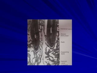
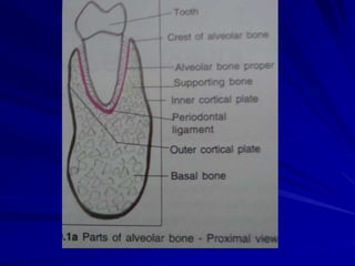
Alveolar Bone
Dr.D.Angelin
Postgraduate student
Dept of oral pathology and microbiology
PMS College of Dental Science and Research
Alveolar process-part of maxilla and
mandible that forms and supports the
sockets of the teeth
Functions of alveolar bone
Houses the roots of teeth
Anchors the root of teeth to the alveoli
Helps to move the teeth for better
occlusion
Helps to absorb and distribute occlusal
forces generated during tooth contact
Supplies vessels to periodontal ligament
Near the end of the 2nd
month of fetal life, mandible
and maxilla form a groove
that is opened toward the
surface of the oral cavity
As tooth germs start to
develop, bony septa form
gradually. The alveolar
process starts developing
strictly during tooth eruption.
FORMATION OF ALVEOLAR BONE
As root develops the alveolar process
increases in height
During growth ,part of the alveolar process
is gradually incorporated into the maxillary
or mandibular body
During period of rapid growth, a tissue
may develop at the alveolar crest that
combines characteristics of cartilage and
bone known as chondroid bone
Alveolar process diminishes in height after
loss of teeth
Structure of the alveolar bone
No distinct boundary between body of
mandible or maxilla and their alveolar
processes
Two parts as a result of adaptation to
function
1. Alveolar bone proper
2. Supporting alveolar bone
Alveolar bone proper
Lamellated bone
Bundle bone
It surrounds the root of the tooth and gives
attachment to principal fibers of the
periodontal ligament.
Thickness of alveolar bone proper-0.1-0.4mm
thick
Lamellated bone
Blood vessel surrounded by concentric
lamellae forms osteons
Some lamellae arranged parallel to
surface of other adjacent marrow spaces
while others form haversian systems
Bundle bone
Bone in which principal fibers of periodontal
ligament are anchored
Bundles of principal fibers continue into bone as
Sharpey’s fibers
Characterised by scarcity of fibrils in intercellular
substance
These fibrils arranged at right angles to
sharpey’s fibers
Lines of rest are seen in bundle bone
Radiographically called as lamina dura- due to
increased opacity due to presence of thick bone
without trabeculations
Alveolar bone proper-perforated by many
openings that carry branches of
interalveolar nerves and blood vessels into
the periodontal ligament-cribriform plate
Interdental and interradicular septa contain
perforating canals of Zuckerkandl and
Hirschfeld
a) outer cortical plates
b) a central spongiosa
SUPPORTING ALVEOLAR BONE
Cortical plates
It is compact bone and forms the outer
and inner plates of alveolar processes
Continuous with compact layers of
maxillary and mandibular body
Thicker in premolar and molar region esp
on the buccal side of lower jaw
Histologically consist of longitudinal
lamellae and haversian systems
Spongy bone
Fills the area between cortical plates and
alveolar bone proper
Trabeculae of lamellar bone surrounded
by marrow
Trabeculae has osteocytes inside and
osteoblasts or osteoclasts on the surface
Classification of spongiosa
based on radiographs
Type I – interdental & interradicular
trabeculae are regular and horizontal in a
ladder like arrangement- seen often in
mandible
Type II – irregularly arranged ,numerous
delicate interdental and interradicular
trabeculae-more common in maxilla
The alveolar crest is found 1.5-2.0 mm below the
level of the CEJ.
If you draw a line connecting the CE junctions of
adjacent teeth, this line should be parallel to the
alveolar crest. If the line is not parallel, then there is
high probability of periodontal disease.
Internal reconstruction of
alveolar bone
Mesial drift and continous tooth eruption
elicit remodelling of alveolar bone
During mesial drift of a tooth bone is
apposed on the distal and resorbed on the
mesial alveolar wall
On mesial alveolar wall howship’s lacuane
with osteoclasts present
On distal wall bundle bone replaced by
lamellar bone by osteoblasts
Age changes
In older individuals
- Alveolar sockets appear jagged and uneven
- Marrow spaces have fatty infiltration
- Alveolar process in edentulous jaws decreases
in size
- Loss of maxillary bone accompanied by increase
in size of the maxillary sinus
- Internal trabecular arrangement is more open
- The distance between crest of alveolar bone and
CEJ increase with age- by 2.81 mm
Clinical considerations
Although one of the hardest tissues in the
human body,bone is biologically a highly
plastic tissue
This enables the orthodontist to move
teeth without disrupting their relations to
the alveolar bone
Bone is resorbed on the side of pressure
and apposed on the side of tension; thus
the entire alveolus is allowed to shift with
the tooth
Lamina dura is an important diagnostic
landmark in determining health of the
periapical tissues
Loss of density usually means
infections,inflammation and resorption of
bone socket
Harmful change in alveolar process is that
associated with periodontal disease
Bone resorption occurs more frequently in
posterior teeth and occurs in episodic
spurts and is both of horizontal and
vertical type and is related to bacterial
plaque and pocket formation
Resorption after tooth loss follows a
predictable pattern
The labial aspect of alveolar crest is the
principal site of resorption which reduces
first in width and later in height
In mandible , the residual alveolar ridge
resorbs downward and outward whereas
in maxilla it is upwards and inwards
Resorption after bone loss is due to disuse
atrophy,decreased blood supply,localised
inflammation or unfavorble prosthesis
pressure

Alveolar bone

  • 1.
    Alveolar Bone Dr.D.Angelin Postgraduate student Deptof oral pathology and microbiology PMS College of Dental Science and Research
  • 2.
    Alveolar process-part ofmaxilla and mandible that forms and supports the sockets of the teeth
  • 3.
    Functions of alveolarbone Houses the roots of teeth Anchors the root of teeth to the alveoli Helps to move the teeth for better occlusion Helps to absorb and distribute occlusal forces generated during tooth contact Supplies vessels to periodontal ligament
  • 4.
    Near the endof the 2nd month of fetal life, mandible and maxilla form a groove that is opened toward the surface of the oral cavity As tooth germs start to develop, bony septa form gradually. The alveolar process starts developing strictly during tooth eruption. FORMATION OF ALVEOLAR BONE
  • 5.
    As root developsthe alveolar process increases in height During growth ,part of the alveolar process is gradually incorporated into the maxillary or mandibular body During period of rapid growth, a tissue may develop at the alveolar crest that combines characteristics of cartilage and bone known as chondroid bone Alveolar process diminishes in height after loss of teeth
  • 6.
    Structure of thealveolar bone No distinct boundary between body of mandible or maxilla and their alveolar processes Two parts as a result of adaptation to function 1. Alveolar bone proper 2. Supporting alveolar bone
  • 9.
    Alveolar bone proper Lamellatedbone Bundle bone It surrounds the root of the tooth and gives attachment to principal fibers of the periodontal ligament. Thickness of alveolar bone proper-0.1-0.4mm thick
  • 10.
    Lamellated bone Blood vesselsurrounded by concentric lamellae forms osteons Some lamellae arranged parallel to surface of other adjacent marrow spaces while others form haversian systems
  • 12.
    Bundle bone Bone inwhich principal fibers of periodontal ligament are anchored Bundles of principal fibers continue into bone as Sharpey’s fibers Characterised by scarcity of fibrils in intercellular substance These fibrils arranged at right angles to sharpey’s fibers Lines of rest are seen in bundle bone Radiographically called as lamina dura- due to increased opacity due to presence of thick bone without trabeculations
  • 14.
    Alveolar bone proper-perforatedby many openings that carry branches of interalveolar nerves and blood vessels into the periodontal ligament-cribriform plate Interdental and interradicular septa contain perforating canals of Zuckerkandl and Hirschfeld
  • 15.
    a) outer corticalplates b) a central spongiosa SUPPORTING ALVEOLAR BONE
  • 16.
    Cortical plates It iscompact bone and forms the outer and inner plates of alveolar processes Continuous with compact layers of maxillary and mandibular body Thicker in premolar and molar region esp on the buccal side of lower jaw Histologically consist of longitudinal lamellae and haversian systems
  • 19.
    Spongy bone Fills thearea between cortical plates and alveolar bone proper Trabeculae of lamellar bone surrounded by marrow Trabeculae has osteocytes inside and osteoblasts or osteoclasts on the surface
  • 20.
    Classification of spongiosa basedon radiographs Type I – interdental & interradicular trabeculae are regular and horizontal in a ladder like arrangement- seen often in mandible Type II – irregularly arranged ,numerous delicate interdental and interradicular trabeculae-more common in maxilla
  • 21.
    The alveolar crestis found 1.5-2.0 mm below the level of the CEJ. If you draw a line connecting the CE junctions of adjacent teeth, this line should be parallel to the alveolar crest. If the line is not parallel, then there is high probability of periodontal disease.
  • 23.
    Internal reconstruction of alveolarbone Mesial drift and continous tooth eruption elicit remodelling of alveolar bone During mesial drift of a tooth bone is apposed on the distal and resorbed on the mesial alveolar wall On mesial alveolar wall howship’s lacuane with osteoclasts present On distal wall bundle bone replaced by lamellar bone by osteoblasts
  • 24.
    Age changes In olderindividuals - Alveolar sockets appear jagged and uneven - Marrow spaces have fatty infiltration - Alveolar process in edentulous jaws decreases in size - Loss of maxillary bone accompanied by increase in size of the maxillary sinus - Internal trabecular arrangement is more open - The distance between crest of alveolar bone and CEJ increase with age- by 2.81 mm
  • 25.
    Clinical considerations Although oneof the hardest tissues in the human body,bone is biologically a highly plastic tissue This enables the orthodontist to move teeth without disrupting their relations to the alveolar bone Bone is resorbed on the side of pressure and apposed on the side of tension; thus the entire alveolus is allowed to shift with the tooth
  • 26.
    Lamina dura isan important diagnostic landmark in determining health of the periapical tissues Loss of density usually means infections,inflammation and resorption of bone socket
  • 27.
    Harmful change inalveolar process is that associated with periodontal disease Bone resorption occurs more frequently in posterior teeth and occurs in episodic spurts and is both of horizontal and vertical type and is related to bacterial plaque and pocket formation
  • 28.
    Resorption after toothloss follows a predictable pattern The labial aspect of alveolar crest is the principal site of resorption which reduces first in width and later in height In mandible , the residual alveolar ridge resorbs downward and outward whereas in maxilla it is upwards and inwards Resorption after bone loss is due to disuse atrophy,decreased blood supply,localised inflammation or unfavorble prosthesis pressure