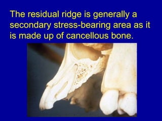
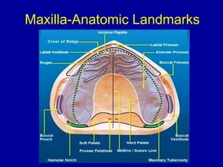
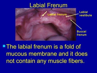
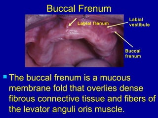
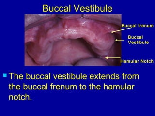
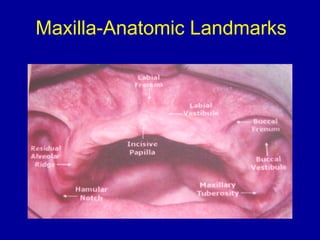
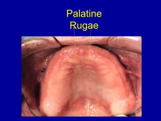
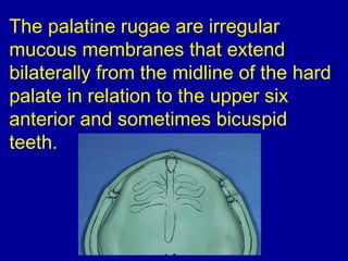
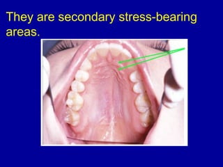
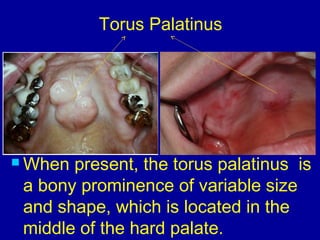
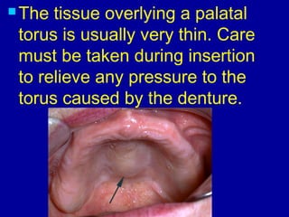
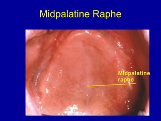
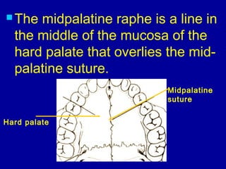
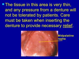
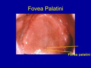
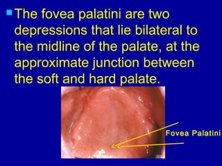
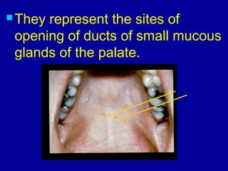
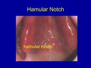
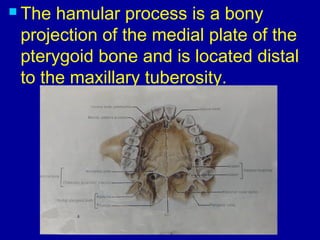
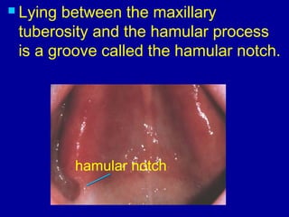
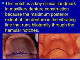
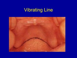
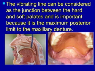
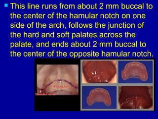
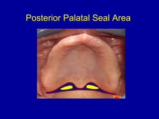
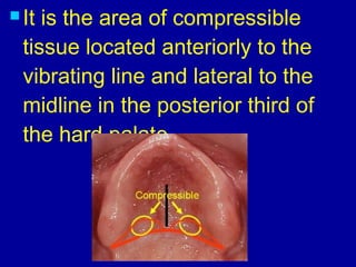
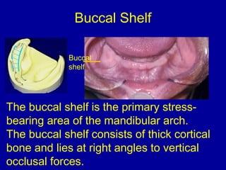
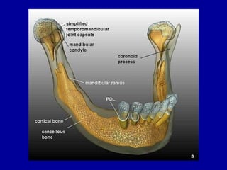
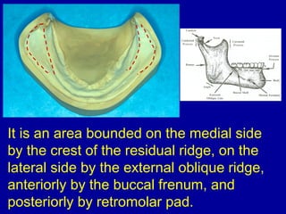
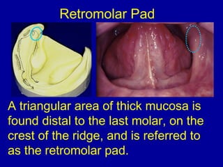
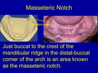
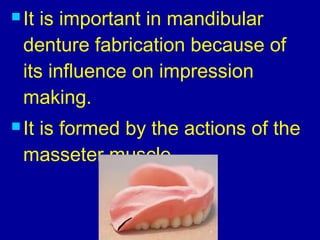
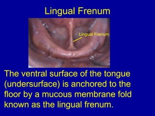
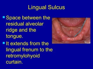
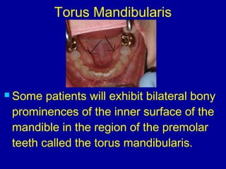
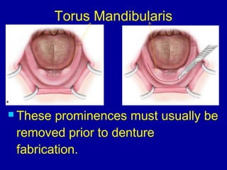
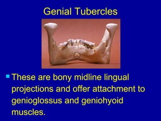
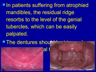
ORAL ANATOMY IN RELATION
TO COMPLETE DENTURE
PROSTHODONTICS
Learning outcomes
1. Describe what are relief areas, support
areas and stress bearing areas.
2. Name the maxillary edentulous
anatomical landmarks.
3. Name the mandibular edentulous
anatomical landmarks.
4. Memorize the primary and secondary
stress bearing areas of maxilla and
mandible.
 In order to properly construct a
denture, one must understand
the anatomy and physiology of
the edentulous patient’s mouth.
 A thorough knowledge of the
origins and functions of the
muscles of mastication, facial
expression, tongue and floor of
the mouth is essential.
When the key anatomic
landmarks and their role with
respect to retention, stability,
support are mastered, dentures
can be fabricated as important
parts of each patient’s oral cavity
and not just mechanical artificial
substitutes.
EDENTULOUS ANATOMY
Relief areas
 The reduction or elimination of
undesirable pressure or force
from a specific region under a
denture base.
Support areas
 Those areas of the maxillary
and mandibular edentulous
ridges that are considered best
suited to carry the forces of
mastication when the dentures
are in function.
Stress bearing areas
 The surfaces of oral
structures that resist forces,
strains, or pressures brought
on them during function.
 Primary stress bearing area
 Secondary stress bearing
area
 The primary stress-bearing areas
generally have thicker mucosa
and /or underlying bone that is
less subject to resorption because
it is cortical bone.
The residual ridge is generally a
secondary stress-bearing area as it
is made up of cancellous bone.
Maxilla-Anatomic Landmarks
Labial frenum
Labial
vestibule
Buccal
frenum
Labial Frenum
 The labial frenum is a fold of
mucous membrane and it does
not contain any muscle fibers.
 The dentures should be relieved away
from the frenum, to avoid irritation to
these folds and to prevent future
instability of the dentures.
Labial frenum
Labial
vestibule
Buccal
frenum
Labial Vestibule
 It runs from one buccal frenum to
the other on the labial side of the
ridge.
Labial frenum
Labial
vestibule
Buccal
frenum
Buccal Frenum
 The buccal frenum is a mucous
membrane fold that overlies dense
fibrous connective tissue and fibers of
the levator anguli oris muscle.
Hamular Notch
Buccal
Vestibule
Buccal Vestibule
 The buccal vestibule extends from
the buccal frenum to the hamular
notch.
Buccal frenum
Maxilla-Anatomic Landmarks
Alveolar (Residual) Ridges
Following full mouth extractions, the
alveolar ridges undergo major bony
changes, with the largest changes
seen on the mandibular arch.
 Studies indicate that the
mandibular ridge resorps
approximately four times as
much as the maxillary arch.
 The direction of mandibular
resorption is downward and
outward, while maxillary
resorption is upward and inward.
Alveolar (Residual) Ridges
Slopes of maxillary residual ridge and
most of hard palate are the primary
stress-bearing areas.
Posterior
Palatal Seal
Area
Tuberosity
Maxillary Tuberosity
The maxillary tuberosity is the
most posterior part of the alveolar
ridge; it lies distal to the position of
the last molar.
The maxillary tuberosity is
important from a denture
standpoint because it is
considered a primary stress-
bearing area.
Incisive papilla
Incisive Papilla
The incisive papilla is a small tubercle
located on the palatal side between
the two central incisors.
It overlies the incisive foramen,
through which the incisive nerve and
blood vessels exit.
Because of the sensitivity of this
structure, care must be taken when
inserting the maxillary denture to
relieve almost all pressure in this area.
The incisive papilla is a good
landmark when arranging teeth
because studies indicate that the
facial surfaces of the natural central
incisors, when present, were
approximately 8-10 mm anterior to the
middle of the incisive papilla,
and the tips of the canines were
approximately in line with the middle
of the incisal papilla.
Palatine
Rugae
The palatine rugae are irregular
mucous membranes that extend
bilaterally from the midline of the hard
palate in relation to the upper six
anterior and sometimes bicuspid
teeth.
They are secondary stress-bearing
areas.
Torus Palatinus
 When present, the torus palatinus is
a bony prominence of variable size
and shape, which is located in the
middle of the hard palate.
 The tissue overlying a palatal
torus is usually very thin. Care
must be taken during insertion
to relieve any pressure to the
torus caused by the denture.
Midpalatine Raphe
Midpalatine
raphe
 The midpalatine raphe is a line in
the middle of the mucosa of the
hard palate that overlies the mid-
palatine suture.
Midpalatine
suture
Hard palate
 The tissue in this area is very thin,
and any pressure from a denture will
not be tolerated by patients. Care
must be taken when inserting the
denture to provide necessary relief.
Midpalatine
raphe
Fovea Palatini
Fovea palatini
 The fovea palatini are two
depressions that lie bilateral to
the midline of the palate, at the
approximate junction between
the soft and hard palate.
Fovea Palatini
 They represent the sites of
opening of ducts of small mucous
glands of the palate.
Hamular Notch
hamular notch
 The hamular process is a bony
projection of the medial plate of the
pterygoid bone and is located distal
to the maxillary tuberosity.
 Lying between the maxillary
tuberosity and the hamular process
is a groove called the hamular notch.
hamular notch
 This notch is a key clinical landmark
in maxillary denture construction
because the maximum posterior
extent of the denture is the vibrating
line that runs bilaterally through the
hamular notches.
Vibrating Line
 The vibrating line is very important to
locate for proper construction of the
maxillary complete denture.
 The vibrating line can be considered
as the junction between the hard
and soft palates and is important
because it is the maximum posterior
limit to the maxillary denture.
 This line runs from about 2 mm buccal to
the center of the hamular notch on one
side of the arch, follows the junction of
the hard and soft palates across the
palate, and ends about 2 mm buccal to
the center of the opposite hamular notch.
Posterior Palatal Seal Area
T- burnisher
 The posterior palatal seal area is
very important in maxillary
complete denture fabrication and
must be identified and evaluated.
 It is the area of compressible
tissue located anteriorly to the
vibrating line and lateral to the
midline in the posterior third of
the hard palate.
 The distal extent of this area is the
vibrating line, while the anterior
border is unclear.
Mandible-Anatomic Landmarks
Histologically and functionally it is the
same as in the maxilla. It is a mucous
membrane without significant muscle
fibers.
Labial Frenum
Labial Frenum
Labial vestibule
It runs from one buccal frenum to the
other on the labial side of the ridge.
Labial Vestibule
Buccal Frenum
 The buccal frenum is a mucous
membrane fold that overlies dense
fibrous connective tissue and fibers of
the depressor anguli oris muscle.
Buccal Frenum
Buccal
Vestibule
Buccal Vestibule
 The buccal vestibule extends
posteriorly from the buccal frenum to
the posterior lateral aspect of the
retromolar pad.
Buccal frenum
Retromolar
pad
The buccal shelf is the primary stress-
bearing area of the mandibular arch.
The buccal shelf consists of thick cortical
bone and lies at right angles to vertical
occlusal forces.
Buccal
shelf
Buccal Shelf
It is an area bounded on the medial side
by the crest of the residual ridge, on the
lateral side by the external oblique ridge,
anteriorly by the buccal frenum, and
posteriorly by retromolar pad.
A triangular area of thick mucosa is
found distal to the last molar, on the
crest of the ridge, and is referred to
as the retromolar pad.
Retromolar Pad
It is composed of:
Loose connective tissue
Glandular tissue
Fibers of the temporalis tendon
Fibers of buccinator muscle
Fibers of superior constrictor
muscle
The pterygomandibular raphe
The retromolar pads should be
covered by the denture, and the
plane of occlusion is generally
located at the level of the middle to
upper-third of this pad.
Just buccal to the crest of the
mandibular ridge in the distal-buccal
corner of the arch is an area known
as the masseteric notch.
Masseteric Notch
Masseteric Notch
Masseteric Notch
 It is important in mandibular
denture fabrication because of
its influence on impression
making.
 It is formed by the actions of the
masseter muscle.
The ventral surface of the tongue
(undersurface) is anchored to the
floor by a mucous membrane fold
known as the lingual frenum.
Lingual Frenum
Lingual Frenum
Lingual Sulcus
 Space between the
residual alveolar
ridge and the
tongue.
 It extends from the
lingual frenum to the
retromylohyoid
curtain.
 Some patients will exhibit bilateral bony
prominences of the inner surface of the
mandible in the region of the premolar
teeth called the torus mandibularis.
Torus Mandibularis
 These prominences must usually be
removed prior to denture
fabrication.
Torus Mandibularis
Alveolar (Residual) Ridges
 The mandibular residual alveolar
ridge is the secondary stress-
bearing area.
Genial Tubercles
 These are bony midline lingual
projections and offer attachment to
genioglossus and geniohyoid
muscles.
 In patients suffering from atrophied
mandibles, the residual ridge
resorbs to the level of the genial
tubercles, which can be easily
palpated.
 The dentures should be trimmed
around the genial tubercles in those
cases.
oral anatomy
oral anatomy

oral anatomy

  • 2.
    ORAL ANATOMY INRELATION TO COMPLETE DENTURE PROSTHODONTICS
  • 3.
    Learning outcomes 1. Describewhat are relief areas, support areas and stress bearing areas. 2. Name the maxillary edentulous anatomical landmarks. 3. Name the mandibular edentulous anatomical landmarks. 4. Memorize the primary and secondary stress bearing areas of maxilla and mandible.
  • 4.
     In orderto properly construct a denture, one must understand the anatomy and physiology of the edentulous patient’s mouth.
  • 5.
     A thoroughknowledge of the origins and functions of the muscles of mastication, facial expression, tongue and floor of the mouth is essential.
  • 6.
    When the keyanatomic landmarks and their role with respect to retention, stability, support are mastered, dentures can be fabricated as important parts of each patient’s oral cavity and not just mechanical artificial substitutes.
  • 7.
  • 8.
    Relief areas  Thereduction or elimination of undesirable pressure or force from a specific region under a denture base.
  • 9.
    Support areas  Thoseareas of the maxillary and mandibular edentulous ridges that are considered best suited to carry the forces of mastication when the dentures are in function.
  • 10.
    Stress bearing areas The surfaces of oral structures that resist forces, strains, or pressures brought on them during function.  Primary stress bearing area  Secondary stress bearing area
  • 11.
     The primarystress-bearing areas generally have thicker mucosa and /or underlying bone that is less subject to resorption because it is cortical bone.
  • 12.
    The residual ridgeis generally a secondary stress-bearing area as it is made up of cancellous bone.
  • 13.
  • 14.
    Labial frenum Labial vestibule Buccal frenum Labial Frenum The labial frenum is a fold of mucous membrane and it does not contain any muscle fibers.
  • 15.
     The denturesshould be relieved away from the frenum, to avoid irritation to these folds and to prevent future instability of the dentures.
  • 16.
    Labial frenum Labial vestibule Buccal frenum Labial Vestibule It runs from one buccal frenum to the other on the labial side of the ridge.
  • 17.
    Labial frenum Labial vestibule Buccal frenum Buccal Frenum The buccal frenum is a mucous membrane fold that overlies dense fibrous connective tissue and fibers of the levator anguli oris muscle.
  • 18.
    Hamular Notch Buccal Vestibule Buccal Vestibule The buccal vestibule extends from the buccal frenum to the hamular notch. Buccal frenum
  • 19.
  • 20.
    Alveolar (Residual) Ridges Followingfull mouth extractions, the alveolar ridges undergo major bony changes, with the largest changes seen on the mandibular arch.
  • 21.
     Studies indicatethat the mandibular ridge resorps approximately four times as much as the maxillary arch.
  • 22.
     The directionof mandibular resorption is downward and outward, while maxillary resorption is upward and inward.
  • 23.
    Alveolar (Residual) Ridges Slopesof maxillary residual ridge and most of hard palate are the primary stress-bearing areas.
  • 24.
    Posterior Palatal Seal Area Tuberosity Maxillary Tuberosity Themaxillary tuberosity is the most posterior part of the alveolar ridge; it lies distal to the position of the last molar.
  • 25.
    The maxillary tuberosityis important from a denture standpoint because it is considered a primary stress- bearing area.
  • 26.
    Incisive papilla Incisive Papilla Theincisive papilla is a small tubercle located on the palatal side between the two central incisors.
  • 27.
    It overlies theincisive foramen, through which the incisive nerve and blood vessels exit.
  • 28.
    Because of thesensitivity of this structure, care must be taken when inserting the maxillary denture to relieve almost all pressure in this area.
  • 29.
    The incisive papillais a good landmark when arranging teeth because studies indicate that the facial surfaces of the natural central incisors, when present, were approximately 8-10 mm anterior to the middle of the incisive papilla,
  • 30.
    and the tipsof the canines were approximately in line with the middle of the incisal papilla.
  • 31.
  • 32.
    The palatine rugaeare irregular mucous membranes that extend bilaterally from the midline of the hard palate in relation to the upper six anterior and sometimes bicuspid teeth.
  • 33.
    They are secondarystress-bearing areas.
  • 34.
    Torus Palatinus  Whenpresent, the torus palatinus is a bony prominence of variable size and shape, which is located in the middle of the hard palate.
  • 35.
     The tissueoverlying a palatal torus is usually very thin. Care must be taken during insertion to relieve any pressure to the torus caused by the denture.
  • 36.
  • 37.
     The midpalatineraphe is a line in the middle of the mucosa of the hard palate that overlies the mid- palatine suture. Midpalatine suture Hard palate
  • 38.
     The tissuein this area is very thin, and any pressure from a denture will not be tolerated by patients. Care must be taken when inserting the denture to provide necessary relief. Midpalatine raphe
  • 39.
  • 40.
     The foveapalatini are two depressions that lie bilateral to the midline of the palate, at the approximate junction between the soft and hard palate. Fovea Palatini
  • 41.
     They representthe sites of opening of ducts of small mucous glands of the palate.
  • 42.
  • 43.
     The hamularprocess is a bony projection of the medial plate of the pterygoid bone and is located distal to the maxillary tuberosity.
  • 44.
     Lying betweenthe maxillary tuberosity and the hamular process is a groove called the hamular notch. hamular notch
  • 45.
     This notchis a key clinical landmark in maxillary denture construction because the maximum posterior extent of the denture is the vibrating line that runs bilaterally through the hamular notches.
  • 46.
  • 47.
     The vibratingline is very important to locate for proper construction of the maxillary complete denture.
  • 48.
     The vibratingline can be considered as the junction between the hard and soft palates and is important because it is the maximum posterior limit to the maxillary denture.
  • 49.
     This lineruns from about 2 mm buccal to the center of the hamular notch on one side of the arch, follows the junction of the hard and soft palates across the palate, and ends about 2 mm buccal to the center of the opposite hamular notch.
  • 50.
  • 51.
    T- burnisher  Theposterior palatal seal area is very important in maxillary complete denture fabrication and must be identified and evaluated.
  • 52.
     It isthe area of compressible tissue located anteriorly to the vibrating line and lateral to the midline in the posterior third of the hard palate.
  • 53.
     The distalextent of this area is the vibrating line, while the anterior border is unclear.
  • 56.
  • 57.
    Histologically and functionallyit is the same as in the maxilla. It is a mucous membrane without significant muscle fibers. Labial Frenum Labial Frenum
  • 58.
    Labial vestibule It runsfrom one buccal frenum to the other on the labial side of the ridge. Labial Vestibule
  • 59.
    Buccal Frenum  Thebuccal frenum is a mucous membrane fold that overlies dense fibrous connective tissue and fibers of the depressor anguli oris muscle. Buccal Frenum
  • 60.
    Buccal Vestibule Buccal Vestibule  Thebuccal vestibule extends posteriorly from the buccal frenum to the posterior lateral aspect of the retromolar pad. Buccal frenum Retromolar pad
  • 61.
    The buccal shelfis the primary stress- bearing area of the mandibular arch. The buccal shelf consists of thick cortical bone and lies at right angles to vertical occlusal forces. Buccal shelf Buccal Shelf
  • 63.
    It is anarea bounded on the medial side by the crest of the residual ridge, on the lateral side by the external oblique ridge, anteriorly by the buccal frenum, and posteriorly by retromolar pad.
  • 64.
    A triangular areaof thick mucosa is found distal to the last molar, on the crest of the ridge, and is referred to as the retromolar pad. Retromolar Pad
  • 65.
    It is composedof: Loose connective tissue Glandular tissue Fibers of the temporalis tendon Fibers of buccinator muscle Fibers of superior constrictor muscle The pterygomandibular raphe
  • 66.
    The retromolar padsshould be covered by the denture, and the plane of occlusion is generally located at the level of the middle to upper-third of this pad.
  • 67.
    Just buccal tothe crest of the mandibular ridge in the distal-buccal corner of the arch is an area known as the masseteric notch. Masseteric Notch Masseteric Notch
  • 68.
  • 69.
     It isimportant in mandibular denture fabrication because of its influence on impression making.  It is formed by the actions of the masseter muscle.
  • 70.
    The ventral surfaceof the tongue (undersurface) is anchored to the floor by a mucous membrane fold known as the lingual frenum. Lingual Frenum Lingual Frenum
  • 71.
    Lingual Sulcus  Spacebetween the residual alveolar ridge and the tongue.  It extends from the lingual frenum to the retromylohyoid curtain.
  • 72.
     Some patientswill exhibit bilateral bony prominences of the inner surface of the mandible in the region of the premolar teeth called the torus mandibularis. Torus Mandibularis
  • 73.
     These prominencesmust usually be removed prior to denture fabrication. Torus Mandibularis
  • 74.
    Alveolar (Residual) Ridges The mandibular residual alveolar ridge is the secondary stress- bearing area.
  • 75.
    Genial Tubercles  Theseare bony midline lingual projections and offer attachment to genioglossus and geniohyoid muscles.
  • 76.
     In patientssuffering from atrophied mandibles, the residual ridge resorbs to the level of the genial tubercles, which can be easily palpated.  The dentures should be trimmed around the genial tubercles in those cases.

Editor's Notes

  • #5 Kinetics: dynamics/movement
  • #6 Kinetics: dynamics/movement
  • #18 LEVATOR ANGULI ORIS - CANINUS
  • #19 LEVATOR ANGULI ORIS - CANINUS
  • #21 LEVATOR ANGULI ORIS - CANINUS
  • #57 The retro molar pad area is the most stable area of the mandibular alveolar ridge because it is an area where there has been no tooth that has weakened the bone in this area. The lingual frenulum plays a very important role in the stability of mandibular denture as it attaches the tongue to the anterior alveolar ridge area and if it is not allowed freedom of movement it will cause the denture to rise during function. The labial vestibule is an area that the flange of the mandibular denture is most commonly under-reduced during the insertion. The patient will return with a large ulcer and the complaint that the denture moves up and down when he eats or speaks. The retromylohyoid fossa plays an important role in the retention of a mandibular denture by resisting the tendency of the denture to move up in the posterior region when a patient bites down on the anterior teeth. The submaxillary caruncles and the lingual tubercle are major areas that become very sore when the denture flanges are left long in this area. The size, shape, and position of the tongue are very important in the stability of the lower denture. The tongue will either work to hold the denture in place or work against it to dislodge it.
  • #61 LEVATOR ANGULI ORIS - CANINUS
  • #75 LEVATOR ANGULI ORIS - CANINUS