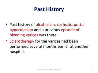
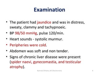
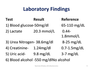
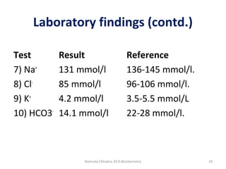
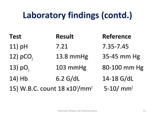
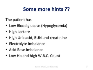
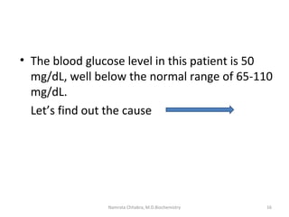
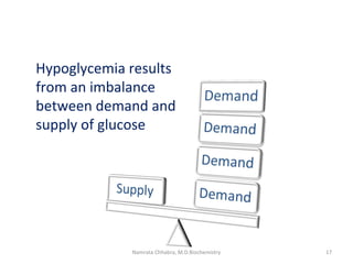
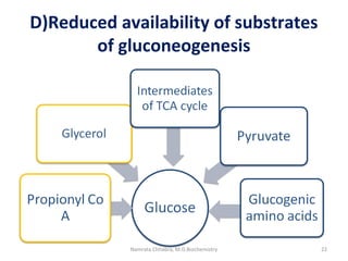
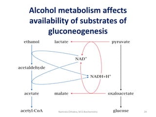
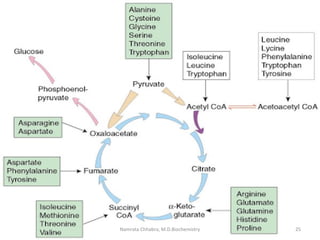
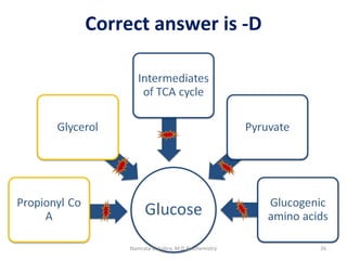
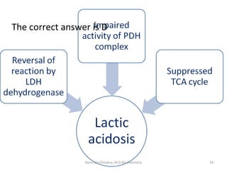
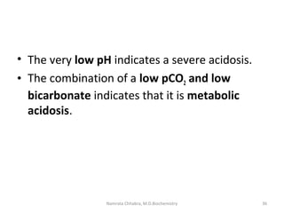
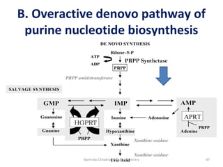
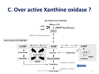
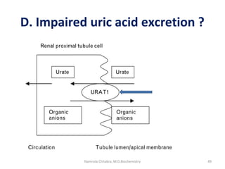
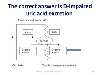
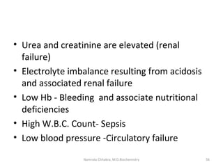
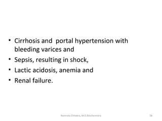
This document describes a case study of a 60-year-old male alcoholic who was brought to the hospital in serious condition. Laboratory tests revealed hypoglycemia, lactic acidosis, hyperuricemia, and other metabolic alterations. The patient's history of alcoholism and cirrhosis indicated that the metabolic derangements were due to alcohol-induced impairments in gluconeogenesis and tricarboxylic acid cycle function, leading to reduced glucose availability, increased lactate production, and impaired uric acid excretion. The patient was diagnosed with cirrhosis, portal hypertension, bleeding varices, sepsis, shock, renal failure, and other complications of long-term heavy alcohol use and malnutrition.
6. Case details
• A 60 year old man was brought to hospital in a
very serious condition.
• The patient complained of
o Constant vomiting containing several hundred
mL of dark brown fluid from the previous two
days plus
o Several episodes of melaena.
6Namrata Chhabra, M.D.Biochemistry
7. Past History
• Past history of alcoholism, cirrhosis, portal
hypertension and a previous episode of
bleeding varices was there.
• Sclerotherapy for the varices had been
performed several months earlier at another
hospital.
7Namrata Chhabra, M.D.Biochemistry
8. Examination
• The patient had jaundice and was in distress,
sweaty, clammy and tachypnoeic.
• BP 98/50 mmHg, pulse 120/min.
• Heart sounds - systolic murmur.
• Peripheries were cold.
• Abdomen was soft and non tender.
• Signs of chronic liver disease were present
(spider naevi, gynecomastia, and testicular
atrophy).
8Namrata Chhabra, M.D.Biochemistry
13. • The patient has multiple problems
• Circulatory failure
• GI bleeding on a background of known
Cirrhosis with Portal hypertension
• Many other ??
Some Hints???
13Namrata Chhabra, M.D.Biochemistry
14. Some more hints ??
The patient has
• Low Blood glucose (Hypoglycemia)
• High Lactate
• High Uric acid, BUN and creatinine
• Electrolyte imbalance
• Acid Base imbalance
• Low Hb and high W.B.C. Count
14Namrata Chhabra, M.D.Biochemistry
16. • The blood glucose level in this patient is 50
mg/dL, well below the normal range of 65-110
mg/dL.
Let’s find out the cause
16Namrata Chhabra, M.D.Biochemistry
18. Which of the following conditions best explains
the underlying cause of hypoglycemia in this
patient?
A. Impaired activity of Glycogen phosphorylase
B. Impaired activity of Glucose-6-Phosphatase
C. Impaired activity of Pyruvate Kinase
D. Reduced availability of substrates of
Gluconeogenesis
18Namrata Chhabra, M.D.Biochemistry
19. A) Impaired activity of Glycogen
phosphorylase?
19Namrata Chhabra, M.D.Biochemistry
20. B) Impaired activity of Glucose-6-
Phosphatase ?
20Namrata Chhabra, M.D.Biochemistry
29. 2) What is the cause of Lactic
Acidosis in this patient ?
A. Reversal of reaction catalyzed by lactate
dehydrogenase
B. Impaired activity of PDH complex
C. Suppressed TCA cycle
D. All of the above.
29Namrata Chhabra, M.D.Biochemistry
30. A) Reversal of reaction caused by
Lactate dehydrogenase?
Pyruvate is converted to lactate to regenerate
NAD +.
30Namrata Chhabra, M.D.Biochemistry
36. • The very low pH indicates a severe acidosis.
• The combination of a low pCO2 and low
bicarbonate indicates that it is metabolic
acidosis.
36Namrata Chhabra, M.D.Biochemistry
37. Determination of Acid base status
pH H+
P CO2 HCO3
-
Normal 7.4 40 mEq/L 40mm Hg 24 mEq/L
Respiratory
acidosis
Respiratory
Alkalosis
Metabolic
acidosis
Metabolic
Alkalosis
R
O
M
E
37Namrata Chhabra, M.D.Biochemistry
39. Normal or high anion gap
metabolic acidosis ?
• The anion gap is 42 indicating the presence of
a high anion gap disorder.
• The lactate level of 20.3mmol/l is extremely
high and this is responsible for causing high
anion gap.
39Namrata Chhabra, M.D.Biochemistry
40. High anion gap acidosis
• High anion gap is also there due to underlying
Ketoacidosis.
• Acetyl co A fails to get utilized in TCA cycle,
and the excess is channeled towards
alternative pathways.
40Namrata Chhabra, M.D.Biochemistry
42. • Gouty arthritis is a common
finding in chronic alcoholics
• Gout results from an increased
body pool of urate with
hyperuricemia.
• It is typically characterized by
episodic acute and chronic
arthritis, due to deposition of
Mono sodium urate crystals in
and around joints.
42Namrata Chhabra, M.D.Biochemistry
44. • In the given patient, serum uric acid
concentration is higher than normal (9.8
mg/dL).
• What is the cause of Hyperuricemia in this
patient?
44Namrata Chhabra, M.D.Biochemistry
45. A. Inhibition of salvage pathway of purine
nucleotide biosynthesis
B. Overactive denovo pathway of purine
nucleotide biosynthesis
C. Overactive xanthine oxidase
D. Impaired excretion of uric acid
45Namrata Chhabra, M.D.Biochemistry
46. A) Inhibition of salvage pathway?
PRPP Synthetase
46Namrata Chhabra, M.D.Biochemistry
47. B. Overactive denovo pathway of
purine nucleotide biosynthesis
PRPP Synthetase
47Namrata Chhabra, M.D.Biochemistry
48. C. Over active Xanthine oxidase ?
PRPP Synthetase
48Namrata Chhabra, M.D.Biochemistry
49. D. Impaired uric acid excretion ?
49Namrata Chhabra, M.D.Biochemistry
54. High purine content in alcoholic
beverages ?
• The higher purine content in some alcoholic
beverages such as beer is also an additional
factor.
54Namrata Chhabra, M.D.Biochemistry
Every organ system is involved, more than 200 diseases have been reported, and not to forget, the traumatic injuries, that are more common than the reported diseases. The incidence of alcohol related diseases is rising day by day.
The major proportion of the alcohol, approximately 80 %, is metabolized by the cytoplasmic alcohol dehydrogenase to form acetaldehyde, which is a toxic product. The acetaldehyde is subsequently metabolized by mitochondrial aldehyde dehydrogenase to a relatively non toxic form which is acetate. Both these dehydrogenases are NAD+ dependent, and during the metabolic processes, a constant supply of NAD + is needed for the optimum functioning of these enzymes.
Three things are worth mentioning here-
1) The alcohol dehydrogenase has a high affinity (low km) for ethanol, due to this reason, ethanol is used as an antidote in life threatening methanol or ethylene glycol poisonings .Both these compounds are metabolized in the same way by the same dehydrogenases, and upon metabolism, their toxicity is rather intensified, Ethanol in these conditions is given as a life saving drug, as it is preferentially binds the enzyme sparing the other alcohols.
2) The rate of alcohol dehydrogenase catalyzed reaction is slower than aldehyde dehydrogenase; as a result acetaldehyde can accumulate to cause the side effects such as nausea, vomiting, flushing, headache and hypotension.
3) The other microsomal pathway becomes important in chronic alcoholics, or when large amount of alcohol is consumed, since the said enzyme has large km or low affinity for its substrate, but that enzyme has its own implications, such as generation of free radicals and the enzyme is induced by alcohol, as a result it affects the metabolism of other drugs also.
Here is the major pathway of ethanol metabolism, highlighting the production of NADH. It is understandable that chronic alcohol consumption will lead to accumulation of NADH, affecting the major pathways. NADH is the major causative factor for alcohol related complications.
An overview of metabolic products of alcohol, that is responsible for all the complications. Apart from these reactive oxygen species are also generated when the microsomal oxidizing system is in action.
NADH, Acetaldehyde and acetate. Acetaldehyde is transiently produced, but has the potential to bind with proteins to form adducts to cause damage and also as I said earlier, can cause the associated clinical symptoms such as nausea, vomiting, flushing etc. Acetate is responsible for altering the lipid metabolism in the liver cells and also disturbs the gene expression at the DNA level by causing histone Acetylation
The case details are with you; this is about a chronic alcoholic who was brought in a serious condition. The patients had complaints of constant vomiting containing several hundreds of dark brown colored fluid and several episodes of melaena, both are indicative of GI bleeding.
The patient is a chronic alcoholic, having history of previously diagnosed liver cirrhosis with portal hypertension. He had undergone Sclerotherapy for the bleeding esophageal varices several months earlier at some other hospital.
The examination revealed the patient had jaundice, rapid breathing, sweaty, cold and clammy skin, low blood pressure, a rapid pulse, Systolic murmur up on auscultation, soft, and non tender abdomen and as expected the signs of chronic liver disease, such as spider naevi, gynecomastia and testicular atrophy were also there. Sign of high estrogen concentration in blood, perhaps due to reduced metabolism of estrogen in the cirrhotic liver.
The laboratory findings were suggestive of Low blood glucose concentration (50mg/dl) as compared to normal, high lactate, high blood urea nitrogen, creatinine towards the higher side, uric acid considerably high and very- very high blood alcohol levels.
In electrolyte study, sodium and chloride were low; potassium was within the normal range, whereas the bicarbonates were almost half of the normal.
Blood gas analysis revealed a low pH, very low pCO2 and a normal pO2.
The Hb was very low, perhaps due to bleeding and nutritional deficiencies, but the W.B.C count was very high, suggestive of sepsis.
Based on the history, examination and laboratory findings, hope you can reach at a diagnosis.
Some hints are there
The patient has low blood pressure and some signs and symptoms of circulatory failure. Gastrointestinal bleeding is also there on a background of Cirrhosis with portal hypertension.
Some more hints from the altered laboratory profile
Hypoglycemia- Low blood glucose
High lactate
High uric acid, BUN and creatinine
Electrolyte imbalance
Acid base imbalance
Low Hb and high WB.C count are suggestive of anemia and sepsis.
Let's dissect the problem and explore the underlying metabolic alterations induced by alcohol.
Firstly Hypoglycemia
The blood glucose level in this patient is well below the normal range. Let's try to find out the cause of hypoglycemia in this patient.
Hypoglycemia results from an imbalance between demand and supply of glucose. In normal health, the sources of glucose include diet, glycogen and gluconeogenesis. Dietary deficiencies are obvious, but let's find out the other causes of reduced supply of glucose.
Which of the following conditions best explains the cause of hypoglycemia in this patient?
A. Impaired activity of Glycogen phosphorylase
B. Impaired activity of Glucose-6-Phosphatase
C. Impaired activity of Pyruvate Kinase
D. Reduced availability of substrates of Gluconeogenesis
Let's have an overview of the reaction catalyzed by Glycogen phosphorylase.
The steps of glycogen degradation have been highlighted. Glycogen phosphorylase is the rate limiting enzyme of glycogen degradation. The activity of this enzyme is not primarily affected by alcohol metabolism. The activity might be lowered due to reduced availability of substrate of this enzyme i.e. Glycogen, which is depleted completely after 12-18 hours of fasting.
Now let's see the problem at the level of Glucose-6-Phosphatase
This enzyme catalyzes the breakdown of glucose-6-Phospahte to form free glucose. Skeletal muscle cells lack this enzyme, thus muscle glycogen does not contribute toward maintenance of blood glucose levels, and glucose-6-P is used for energy production through glycolysis. Lactate is produced during intense muscular activity due to relative hypoxia. Lactate accumulation causes fatigue. Lactate is a substrate for gluconeogenesis (Cori's cycle).
Well, there is no problem at the level of this enzyme also, the under activity might be due to reduced availability of the substrate.
C) Impaired activity of pyruvate kinase?
Pyruvate kinase catalyzes the last step of glycolysis. Three things are worth mentioning here, it is the third irreversible reaction of glycolysis, ATP is formed by substrate level phosphorylation, and it is also the regulatory enzyme of glycolysis. In cells lacking mitochondria or under conditions of hypoxia, glycolysis does not end here; pyruvate is converted to lactate to regenerate NAD+, which is required for continuation of glycolysis.
Its impaired activity can lead to non formation of pyruvate, primarily it will affect glycolysis, and the deficiency is known to cause hemolytic anemia, the red cells are deprived of energy, glucose is the only source of energy for the red blood cells and the cells lacking mitochondria. The other cells can utilize alternative fuel molecules such as ketone bodies and fatty acids.
As regards gluconeogenesis, there are alternative sources of pyruvate available, and there are alternative substrates of gluconeogenesis also available, that can help in glucose synthesis.
Let us see the next option
Gluconeogenesis is the synthesis of glucose from non carbohydrate substances. The substrates or precursors are highlighted here, pyruvate/lactate, glucogenic amino acids, intermediated of TCA cycle, beyond alpha keto glutarate, propionyl coA, as its the product of metabolism of methionine, odd chain fatty acids and side chain of cholesterol. Glycerol is also a substrate; fats contribute towards glucose production through glycerol and propionyl CoA.
Can alcohol metabolism affect the availability of substrates?
Please go through the options, discuss and let me know the answer.
May I know the answer?
Before knowing the right option, an overview of alcohol metabolism, that might make the things clearer,
Alcohol metabolism leads to accumulation of NADH, that shifts the equilibriums of many reactions, as have been shown here, pyruvate is converted to lactate and oxaloacetate is converted to malate.
When pyruvate and oxaloacetate are not available, gluconeogenesis becomes ineffective, because all other substrates (green colored) are channeled to the main pathway through formation of oxaloacetate, non availability of Oxalo acetate limits glucose production. Only glycerol (not shown here) can be used, as that enters the pathway at the level of dihydroxy acetone -P perhaps that is the only substrate to sustain life.
To conclude,
The reduced availability of substrates is the correct answer. The supply of glucose is limited, as there is insufficient dietary intake, glycogen stores are also depleted, and the impaired gluconeogenesis, by adding to the existing imbalance between demand and supply precipitates hypoglycemia in chronic alcoholics.
Let's analyze the basis of second metabolic alteration, which is lactic acidosis
Not to forget, accumulation of lactic acid to the extent of causing lactic acidosis occurs, either due to excess Lactic acid production, impaired utilization or as a result of both excess production and impaired utilization.
Let us find out the cause of lactic acidosis in this alcoholic patient? Is it because of :
Reversal of reaction catalyzed by lactate dehydrogenase
Impaired activity of PDH complex
Suppressed TCA cycle
All of the above.
Is it due to excess lactate production as a result of reversal of reaction catalyzed by lactate dehydrogenase?
Lactate dehydrogenase catalyzes the interconversion of pyruvate and lactate. This reaction is a normal process to regenerate NAD + in cells lacking mitochondria, as they lack respiratory chain, or when glycolysis operates under hypoxic conditions, lactate is the end product of glycolysis. Somewhat similar scenario is there in alcoholism, hope you can recollect, there is excess production of NADH resulting from alcohol metabolism. May be the reaction is occurring at a faster rate than normal; to regenerate NAD+ for alcohol metabolism to continue, as a result there is excess lactate production.
Is it due to impaired activity of PDH complex?
PDH complex is a multienzyme complex, comprising of 3 catalytic and 2 regulatory enzymes, and 5 coenzymes. TPP, Thiamine pyrophosphate is one of the 5 coenzymes, required at the first step. TPP deficiency is very common in chronic alcoholics, can this be the reason for its impaired activity and due to this, and the excess pyruvate is channeled towards lactate production?
Another thing of importance is the need for NAD +, since, there is depletion of NAD +pool as a result of alcohol metabolism, and it might also affect the functioning of PDH complex.
It might be due to suppressed activities of TCA cycle
TCA cycle is the place, where lactate, can utilized, through formation of pyruvate and subsequent conversion to Acetyl CoA through PDH complex.
TCA cycle is in a state of suppression?
PDH complex is suppressed, TCA cycle is suppressed as there are three NAD +dependent enzymes , which cannot function in the absence of NAD +, and another factor, which should not be forgotten is depletion of oxaloacetate, that has been converted to malate due to shift of reactions as discussed earlier.
So, what are the right and the most appropriate answer?
May I know the answer?
The correct answer is D- All of the above. Lactic acidosis is as a result of excess production due to reversal of reaction catalyzed by LDH and Impaired activity of PDH complex, and it is also the result of non utilization due to suppression of TCA cycle.
Let's assess the acid base status of this patient also
A combination of low pH, low p CO2 and low bicarbonate indicate, that it is metabolic acidosis.
How to determine the acid base status.
I have shown the primary acid base defects, but in clinical practice, mixed acid base defects can also be there.
The things to pay attention are pH, pCO2 and bicarbonate levels. Hydrogen ion concentration is just the reverse of pH, which represents the negative logarithm of hydrogen ion concentration; you can even ignore it, if you know the pH value.
Respiratory acidosis is also called ‘Primary [H2CO3] excess’. The underlying abnormality is increase in H2CO3 content in the blood which follows decreased elimination of CO2 in the pulmonary alveoli (High pCO2). In acute respiratory acidosis, there is an immediate compensatory elevation (due to cellular buffering mechanisms) in HCO3–, which increases 1 mmol/L for every 10-mmHg increase in PaCO2. In chronic respiratory acidosis (>24 h), renal adaptation increases the [HCO3–] by 4 mmol/L for every 10-mmHg increase in PaCO2.
Respiratory alkalosis is a primary decrease in PCO2 with or without compensatory decrease in HCO3 −; pH may be high or near normal.
Metabolic acidosis is a primary decrease in serum HCO3 - concentration. As a compensatory mechanism, metabolic acidosis leads to alveolar hyperventilation due to stimulation of respiratory centre causing deep and rapid (Kussmaul) breathing. This increased ventilation results in CO2 loss and a fall in PaCO2. As can be seen in this patient also, the patient has manifested with tachypnea.
Metabolic alkalosis is primary increase in HCO3 − with or without compensatory increase in PCO2; pH may be high or nearly normal.
Remember ROME
All respiratory defects have opposite findings- RO
Low pH- high PCO2 and high bicarbonate
High pH- low p CO2 and low bicarbonate
All metabolic defect have equal findings -ME
Metabolic acidosis- low pH, low pCO2 and low bicarbonate
Metabolic alkalosis- High p H, high pco2 and high bicarbonate
Plasma, like any other body fluid compartment, is neutral; total anions match total cations. The major plasma cation is Na+, and major plasma anions are Cl- and HCO3 -. Extracellular anions present in lower concentrations include phosphate, sulfate, and some organic anions, while other cations present include K+, Mg2+, and Ca2+. The anion gap (AG) is the difference between the concentration of the major measured cation Na+ and the major measured anions Cl- and HCO3 -. Normal values for those ions are 140, 108, and 24 mEq/L, respectively, and the gap is usually between 6 and 12 mEq/L.
Metabolic acidosis is classified on the basis of AG into normal- (also called non-AG or hyperchloremic metabolic acidosis) and high-AG metabolic acidosis.
In this patient the anion gap is 42, indicating high anion gap metabolic acidosis.
It might be due to high lactate concentration.
High anion gap is also due to keto acidosis, acetyl co Fails to get oxidized in TCA and is alternatively channeled towards pathway of ketogenesis
Coming to the 3 rd metabolic alteration
Hyperuricemia- let's find out the basis for hyperuricemia.
Gouty arthritis is very common in chronic alcoholics
Gout is characterized by hyperuricemia, and episodes of acute and chronic arthritis resulting from the inflammation caused by the mono sodium urate crystals, that are deposited in and around the joints.
Hyperuricemia occurs due to excess uric acid production, impaired excretion or as a result of both excess production and impaired excretion
In the given case the patient has high uric acid level (9.8 mg/dl). Let's see what the cause of hyperuricemia is in chronic alcoholism.
A. Inhibition of salvage pathway of purine nucleotide biosynthesis
B. Overactive denovo pathway of purine nucleotide biosynthesis
C. Overactive xanthine oxidase
D. Impaired excretion of uric acid.
Let me just give you an overview of each of the options
Firstly - Inhibition of salvage pathway of purine nucleotide biosynthesis
Uric acid is the end product of purine metabolism. Excess purine synthesis more than the requirement also leads to more degradation to form uric acid.
There are two ways by which purine nucleotides are synthesized- denovo- from the precursor molecules and salvage pathway for the recycling of the purines obtained from the breakdown of nucleotides.
AS shown in this Hypoxanthine is converted to IMP, and Guanine is converted to GMP, by HGPRT enzyme and there is a separate enzyme for the synthesis of AMP from Adenine.
Can this pathway be non functional, so that hypoxanthine and guanine are broken to uric acid, instead of reutilization?
Or it may be due to overactive denovo pathway leading to excess uric production; it might be due to overactive PRPP synthetase?
It may be due to overactive xanthine oxidase
Xanthine oxidase catalyzes the last step of oxidation of hypoxanthine to form xanthine and then subsequent oxidation to form uric acid. Can it be overactive?
Or it may be due to impaired uric acid excretion?
Uric acid is excreted from the renal tubules through a transporter that allows the entry of organic anions in exchange for uric acid, antiport, it might get defective, so as to retain uric acid in blood, to cause hyperuricemia.
Think about your answer?
The correct answer is D- Impaired uric acid excretion. Lactate and ketone bodies that are in excess are excreted out and uric acid is retained, in blood causing hyperuricemia.
Additionally there are other factors also responsible for excess uric acid production.
Excess purine nucleotide degradation is another contributing factor. Acetate resulting from Alcohol metabolism through either of the pathways, is converted to Acetyl co A that leads to expenditure of ATP, the AMP thus released is degraded to form uric acid.
High purine content in certain alcoholic beverages, such as beer also contributes to hyperuricemia in alcoholics.
Let's see details of the other findings
Urea and creatinine are elevated ( sign of renal failure)
Electrolyte imbalance resulting from acidosis and associated renal failure
Low Hb - Bleeding and associate nutritional deficiencies
High W.B.C. Count- Sepsis
Low blood pressure -Circulatory failure
We have gone through all the clinical problems, let us conclude by reaching at a specific clinical diagnosis, although in parts I have mentioned at many places.
Cirrhosis and portal hypertension with bleeding varices and
Sepsis, resulting in shock,
Lactic acidosis, anemia and
Renal failure.
Some high yield facts
NADH causes alteration of carbohydrate metabolism- shown in green, impairing glycolysis, PDH complex, TCA cycle and Gluconeogenesis.
It also affects lipid metabolism shown in yellow, by inhibiting fatty acid oxidation, increasing fatty acid synthesis and reverse the reaction catalyzed by cytosolic Glycerol-3-P dehydrogenase.
Implications of excess Acetate
It mainly affects Lipid metabolism.
Acetate is converted to acetyl CoA that is responsible for ketosis, hypercholesterolemia and fatty acid synthesis. Increased TGs in liver cells cause fatty liver that progresses to stages of hepatitis and then cirrhosis.
Thus, all the metabolic pathways are affected; multiorgan dysfunction sets in as a result of two simple reactions.