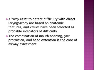
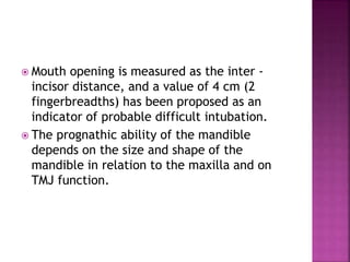
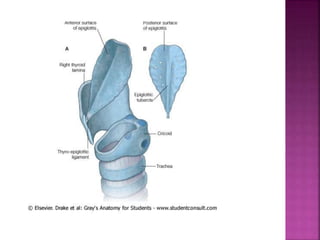
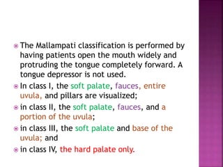
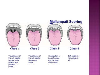
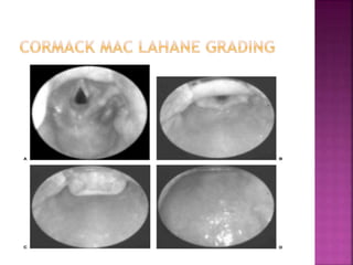
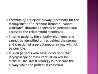
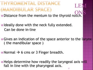
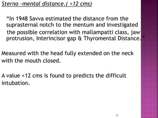
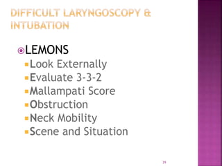
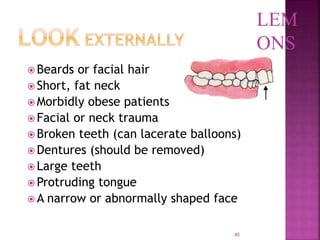
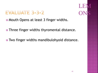
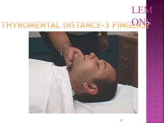
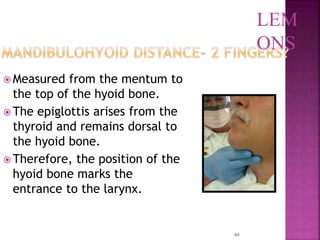
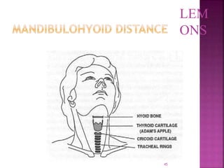
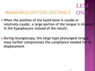
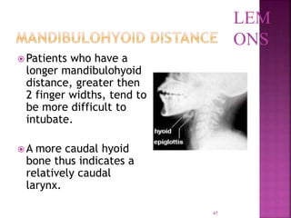
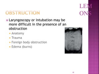
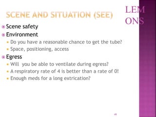
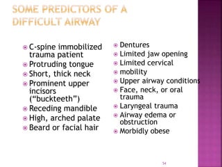
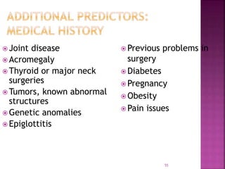
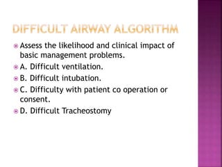
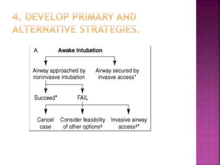
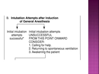
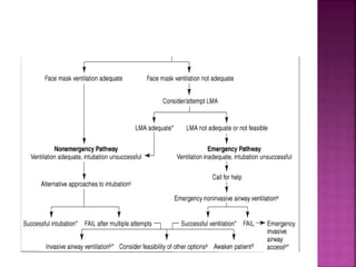
Presenter: Dr. Sharath K.
Moderator: Dr. Suresh Kumar
 Three basic decisions needed before
induction of anesthesia in every patient are
whether
 To use awake endotracheal intubation
 To use a percutaneous technique
 To maintain spontaneous ventilation
 Airway tests to detect difficulty with direct
laryngoscopy are based on anatomic
features, and values have been selected as
probable indicators of difficulty.
 The combination of mouth opening, jaw
protrusion, and head extension is the core of
airway assessment
 Mouth opening is measured as the inter -
incisor distance, and a value of 4 cm (2
fingerbreadths) has been proposed as an
indicator of probable difficult intubation.
 The prognathic ability of the mandible
depends on the size and shape of the
mandible in relation to the maxilla and on
TMJ function.
 Prognathic inability of the mandible (the
mandibular incisors cannot be brought in line
with the maxillary incisors) is associated with
difficult intubation.
 Limited head (occipito-atlanto-axial)
extension impairs direct laryngoscopy
 The Mallampati test (visibility of pharyngeal
structures) is of limited value on its own but
can be combined with an assessment of
dentition.
 The thyromental distance is of limited value
as a predictor of difficult laryngoscopy,
 Examination ensures that the laryngeal
cartilage is palpated and
 Submandibular compliance assessed.
 The larynx consists of a framework of
articulating cartilage connected by fascia,
muscles, and ligaments.
 It is suspended from the hyoid bone by the
thyro-hyoid membrane.
 The principal cartilages are the thyroid,
cricoid, and posterior (arytenoid,
corniculate, and cuneiform) cartilage and
the epiglottis.
 The cricoid cartilage is a complete ring that
articulates with the thyroid and arytenoid
cartilage.
 The arytenoid cartilage sits on the postero-
lateral border of the cricoid, from where it
can be dislocated during airway
management.
 The laryngeal inlet is bounded by the
epiglottis, aryepiglottic folds, posterior
cartilage, and interarytenoid notch.
 The vocal cords run between the vocal
processes of the arytenoid cartilage and the
posterior surface of the thyroid cartilage.
 The lower end of the leaf-shaped epiglottis is
attached to the middle of the posterior
surface of the thyroid cartilage.
 The anterior surface is connected to the
hyoid bone by the hyo-epiglottic ligament
and to the tongue by the median glosso-
epiglottic fold.
 The valleculae (often called vallecula) are
depressions between the median and lateral
glossoepiglottic folds that connect the lateral
edges of the epiglottis to the base of the
tongue.
 The Macintosh technique of laryngoscopy
involves insertion of the tip of the
laryngoscope into the vallecula, where it
tensions the hyo-epiglottic ligament to
achieve indirect elevation of the epiglottis.
 The pharynx is a fibromuscular tube that
extends from the base of the skull to the
lower border of the cricoid cartilage.
 It joins the nasal and oral cavities above with
the larynx and esophagus below.
 Both the pharynx and esophagus can be
perforated by blind attempts at tracheal
intubation
 The examination described by El-Ganzouri
and colleagues
 assessment of mouth opening,
 prognathic ability,
 head extension,
 thyromental distance,
 and Mallampati test has been used with
minor modification by others.
 The ability to achieve good mouth opening is
important for many airway procedures.
 Initial mouth opening is achieved by rotation
within the temporomandibular joint (TMJ)
and subsequent opening by sliding of the
condyles of the mandible within the TMJ.
 Also known as protrusion, translocation, or
subluxation.
17
 Mask Seal
 Obesity or Obstruction
 Age > 55
 No Teeth
 Stiff
18
 Global airway assessment.
(history & physical examination, airway compromising conditions)
 Regional anatomical assessment
(Teeth, oral cavity, mandibular space and neck)
 LEMON Airway assessment method.
 Radiological assessment.
( Skeletal films, fluoroscopy, USG, CT/MRI )
 Wilson’s rule for predicting difficult intubation.
 Quick assessment of airway “the rule of 1-2-3”
Teeth  inter-incisor distance
“ It is the distance between the upper & lower
incisors when the mouth is widely open”
Normal  4.6 cm or more
Less than 3.8 cm predicts difficult airway.
 The Mallampati classification is performed by
having patients open the mouth widely and
protruding the tongue completely forward. A
tongue depressor is not used.
 In class I, the soft palate, fauces, entire
uvula, and pillars are visualized;
 in class II, the soft palate, fauces, and a
portion of the uvula;
 in class III, the soft palate and base of the
uvula; and
 in class IV, the hard palate only.
 Relates to tongue size to pharyngeal size.
 Performed with patient in a sitting position,
head neutral, mouth open wide and tongue
protruding to the maximum.
 The evaluation should also document the
status of teeth,
 range of motion of the neck, neck
circumference (increasing size predicts
difficulty with laryngoscopy),
 thyromental distance,
 body habitus, and
 pertinent deformities.
 Ventilation via mask requires the ability to
achieve a seal between the mask and face
and to overcome upper airway obstruction.
 Limited mandibular protrusion, abnormal
neck anatomy, sleep apnea, snoring, and
obesity are independent predictors of
moderate or severe difficulty with mask
ventilation.
 Snoring and a thyromental distance of less
than 6 cm are independent predictors of
severe difficulty
 Airway assessment cannot detect some
serious problems, including
 asymptomatic lesions in the vicinity of the
larynx,
 skeletal factors, and some
 varieties of TMJ dysfunction.
 The problem with airway assessment is that
the risk of difficulty is overestimated and not
all cases of difficult airway management can
be predicted.
 Airway evaluation gives some indication of
potential difficulty and should always be
performed.
 The anesthesiologist must then make a
judgment of whether direct laryngoscopy,
mask ventilation, and percutaneous rescue
are likely to be successful.
 The limitations of airway assessment mean
that preparation of an airway strategy for
the management of unanticipated difficulty
is the ultimate key to safe practice.
 Because of the relatively high incidence of
dental injuries during anesthesia, thorough
documentation of preexisting tooth
abnormalities is useful.
 Grade 1: Full aperture visible
 Grade 2: Lower part of cords visible
 Grade 3: Only epiglottis visible
 Grade 4: Epiglottis not visible
 Length of the upper incisors
 Condition of the teeth
 Relationship of the upper (maxillary) incisors
to the lower (mandibular) incisors
 Ability to protrude or advance the lower
(mandibular) incisors in front of the upper
(maxillary) incisors
 Inter-incisor or intergum (if edentulous)
distance
 Tongue size
 Visibility of the uvula
 Presence of heavy facial hair
 Compliance of the mandibular space
 Thyromental distance with the head in
maximum extension
 Length of the neck
 Thickness or circumference of the neck
 Range of motion of the head and neck
 Creation of a surgical airway (necessary for the
management of a “cannot intubate, cannot
ventilate” situation) depends on percutaneous
access to the cricothyroid membrane.
 In some patients the cricothyroid membrane
cannot be identified or lies behind the sternum,
and creation of a percutaneous airway will not
be possible.
 In such patients who have indications that
laryngoscopy or mask ventilation will be
difficult, the safest strategy is to secure the
airway while the patient is conscious.
35
Atlanto –occipital joint extension normal: 35
degrees.
Any reduction in extension is expressed as grades’
Grade I  >35 degree
Grade II  22 – 34 degree
Grade III 12 – 21 degree
Grade IV <12 degree
LEM
ONS
37
 Distance from the mentum to the thyroid notch.
 Ideally done with the neck fully extended.
Can be done in-line
 Gives an indication of the space anterior to the larynx
( the mandibular space )
 Normal  6 cms or 3 Finger breadth.
 Helps determine how readily the laryngeal axis will
fall in line with the pharyngeal axis.
LEM
ONS
38
Sterno -mental distance.( >12 cms)
“In 1948 Savva estimated the distance from the
suprasternal notch to the mentum and investigated
the possible correlation with mallampatti class, jaw
protrusion, Interincisor gap & Thyromental Distance.”
Measured with the head fully extended on the neck
with the mouth closed.
A value <12 cms is found to predicts the difficult
intubation.
39
LEMONS
Look Externally
Evaluate 3-3-2
Mallampati Score
Obstruction
Neck Mobility
Scene and Situation
40
 Beards or facial hair
 Short, fat neck
 Morbidly obese patients
 Facial or neck trauma
 Broken teeth (can lacerate balloons)
 Dentures (should be removed)
 Large teeth
 Protruding tongue
 A narrow or abnormally shaped face
LEM
ONS
41
 Mouth Opens at least 3 finger widths.
 Three finger widths thyromental distance.
 Two finger widths mandibulohyoid distance.
LEM
ONS
42
LEM
ONS
43
LEM
ONS
44
 Measured from the mentum to
the top of the hyoid bone.
 The epiglottis arises from the
thyroid and remains dorsal to
the hyoid bone.
 Therefore, the position of the
hyoid bone marks the
entrance to the larynx.
LEM
ONS
45
LEM
ONS
46
 When the position of the hyoid bone is caudal or
relatively caudal, a large portion of the tongue is situated
in the hypopharynx instead of the mouth.
 During laryngoscopy, this large hypo pharyngeal tongue
mass further compromises the compliance needed for its
displacement
LEM
ONS
47
 Patients who have a
longer mandibulohyoid
distance, greater then
2 finger widths, tend to
be more difficult to
intubate.
 A more caudal hyoid
bone thus indicates a
relatively caudal
larynx.
LEM
ONS
48
 Laryngoscopy or intubation may be
more difficult in the presence of an
obstruction
 Anatomy
 Trauma
 Foreign body obstruction
 Edema (burns)
LEM
ONS
49
 Scene safety
 Environment
 Do you have a reasonable chance to get the tube?
 Space, positioning, access
 Egress
 Will you be able to ventilate during egress?
 A respiratory rate of 4 is better than a rate of 0!
 Enough meds for a long extrication?
LEM
ONS
50
Risk factor. Level score.
Weight. 90kg 0
90-110kg 1
>110kg 2
Head & neck above 90 0
Movement: about 90 1
below 90 2
Jaw movement IG>5cm 0
IG<5cm 1
IG<5cm 2
Receding mandible normal 0
moderate 1
severe 2
Buck teeth normal 0
moderate 1
severe 2
51
 Subtle bony abnormalities of mandible,
maxilla & cervical spine difficulty in
visualization of larynx
 White & kander made several measurements
on the radiographs  lateral, PA &
Submento-vertical view.
1. Posterior depth of the mandible
2. Effective mandibular length.
3. Gap between the occiput & spine of the
first cervical vertebra
52
Posterior depth of the mandible.
The distance between the alveolus immediately behind the
3rd molar tooth to the lower border of the mandible
Effective mandibular length
The distance from the tip of the lower incisors to the
tempero-mandibular joint
“ direct laryngoscopy was difficult when the effective
mandibular length was less than 3.6 times the posterior
depth of the mandible”
53
54
 C-spine immobilized
trauma patient
 Protruding tongue
 Short, thick neck
 Prominent upper
incisors
(“buckteeth”)
 Receding mandible
 High, arched palate
 Beard or facial hair
 Dentures
 Limited jaw opening
 Limited cervical
 mobility
 Upper airway conditions
 Face, neck, or oral
trauma
 Laryngeal trauma
 Airway edema or
obstruction
 Morbidly obese
55
 Joint disease
 Acromegaly
 Thyroid or major neck
surgeries
 Tumors, known abnormal
structures
 Genetic anomalies
 Epiglottitis
 Previous problems in
surgery
 Diabetes
 Pregnancy
 Obesity
 Pain issues
 Assess the likelihood and clinical impact of
basic management problems.
 A. Difficult ventilation.
 B. Difficult intubation.
 C. Difficulty with patient co operation or
consent.
 D. Difficult Tracheostomy
2. Actively Pursue opportunities to deliver
supplemental oxygen throughout the process
of difficult airway management.
3. Consider the relative merits and feasibility
of basic management choices.
A. Awake intubation Vs. Intubation attempts
after induction of GA.
B. Non invasive technique for initial approach
to intubation Vs. invasive technique for
initial approach to intubation.
C. Preservation of spontaneous ventilation Vs.
Ablation of Spontaneous Ventilation.
 Millers Anesthesia- 7th Edition.
 Morgan’s Clinical Anesthesiology- 4th Edition.
 Indian Journal of Anesthesiology- 2005 49(4)
 Airway assessment- Predictors of difficult
airway.
Airway assessment

Airway assessment

  • 1.
    Presenter: Dr. SharathK. Moderator: Dr. Suresh Kumar
  • 2.
     Three basicdecisions needed before induction of anesthesia in every patient are whether  To use awake endotracheal intubation  To use a percutaneous technique  To maintain spontaneous ventilation
  • 3.
     Airway teststo detect difficulty with direct laryngoscopy are based on anatomic features, and values have been selected as probable indicators of difficulty.  The combination of mouth opening, jaw protrusion, and head extension is the core of airway assessment
  • 4.
     Mouth openingis measured as the inter - incisor distance, and a value of 4 cm (2 fingerbreadths) has been proposed as an indicator of probable difficult intubation.  The prognathic ability of the mandible depends on the size and shape of the mandible in relation to the maxilla and on TMJ function.
  • 5.
     Prognathic inabilityof the mandible (the mandibular incisors cannot be brought in line with the maxillary incisors) is associated with difficult intubation.  Limited head (occipito-atlanto-axial) extension impairs direct laryngoscopy
  • 6.
     The Mallampatitest (visibility of pharyngeal structures) is of limited value on its own but can be combined with an assessment of dentition.  The thyromental distance is of limited value as a predictor of difficult laryngoscopy,  Examination ensures that the laryngeal cartilage is palpated and  Submandibular compliance assessed.
  • 7.
     The larynxconsists of a framework of articulating cartilage connected by fascia, muscles, and ligaments.  It is suspended from the hyoid bone by the thyro-hyoid membrane.  The principal cartilages are the thyroid, cricoid, and posterior (arytenoid, corniculate, and cuneiform) cartilage and the epiglottis.
  • 8.
     The cricoidcartilage is a complete ring that articulates with the thyroid and arytenoid cartilage.  The arytenoid cartilage sits on the postero- lateral border of the cricoid, from where it can be dislocated during airway management.  The laryngeal inlet is bounded by the epiglottis, aryepiglottic folds, posterior cartilage, and interarytenoid notch.
  • 9.
     The vocalcords run between the vocal processes of the arytenoid cartilage and the posterior surface of the thyroid cartilage.  The lower end of the leaf-shaped epiglottis is attached to the middle of the posterior surface of the thyroid cartilage.  The anterior surface is connected to the hyoid bone by the hyo-epiglottic ligament and to the tongue by the median glosso- epiglottic fold.
  • 10.
     The valleculae(often called vallecula) are depressions between the median and lateral glossoepiglottic folds that connect the lateral edges of the epiglottis to the base of the tongue.  The Macintosh technique of laryngoscopy involves insertion of the tip of the laryngoscope into the vallecula, where it tensions the hyo-epiglottic ligament to achieve indirect elevation of the epiglottis.
  • 14.
     The pharynxis a fibromuscular tube that extends from the base of the skull to the lower border of the cricoid cartilage.  It joins the nasal and oral cavities above with the larynx and esophagus below.  Both the pharynx and esophagus can be perforated by blind attempts at tracheal intubation
  • 15.
     The examinationdescribed by El-Ganzouri and colleagues  assessment of mouth opening,  prognathic ability,  head extension,  thyromental distance,  and Mallampati test has been used with minor modification by others.
  • 16.
     The abilityto achieve good mouth opening is important for many airway procedures.  Initial mouth opening is achieved by rotation within the temporomandibular joint (TMJ) and subsequent opening by sliding of the condyles of the mandible within the TMJ.  Also known as protrusion, translocation, or subluxation.
  • 17.
    17  Mask Seal Obesity or Obstruction  Age > 55  No Teeth  Stiff
  • 18.
    18  Global airwayassessment. (history & physical examination, airway compromising conditions)  Regional anatomical assessment (Teeth, oral cavity, mandibular space and neck)  LEMON Airway assessment method.  Radiological assessment. ( Skeletal films, fluoroscopy, USG, CT/MRI )  Wilson’s rule for predicting difficult intubation.  Quick assessment of airway “the rule of 1-2-3”
  • 20.
    Teeth  inter-incisordistance “ It is the distance between the upper & lower incisors when the mouth is widely open” Normal  4.6 cm or more Less than 3.8 cm predicts difficult airway.
  • 21.
     The Mallampaticlassification is performed by having patients open the mouth widely and protruding the tongue completely forward. A tongue depressor is not used.  In class I, the soft palate, fauces, entire uvula, and pillars are visualized;  in class II, the soft palate, fauces, and a portion of the uvula;  in class III, the soft palate and base of the uvula; and  in class IV, the hard palate only.
  • 23.
     Relates totongue size to pharyngeal size.  Performed with patient in a sitting position, head neutral, mouth open wide and tongue protruding to the maximum.
  • 24.
     The evaluationshould also document the status of teeth,  range of motion of the neck, neck circumference (increasing size predicts difficulty with laryngoscopy),  thyromental distance,  body habitus, and  pertinent deformities.
  • 25.
     Ventilation viamask requires the ability to achieve a seal between the mask and face and to overcome upper airway obstruction.  Limited mandibular protrusion, abnormal neck anatomy, sleep apnea, snoring, and obesity are independent predictors of moderate or severe difficulty with mask ventilation.  Snoring and a thyromental distance of less than 6 cm are independent predictors of severe difficulty
  • 26.
     Airway assessmentcannot detect some serious problems, including  asymptomatic lesions in the vicinity of the larynx,  skeletal factors, and some  varieties of TMJ dysfunction.
  • 27.
     The problemwith airway assessment is that the risk of difficulty is overestimated and not all cases of difficult airway management can be predicted.  Airway evaluation gives some indication of potential difficulty and should always be performed.
  • 28.
     The anesthesiologistmust then make a judgment of whether direct laryngoscopy, mask ventilation, and percutaneous rescue are likely to be successful.  The limitations of airway assessment mean that preparation of an airway strategy for the management of unanticipated difficulty is the ultimate key to safe practice.
  • 29.
     Because ofthe relatively high incidence of dental injuries during anesthesia, thorough documentation of preexisting tooth abnormalities is useful.
  • 31.
     Grade 1:Full aperture visible  Grade 2: Lower part of cords visible  Grade 3: Only epiglottis visible  Grade 4: Epiglottis not visible
  • 32.
     Length ofthe upper incisors  Condition of the teeth  Relationship of the upper (maxillary) incisors to the lower (mandibular) incisors  Ability to protrude or advance the lower (mandibular) incisors in front of the upper (maxillary) incisors  Inter-incisor or intergum (if edentulous) distance  Tongue size
  • 33.
     Visibility ofthe uvula  Presence of heavy facial hair  Compliance of the mandibular space  Thyromental distance with the head in maximum extension  Length of the neck  Thickness or circumference of the neck  Range of motion of the head and neck
  • 34.
     Creation ofa surgical airway (necessary for the management of a “cannot intubate, cannot ventilate” situation) depends on percutaneous access to the cricothyroid membrane.  In some patients the cricothyroid membrane cannot be identified or lies behind the sternum, and creation of a percutaneous airway will not be possible.  In such patients who have indications that laryngoscopy or mask ventilation will be difficult, the safest strategy is to secure the airway while the patient is conscious.
  • 35.
    35 Atlanto –occipital jointextension normal: 35 degrees. Any reduction in extension is expressed as grades’ Grade I  >35 degree Grade II  22 – 34 degree Grade III 12 – 21 degree Grade IV <12 degree LEM ONS
  • 37.
    37  Distance fromthe mentum to the thyroid notch.  Ideally done with the neck fully extended. Can be done in-line  Gives an indication of the space anterior to the larynx ( the mandibular space )  Normal  6 cms or 3 Finger breadth.  Helps determine how readily the laryngeal axis will fall in line with the pharyngeal axis. LEM ONS
  • 38.
    38 Sterno -mental distance.(>12 cms) “In 1948 Savva estimated the distance from the suprasternal notch to the mentum and investigated the possible correlation with mallampatti class, jaw protrusion, Interincisor gap & Thyromental Distance.” Measured with the head fully extended on the neck with the mouth closed. A value <12 cms is found to predicts the difficult intubation.
  • 39.
    39 LEMONS Look Externally Evaluate 3-3-2 MallampatiScore Obstruction Neck Mobility Scene and Situation
  • 40.
    40  Beards orfacial hair  Short, fat neck  Morbidly obese patients  Facial or neck trauma  Broken teeth (can lacerate balloons)  Dentures (should be removed)  Large teeth  Protruding tongue  A narrow or abnormally shaped face LEM ONS
  • 41.
    41  Mouth Opensat least 3 finger widths.  Three finger widths thyromental distance.  Two finger widths mandibulohyoid distance. LEM ONS
  • 42.
  • 43.
  • 44.
    44  Measured fromthe mentum to the top of the hyoid bone.  The epiglottis arises from the thyroid and remains dorsal to the hyoid bone.  Therefore, the position of the hyoid bone marks the entrance to the larynx. LEM ONS
  • 45.
  • 46.
    46  When theposition of the hyoid bone is caudal or relatively caudal, a large portion of the tongue is situated in the hypopharynx instead of the mouth.  During laryngoscopy, this large hypo pharyngeal tongue mass further compromises the compliance needed for its displacement LEM ONS
  • 47.
    47  Patients whohave a longer mandibulohyoid distance, greater then 2 finger widths, tend to be more difficult to intubate.  A more caudal hyoid bone thus indicates a relatively caudal larynx. LEM ONS
  • 48.
    48  Laryngoscopy orintubation may be more difficult in the presence of an obstruction  Anatomy  Trauma  Foreign body obstruction  Edema (burns) LEM ONS
  • 49.
    49  Scene safety Environment  Do you have a reasonable chance to get the tube?  Space, positioning, access  Egress  Will you be able to ventilate during egress?  A respiratory rate of 4 is better than a rate of 0!  Enough meds for a long extrication? LEM ONS
  • 50.
    50 Risk factor. Levelscore. Weight. 90kg 0 90-110kg 1 >110kg 2 Head & neck above 90 0 Movement: about 90 1 below 90 2 Jaw movement IG>5cm 0 IG<5cm 1 IG<5cm 2 Receding mandible normal 0 moderate 1 severe 2 Buck teeth normal 0 moderate 1 severe 2
  • 51.
    51  Subtle bonyabnormalities of mandible, maxilla & cervical spine difficulty in visualization of larynx  White & kander made several measurements on the radiographs  lateral, PA & Submento-vertical view. 1. Posterior depth of the mandible 2. Effective mandibular length. 3. Gap between the occiput & spine of the first cervical vertebra
  • 52.
    52 Posterior depth ofthe mandible. The distance between the alveolus immediately behind the 3rd molar tooth to the lower border of the mandible Effective mandibular length The distance from the tip of the lower incisors to the tempero-mandibular joint “ direct laryngoscopy was difficult when the effective mandibular length was less than 3.6 times the posterior depth of the mandible”
  • 53.
  • 54.
    54  C-spine immobilized traumapatient  Protruding tongue  Short, thick neck  Prominent upper incisors (“buckteeth”)  Receding mandible  High, arched palate  Beard or facial hair  Dentures  Limited jaw opening  Limited cervical  mobility  Upper airway conditions  Face, neck, or oral trauma  Laryngeal trauma  Airway edema or obstruction  Morbidly obese
  • 55.
    55  Joint disease Acromegaly  Thyroid or major neck surgeries  Tumors, known abnormal structures  Genetic anomalies  Epiglottitis  Previous problems in surgery  Diabetes  Pregnancy  Obesity  Pain issues
  • 56.
     Assess thelikelihood and clinical impact of basic management problems.  A. Difficult ventilation.  B. Difficult intubation.  C. Difficulty with patient co operation or consent.  D. Difficult Tracheostomy
  • 57.
    2. Actively Pursueopportunities to deliver supplemental oxygen throughout the process of difficult airway management. 3. Consider the relative merits and feasibility of basic management choices. A. Awake intubation Vs. Intubation attempts after induction of GA. B. Non invasive technique for initial approach to intubation Vs. invasive technique for initial approach to intubation. C. Preservation of spontaneous ventilation Vs. Ablation of Spontaneous Ventilation.
  • 61.
     Millers Anesthesia-7th Edition.  Morgan’s Clinical Anesthesiology- 4th Edition.  Indian Journal of Anesthesiology- 2005 49(4)  Airway assessment- Predictors of difficult airway.