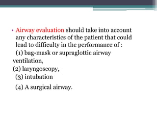
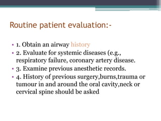
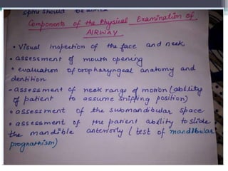
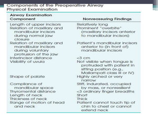
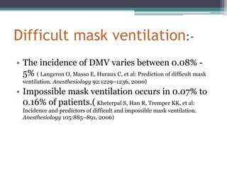
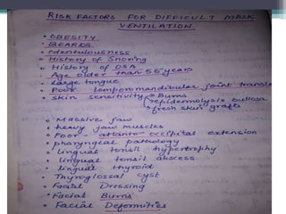
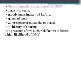
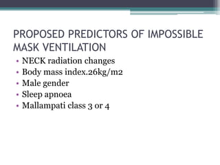
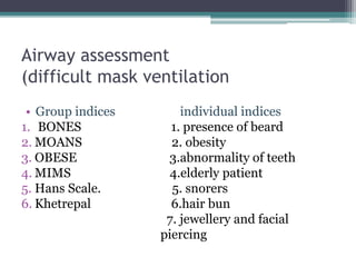
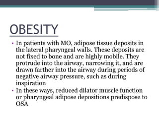
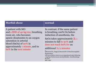
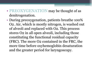
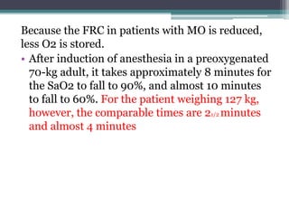
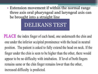
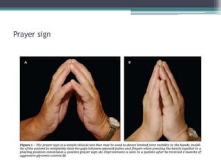
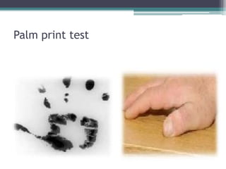
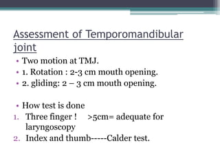
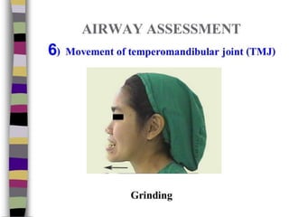
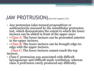
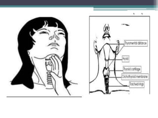
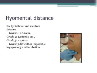
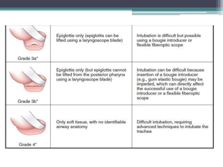
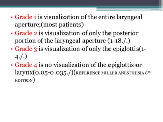
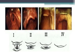
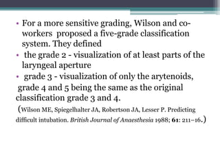
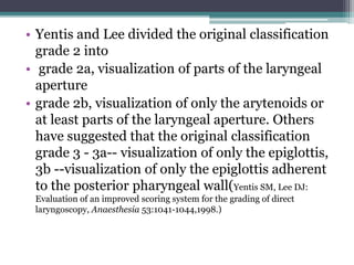
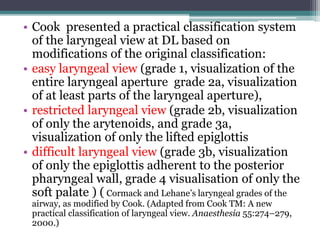
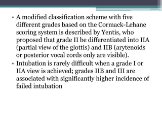
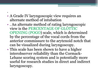
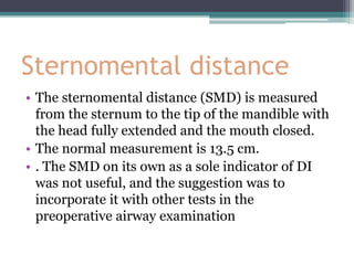
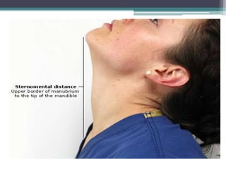
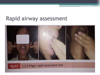
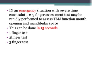
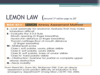
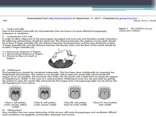
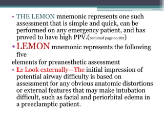
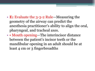
This document discusses methods for assessing a patient's airway for difficulty with mask ventilation, laryngoscopy, intubation, and a surgical airway. It describes various physical exam findings and grading scales that can help predict challenges, such as neck circumference, mouth opening, jaw protrusion, Mallampati score, thyromental distance, and laryngoscopic view with intubation. Factors like obesity, beard, lack of teeth, older age, and snoring increase risk. Proper airway assessment is important for anesthesia planning and preparing for potential difficulty.
Airway Assessment Reference Guide for Difficult Mask Ventilation & Intubation
1. AIRWAY
ASSESSMENT
REFERENCE:-
1.BENUMOF AND HAGBERG’S AIRWAY MANAGEMENT
3RD EDITION
2.PAUL G. BARASH CLINICAL ANESTHESIA
3.MORGAN AND MIKHAIL’S CLINICAL
ANESTHESIOLOGY 5TH EDITION
4.ATLAS OF AIRWAY MANAGEMNT STEVEN
L.OREBAUGH PAUL BIGELSIEN
5.RASHID M KHAN AIRWAY MANAGEMENT 5TH
EDITION
2. • Airway evaluation should take into account
any characteristics of the patient that could
lead to difficulty in the performance of :
(1) bag-mask or supraglottic airway
ventilation,
(2) laryngoscopy,
(3) intubation
(4) A surgical airway.
3. Routine patient evaluation:-
• 1. Obtain an airway history
• 2. Evaluate for systemic diseases (e.g.,
respiratory failure, coronary artery disease.
• 3. Examine previous anesthetic records.
• 4. History of previous surgery,burns,trauma or
tumour in and around the oral cavity,neck or
cervical spine should be asked
4.
5.
6. Difficult mask ventilation:-
• The incidence of DMV varies between 0.08% -
5% ( Langeron O, Masso E, Huraux C, et al: Prediction of difficult mask
ventilation. Anesthesiology 92:1229–1236, 2000)
• Impossible mask ventilation occurs in 0.07% to
0.16% of patients.( Kheterpal S, Han R, Tremper KK, et al:
Incidence and predictors of difficult and impossible mask ventilation.
Anesthesiology 105:885–891, 2006)
7.
8. • Five independent criteria predict DMV
• 1.age >55 years,
• 2.body mass index >26 kg/m2,
• 3.lack of teeth
• ,4. presence of mustache or beard,
• 5. history of snoring
the presence of two such risk factors indicates
a high likelihood of DMV
9. PROPOSED PREDICTORS OF IMPOSSIBLE
MASK VENTILATION
• NECK radiation changes
• Body mass index.26kg/m2
• Male gender
• Sleep apnoea
• Mallampati class 3 or 4
10. Airway assessment
(difficult mask ventilation
• Group indices individual indices
1. BONES 1. presence of beard
2. MOANS 2. obesity
3. OBESE 3.abnormality of teeth
4. MIMS 4.elderly patient
5. Hans Scale. 5. snorers
6. Khetrepal 6.hair bun
7. jewellery and facial
piercing
11. Presence of beard
• Presence of beard creates diificulty in creating
effective seal by mask leading to loss of
ventilated volume
• Shaving or vaseline
12.
13. OBESITY
• In patients with MO, adipose tissue deposits in
the lateral pharyngeal walls. These deposits are
not fixed to bone and are highly mobile. They
protrude into the airway, narrowing it, and are
drawn farther into the airway during periods of
negative airway pressure, such as during
inspiration
• In these ways, reduced dilator muscle function
or pharyngeal adipose depositions predispose to
OSA
14. Morbid obese normal
A patient with MO
and a BMI of 40 kg/m2, breathing
room air, who becomes
apneic desaturates to an oxygen
saturation in arterial
blood (SaO2) of 90% in
approximately 1 minute, and to
60% in the next minute
In contrast, if the same patient
is breathing 100% O2 before
induction of anesthesia, the
SaO2 takes approximately 21/2
minutes to fall to 90% and
does not reach 60% for an
additional 11/2 minutes
(Benumof JL, Dagg R, Benumof R: Critical hemoglobin
desaturation
will occur before return to an unparalyzed state following
1 mg/kg of intravenous succinylcholine. Anesthesiology
87:979–
982, 1997
15. • PREOXYGENATION may be thought of as
denitrogenation.
• During preoxygenation, patients breathe 100%
O2. Air, which is mostly nitrogen, is washed out
of alveoli and replaced with O2. This process
stores O2 in all open alveoli, including those
constituting the functional residual capacity
(FRC). The more O2 contained in the FRC, the
more time before oxyhemoglobin desaturation
and the greater period for laryngoscopy.
16. Because the FRC in patients with MO is reduced,
less O2 is stored.
• After induction of anesthesia in a preoxygenated
70-kg adult, it takes approximately 8 minutes for
the SaO2 to fall to 90%, and almost 10 minutes
to fall to 60%. For the patient weighing 127 kg,
however, the comparable times are 21/2 minutes
and almost 4 minutes
17. • A NECK CIRCUMFERENCE greater than
43 cm (17 inches) is associated with difficulty with
tracheal intubation Brodsky showed that a large
neck circumference is, in fact, more predictive of
difficulty with endotracheal intubation than a high
body mass index (BMI).
•TOOTH-
• Poor dentition is at risk for damage as the mouth is
opened and as the laryngoscope blade is employed.
• Prominent maxillary incisors complicate
laryngoscopy in another way. They protrude into the
mouth and block the line of sight to the larynx
18. • ARTIFICIAL DENTURES
• FIXED if they are loose
• Left in place than after patient
well oxygenated and
and prior to laryngo-
scopy and intubation
they are removed
EDENTULOUS PATIENT
problem in fixing the mask place the
unfolde gauze fluffed and compressed inside the
mouth along the buccal pouches restore the cheek
fullness optimal mask seal
19. BONES
B-bearded individual
O-obesity
N-no teeth
E-elderly patient >55 years
S-snorer
MOANS
M-reciding mandible
O-obese
A-advanced age-
N-no teeth
S-snorer
OBESE
O-obese
B-bearded
E-elderly
S-snorer
E-edentulous
MIMS(GIVEN BY YILDES ET AL)
M-male sex
I-increasing age
M-mallampati class4
S-snorer
KHETREPAL ET AL
1.Body mass index
2.Snoring
3.OSA
4.thick/obese neck
5.Limited mandibular
protrusion(Kheterpal S, Martin L, Shanks
AM, et al: Prediction of outcomes
of impossible mask ventilation: A review of
20. • From Han R, Tremper KK, Kheterpal S, O’Reilly M: Grading scale
for mask ventilation. Anesthesiology 101:267, 2004.
21. Physical examination indices
• Assessment of cervical and atlanto occipital joint
function
• DIRECT ASSESSMENT;-
• Laryngoscopic view becomes easier when the neck is
flexed on the chest by 25-35 degree and the atlanto
occipital is well extended (85degree)-sniffing
ormagill position
• Touch his manubri sterni with his chin
• Look ceiling without raising eyebrows
22.
23.
24.
25. • Extension movement if within the normal range
three axis oral pharyngeal and laryngeal axis can
be brought into a straight line
DELIKANS TEST
28. • Grade 0: all phalangeal area visible
• Grade 1: deficiency in the inter phalangeal areas
of 4th and/or 5th digit.
• Grade 2: deficiency in the inter phalangeal area
of 2nd to 5th digit.
• Grade 3: only tips of digits are seen.
• Higher grade~ higher chance of difficult
intubation.
29. Assessment of Temporomandibular
joint
• Two motion at TMJ.
• 1. Rotation : 2-3 cm mouth opening.
• 2. gliding: 2 – 3 cm mouth opening.
• How test is done
1. Three finger ! >5cm= adequate for
laryngoscopy
2. Index and thumb-----Calder test.
30.
31.
32. JAW PROTRUSION(benumof pageno.212)
• Jaw protrusion (also termed prognathism or
subluxation)is assessed by the mandibular protrusion
test, which demonstrates the extent to which the lower
incisors can be slided in front of the upper ones:
• • Class A: The lower incisors can be protruded anterior
to the upper incisors.
• • Class B: The lower incisors can be brought edge-to-
edge with the upper incisors.
Class C: The lower incisors cannot reach the top
incisors.
• Class C protrusion was associated with difficult
laryngoscopy and Difficult mask ventilation, whereas
class A protrusion rarely produced any difficulty.
39. Assessment of Submandibular space
•Thyromental distance
• The patient’s head is extended at the atlanto-occipital joint.
The mentum of the mandible and the thyroid cartilage are
identified. The “Adam’s apple” (thyroid notch) is the most
superficial structure in the neck and serves as a good
landmark for the thyroid cartilage.
• The vocal cords lie just caudal to the thyroid notch. The
distance between the thyroid cartilage and the mentum
(thyromental distance, or TMD) is measured in one of three
ways:
• with a set of spacers,
• with a small pocket ruler,
• or with the observer’s fingers
40. • . The normal TMD is 6.5 cm.
• A TMD greater than 6 cm is predictive of easy
intubation
• a TMD of less than 6 cm is suggestive of DI.
• If a ruler is not present at the bedside,
practitioners can judge the TMD with their own
fingers.
• The width of one’s middle three fingers
frequently approximates 6 cm, and the TMD can
be compared with the fingers’ span.
41.
42. Hyomental distance
btw hyoid bone and mentum
distance.
Grade 1: >6.0 cm,
Grade 2: 4.0 to 6.0 cm ,
Grade 3: < 4.0 cm
Grade 3 difficult or impossible
laryngoscopy and intubation
43. Assessing the adequacy of oropharynx
for laryngoscopy and intubation(benumof
212)
• MALLAMPATI GRADING:-
• 1983, Mallampati and associates described a clinical sign to
predict difficult tracheal intubation based on the size of the
base of the tongue.( Mallampati SR: Clinical sign to predict difficult
tracheal intubation (hypothesis), Can Anaesth Soc J 30:316-317, 1983)
• A Mallampati classification of I to III is assigned, based on
the visibility of the faucial pillars, uvula, and soft palate when
the patient is seated upright with the head neutral, the mouth
open, the tongue protruded, and no phonation.(Mashour GA,
Kheterpal S, Vanaharam V, et al: The extended Mallampati score and a
diagnosis of diabetes mellitus are predictors of difficult laryngoscopy in
the morbidly obese, Anesth Analg 107:1919-1923, 2008)
44. • Higher scores on the Mallampati
classification indicate poor visibility
of the oropharyngeal structures
attributable to a large tongue
relative to the size of the
oropharyngeal space, and,
subsequently, a more difficult
laryngoscopy.
45. • The modified Mallampati classification
described by Samsoon and Young(Samsoon GL,
Young JR: Anaesthesia 42:487, 1987 )which adds a fourth
classification, is the most commonly used airway
assessment test in current anesthesia practice
and is defined as follows:-
• • Class I: Faucial pillars, uvula, and soft palate
are visualized.
• • Class II: Base of the uvula and soft palate are
visualized.
• • Class III: Soft palate only is visualized.
• • Class IV: Hard palate only is visualized.
46.
47.
48. • As a stand-alone test, the modified Mallampati
classification is insufficient for accurate
prediction of difficult intubation however, it may
have clinical utility in combination with other
difficult airway predictors
• extended Mallampati score with the head in full
extension to improve the predictive value of the
test.
49. • A Mallampati zero classification has been
proposed when the epiglottis can be visualized
during examination of the oropharynx; this
finding is usually associated with easy
laryngoscopy, although difficulty with airway
management attributable to a large, floppy
epiglottis in patients with a Mallampati zero
classification can occur. ( White A, Kander PL: Anatomic
factors in difficult direct laryngoscopy. Br J Anaesth 47:468–474, 1975)
50. ASSESSMENT FOR QUALITY OF GLOTTIC
VIEWING DURING LARYNGOSCOPY
• DIRECT LARYNGOSCOPIC VIEW:-
• CORMACK AND LEHANE
• Cormack and Lehane developed a grading scale in
1984 to describe laryngoscopic views. The grades
range from I to IV, beginning with
• grade I (the best view),-in which the epiglottis and
vocal cords are in complete view, and culminating
with grade IV (the most difficult view), in which the
epiglottis or larynx is not visualized(difficult tracheal
intubation in obstetrics 1984 Anaesthesia, 1984, Volume 39,
pages 1105-1 I 1 1 r.s.cormack and j.lehane)
51.
52.
53.
54. • Grade 1 is visualization of the entire laryngeal
aperture;(most patients)
• Grade 2 is visualization of only the posterior
portion of the laryngeal aperture (1-18./.)
• Grade 3 is visualization of only the epiglottis(1-
4./.)
• Grade 4 is no visualization of the epiglottis or
larynx(0.05-0.035./)(REFERENCE MILLER ANESTHESIA 8TH
EDITION)
55.
56. • For a more sensitive grading, Wilson and co-
workers proposed a five-grade classification
system. They defined
• the grade 2 - visualization of at least parts of the
laryngeal aperture
• grade 3 - visualization of only the arytenoids,
grade 4 and 5 being the same as the original
classification grade 3 and 4.
(Wilson ME, Spiegelhalter JA, Robertson JA, Lesser P. Predicting
difficult intubation. British Journal of Anaesthesia 1988; 61: 211–16.)
57. • Yentis and Lee divided the original classification
grade 2 into
• grade 2a, visualization of parts of the laryngeal
aperture
• grade 2b, visualization of only the arytenoids or
at least parts of the laryngeal aperture. Others
have suggested that the original classification
grade 3 - 3a-- visualization of only the epiglottis,
3b --visualization of only the epiglottis adherent
to the posterior pharyngeal wall(Yentis SM, Lee DJ:
Evaluation of an improved scoring system for the grading of direct
laryngoscopy, Anaesthesia 53:1041-1044,1998.)
58. • Cook presented a practical classification system
of the laryngeal view at DL based on
modifications of the original classification:
• easy laryngeal view (grade 1, visualization of the
entire laryngeal aperture grade 2a, visualization
of at least parts of the laryngeal aperture),
• restricted laryngeal view (grade 2b, visualization
of only the arytenoids, and grade 3a,
visualization of only the lifted epiglottis
• difficult laryngeal view (grade 3b, visualization
of only the epiglottis adherent to the posterior
pharyngeal wall, grade 4 visualisation of only the
soft palate ) ( Cormack and Lehane’s laryngeal grades of the
airway, as modified by Cook. (Adapted from Cook TM: A new
practical classification of laryngeal view. Anaesthesia 55:274–279,
2000.)
59. • A modified classification scheme with five
different grades based on the Cormack-Lehane
scoring system is described by Yentis, who
proposed that grade II be differentiated into IIA
(partial view of the glottis) and IIB (arytenoids
or posterior vocal cords only are visible).
• Intubation is rarely difficult when a grade I or
IIA view is achieved; grades IIB and III are
associated with significantly higher incidence of
failed intubation
60. .
• A Grade IV laryngoscopic view requires an
alternate method of intubation
• . An alternate method of rating laryngoscopic
view is the PERCENTAGE OF GLOTTIC
OPENING (POGO) scale, which is determined
by the percentage of the vocal cords from the
anterior commissure to the arytenoid notch that
can be visualized during laryngoscopy.
• This scale has been shown to have a higher
interobserver reliability than the Cormack-
Lehane scoring system and is potentially more
useful for research studies in direct and indirect
laryngoscopy.
61. Sternomental distance
• The sternomental distance (SMD) is measured
from the sternum to the tip of the mandible with
the head fully extended and the mouth closed.
• The normal measurement is 13.5 cm.
• . The SMD on its own as a sole indicator of DI
was not useful, and the suggestion was to
incorporate it with other tests in the
preoperative airway examination
64. • IN an emergency situation with severe time
constraint 1-2-3 finger assessment test may be
rapidly performed to assess TMJ function mouth
opening and mandibular space
• This can be done in 15 seconds
• 1 finger test
• 2finger test
• 3 finger test
67. • THE LEMON mnemonic represents one such
assessment that is simple and quick, can be
performed on any emergency patient, and has
proved to have high PPV.(benumof page no.767)
•LEMON mnemonic represents the following
five
elements for preanesthetic assessment
• L: Look externally—The initial impression of
potential airway difficulty is based on
assessment for any obvious anatomic distortions
or external features that may make intubation
difficult, such as facial and periorbital edema in
a preeclamptic patient.
68. • E: Evaluate the 3-3-2 Rule—Measuring the
geometry of the airway can predict the
anesthesia practitioner’s ability to align the oral,
pharyngeal, and tracheal axes.
• • Mouth opening—The interincisor distance
between the patient’s incisor teeth or the
mandibular opening in an adult should be at
least 4 cm or 3 fingerbreadths
69. • • The distance from the hyoid cartilage to the
chin should be at least 3 cm or 2 finger breadths
(under the chin).
• The ability of the mandible to accommodate the
tongue can be estimated between the mentum
and the hyoid bone.
70. • A smaller mandible is less likely to
accommodate the tongue, which can impair
visualization during laryngoscopy. An unusually
large mandible can elongate the oral axis
• The distance between the thyroid notch and the
floor of the mandible (top of the neck) should be
at least 2 finger breadths
71. • M: MALLAMPATI SCORE—The degree to
which the tongue obstructs the visualization of
the posterior pharynx has some correlation with
the ability to visualize theglottis.
• The MP score can be estimated by having the
patient, in a sitting position, open the mouth
fully and protrude the tongue as far as possible
without phonation.
72. • The relationship of the base of the tongue to the
oropharyngeal structures—uvula and tonsillar
pillars and fauces—is assessed as follows:
• • Class I : Soft palate, uvula, faucial, and tonsillar
• pillars visible (easy intubation)
• • Class II: Soft palate, uvula visible
• • Class III: Soft palate, base of uvula
• Class IV: Hard palate only visible (DI)
73. • O: Obstruction—Pathologic conditions such as
edema,glottic tumor, lingular tonsil, hyperplasia,
and trauma can cause obstruction and can make
laryngoscopy and ventilation difficult.
74. • N: Neck mobility—This is a vital requirement for
successful intubation. The sniffing position is the
optimal,classic position of the head and neck for
facilitating intubation.
• The extension of the atlanto-occipital (A-O)
joint on the cervical spine so as to be able to
align the three axes (oral, pharyngeal, and
laryngeal)during laryngoscopy enhances the
ease of laryngoscopy and tracheal intubation.
75. • Normal A-O joint extension of the head over the
neck is 35 degrees. It can be assessed easily by
getting the patient to place the chin down on the
chest and tilt the head backward as far as
possible.
• A reduction in A-O joint extension of 12 degrees
(33%) or more correlates with intubation
difficulty complete joint immobility significantly
compromises the laryngeal view
76. • Limited A-O joint extension is present in certain
pathologic states such as spondylosis,
rheumatoid arthritis, and cervical spine stenosis,
resulting in symptoms of nerve compression
with cervical extension.
• Complete A-O joint immobility (e.g., hard-collar
neck immobilization)can compromise the view
of the glottis during laryngoscopy