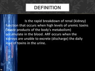
This document provides information on acute kidney failure (ARF), including its definition, risk factors, pathophysiology, diagnosis, and nursing care considerations. ARF occurs when the kidneys are unable to excrete waste from the body due to high levels of toxins. It is characterized by three phases: onset, maintenance, and recovery. Nursing interventions focus on monitoring fluid balance, electrolytes, output, diet, and preventing infections to support the patient's recovery.
Chronic kidney disease (CKD) consists of a spectrum of different pathophysiologic processes associated with abnormal kidney function, and a progressive decline in glomerular filtration rate (GFR).
Urinary Tract Infection with Nursing ManagementSwatilekha Das
Urinary Tract Infection introduction, definition, common microorganisms, classification, predisposing factors, clinical manifestations, pathophysiology, diagnostic studies, medical management and nursing management along with assessment, nursing diagnosis, goal, nursing interventions and expected outcome after the intervention.
Introduction, anatomy of GI tract, definition, cause & risk factors, pathophysiology, types, clinical manifestations, diagnostic tests, medical management, surgical management and nursing management, complications of Regional Enteritis/Crohn's Disease.
Chronic renal failure is a gradual loss of kidney function over time that cannot be reversed. It affects approximately 290,000 people in the United States. The kidneys gradually lose their ability to filter waste and excess fluid from the blood. Chronic renal failure is caused by conditions like hypertension, diabetes, kidney infections, and can lead to complications that affect many organ systems if not managed properly through treatments like dialysis and kidney transplantation. Proper management includes monitoring fluid and electrolytes, reducing metabolic rate, preventing infections, and providing nutritional therapy and skin care.
Hemodialysis is a medical procedure that uses a machine to filter waste and excess fluid from the blood of patients with kidney failure or injury. During hemodialysis, the patient's blood is pumped through a dialyzer filter to remove toxins and regulate electrolyte and mineral levels before being returned. It helps control symptoms but is not a cure for kidney disease. Vascular access is required, either through an arteriovenous fistula, graft, or temporary catheter placed in the subclavian, jugular, or femoral vein. Precise regulation of dialysate solutions, blood flow rates, and treatment time is needed to safely remove waste while avoiding complications.
This document provides an overview of cirrhosis of the liver, including defining it as chronic scarring of the liver from long-term injury commonly caused by alcohol abuse. It discusses the pathophysiology where scar tissue replaces normal liver tissue, blocking blood flow and disturbing function. Types of cirrhosis and their causes are identified. Signs, symptoms, diagnosis, treatment, complications, nursing diagnoses, and nursing interventions are described to educate nursing students on cirrhosis of the liver.
Acute renal failure occurs when the kidneys are unable to excrete waste products from the body, causing them to accumulate in the blood. It can be caused by conditions that decrease renal blood flow or damage the kidneys. Patients are classified as oliguric, excreting less than 500mL of urine per day, or nonoliguric, excreting more than 500mL daily. Risk factors include advanced age, diabetes, heart or liver disease. Diagnosis involves urine and blood tests to check kidney function and imaging tests in some cases. Treatment focuses on treating the underlying cause, managing fluid balance and electrolytes, and dialysis in severe cases.
This document provides information on nursing care for patients undergoing dialysis. It discusses protocols for hemodialysis and peritoneal dialysis. Hemodialysis requires vascular access, a dialysis machine, dialysate solution, and takes place over 12 hours per week divided into 3 sessions. Complications can include infection, blood clotting, and hypotension. Nursing considerations include medication management, access site care, monitoring for complications, and providing education and psychological support. Peritoneal dialysis can cause discomfort and complications like peritonitis. Special care is needed for hospitalized dialysis patients to prevent infections and control electrolyte levels.
Liver cirrhosis is a chronic, progressive disease of the liver characterized by diffused damage to liver cells with fibrosis and nodular regeneration. It is caused by many forms of liver disease and conditions like hepatitis and chronic alcohol abuse. This leads to scarring of the liver and loss of liver function. Common complications of cirrhosis include ascites, bruising/bleeding, jaundice, and portal hypertension which can lead to esophageal or gastric varices. Management involves promoting rest, improving nutrition, preventing infection, and treating complications medically or through procedures like TIPS or liver transplantation.
This powerpoint presentation covers Nephrotic Syndrome. It defines Nephrotic Syndrome as a clinical disorder characterized by proteinuria, hypoalbuminemia, edema, and hyperlipidemia. It discusses the etiology, pathophysiology, clinical manifestations, diagnosis, management, complications, and nursing management of Nephrotic Syndrome. It specifically examines the use of chlorambucil and corticosteroids in treating Nephrotic Syndrome and their potential behavioral side effects.
This document discusses renal calculi (kidney stones). It defines renal calculi and reviews the anatomy and physiology of the renal system. It examines the etiology, risk factors, and pathogenesis of renal calculi. It also describes the clinical manifestations, diagnostic studies, medical and surgical management, nursing management including nursing diagnoses, and prevention of renal calculi.
It is the removal of solutes and water from body across a semipermeable membrane (dialyzer)
care during and after the dialysis is very important to prevent the entry of pathogens in to the body.
Intestinal obstruction occurs when the intestine is blocked, preventing normal movement of its contents. It can be caused by mechanical blockages such as tumors or hernias, or functional issues like neuromuscular problems. Symptoms include abdominal pain, vomiting, distension and constipation. Treatment involves fluid resuscitation, antibiotics, pain management, and sometimes surgery to remove the obstruction. Proper diagnosis and timely treatment are important to prevent complications like perforation and sepsis.
Acute renal failure and chronic renal failureNEHA BHARTI
This document provides information about acute renal failure. It begins by defining renal failure and its two types - acute and chronic. Acute renal failure is described as a sudden loss of kidney function over hours to weeks. Risk factors, phases, clinical manifestations, diagnostic evaluations, and management including pharmacologic therapy, fluid/electrolyte replacement, nutrition, and dialysis are summarized. Chronic renal failure is defined as a gradual loss of kidney function over months to years that progresses to end-stage renal disease requiring dialysis or transplant. Stages and uremic symptoms affecting multiple organ systems are outlined.
Glomerulonephritis is inflammation of the tiny filters in your kidneys (glomeruli). Glomeruli remove excess fluid, electrolytes and waste from your bloodstream and pass them into your urine.
1. The document outlines the nursing management of patients undergoing hemodialysis, including assessments and care before, during, and after dialysis.
2. Key steps before dialysis include assessing vital signs and weight to determine fluid status. During dialysis, nurses monitor the patient for comfort, blood flow and machine alarms, fluid/electrolyte changes, and potential complications like infection or bleeding.
3. After dialysis, nurses check medications, apply a pressure dressing, monitor for hypotension, reinforce diet/fluid instructions, and schedule the next treatment. Care of vascular access sites, including catheters and fistulas/grafts, is also reviewed.
Acute renal failure is a sudden reduction in kidney function that results in waste accumulating in the blood. It can be caused by problems affecting blood flow to the kidneys like dehydration, blood loss from major surgery or burns, or kidney problems preventing proper filtration. Symptoms include reduced urination, swelling, fatigue, and shortness of breath. Treatment focuses on fluid balance, electrolytes, nutrition, and preventing complications like infection that can further stress the kidneys.
Hepatic encephalopathy is one of the deadly complication of liver diseases, occurs due to profound liver failure and from accumulation of ammonia and other toxic metabolites in blood.
Hepatic coma is advanced stage of hepatic encephalopathy.
The prostate gland is located below the bladder in males and produces fluid for semen. Benign prostatic hyperplasia is a common condition where the prostate enlarges with age, compressing the urethra and impairing urinary flow. Symptoms include frequent urination and weak urine stream. Treatment options include medications to shrink the prostate, heat therapies, and surgeries like transurethral resection of the prostate, which uses an electric knife to remove excess prostate tissue through the urethra.
The document discusses pancreatitis, including defining acute pancreatitis as an inflammation of the pancreas that can range from mild edema to severe hemorrhagic necrosis. It outlines causes such as gallstones and alcohol, pathophysiology, clinical manifestations like abdominal pain and vomiting, potential complications, diagnostic tests, treatment including pain management and nutritional support, and discusses chronic pancreatitis.
Gallstones are hardened deposits of bile that can form in your gallbladder. Bile is a digestive fluid produced in your liver and stored in your gallbladder. When you eat, your gallbladder contracts and empties bile into your small intestine (duodenum)
This document provides information about colostomies, including:
- A colostomy is a surgically created opening in the abdomen that connects the colon to the outside of the body. It can be temporary or permanent.
- Indications for a colostomy include birth defects, infections, inflammatory bowel disease, injuries, blockages, and cancers of the colon or rectum.
- There are different types of colostomies including loop, end, and double barrel.
- Post-operative nursing care includes skin care, psychosocial support, nutrition, patient education on colostomy management, and monitoring for potential complications.
.nephrotic syndrome- B.Sc. Nursing III yr Rahul Dhaker
This document provides an overview of nephrotic syndrome, including its definition, incidence, etiology, clinical manifestations, diagnostic evaluation, management, and complications. Nephrotic syndrome is characterized by proteinuria, hypoalbuminemia, hyperlipidemia, and edema. It most commonly affects children ages 2-6 and has many potential causes, either primarily affecting the kidneys or secondarily from other conditions. Diagnosis involves urine and blood tests showing proteinuria and low albumin levels. Treatment focuses on controlling edema, promoting nutrition, and in some cases using corticosteroids, diuretics, or immunosuppressants.
CHRONIC RENAL FAILURE (CRF) or CHRONIC KIDNEY DISEASE (CKD)
Chronic or irreversible renal failure is a progressive reduction of functioning of renal tissue such that the remaining kidney mass can no longer maintain the body’s internal environment.
This document discusses renal calculi (kidney stones). It defines kidney stones as solid masses that form in the kidneys from mineral deposits in urine. The main types are calcium oxalate, calcium phosphate, struvite, uric acid, and cystine stones. Risk factors include certain foods, dehydration, and metabolic disorders. Symptoms include severe flank or abdominal pain. Diagnosis involves tests like ultrasound, IVU, or CT scan. Treatment includes pain medication, increased fluid intake, surgery like lithotripsy or percutaneous nephrolithotomy, and preventing recurrences through diet and lifestyle changes.
1) Acute renal failure is a sudden reduction in kidney function that results in waste accumulating in the blood and is categorized as pre-renal, renal, or post-renal based on the underlying cause.
2) Pre-renal acute renal failure is caused by problems affecting blood flow to the kidneys such as dehydration, blood loss, or heart issues. Renal acute renal failure involves direct damage to the kidney itself from issues like acute tubular necrosis. Post-renal acute renal failure is caused by problems blocking urine flow out of the kidneys.
3) Symptoms can include weakness, fatigue, edema, and electrolyte imbalances. Treatment involves addressing the underlying cause, maintaining
This document provides information on acute and chronic renal failure, including causes, pathophysiology, assessment, diagnosis, complications, nursing diagnoses, and nursing care. Acute renal failure can be pre-renal, intra-renal, or post-renal and is caused by decreased blood flow or obstruction. Chronic renal failure is a progressive loss of kidney function over time due to various injuries and diseases. Common complications include fluid imbalance, electrolyte abnormalities, nutritional deficits, and increased risk of infection or cardiovascular issues. Nursing focuses on monitoring fluid status, diet, nutrition, and treating related symptoms and complications.
This document discusses acute kidney injury (AKI), also known as acute renal failure. It defines AKI as an abrupt loss of kidney function occurring within 7 days, characterized by an increase in serum creatinine and potentially leading to complications affecting other organ systems. The document outlines causes, risk factors, stages, diagnosis, and management of AKI, including identifying and treating the underlying cause, avoiding nephrotoxic substances, optimizing renal hemodynamics, treating complications, and considering renal replacement therapy such as hemodialysis if needed.
Genitourinary disorders are conditions that affect the genitourinary system, which includes the urinary and reproductive systems. Some are congenital, and others are acquired later in life.
Large numbers of patients suffer from a variety of diseases in the genitourinary system, which is composed of kidneys, ureters, bladder, urethra, and genital organs. Genitourinary diseases include congenital abnormalities, iatrogenic injuries, and disorders such as cancer, trauma, infection, and inflammation.
This document discusses acute kidney injury (AKI), including defining AKI, explaining the causes and pathophysiology, differentiating between the three types (prerenal, intrarenal, postrenal), describing diagnostic tests and clinical manifestations, discussing management, and listing nursing diagnoses. It provides objectives for understanding AKI, its causes, urine production in AKI, and differentiating between types of AKI based on history, exams, labs, and tests. Critical topics covered include the effects of critical illness, heart failure, respiratory failure, sepsis, and trauma on AKI as well as management strategies focused on fluid balance, electrolytes, nutrition, and renal replacement therapy.
Renal failure occurs when the kidneys are unable to remove waste and regulate fluids and electrolytes. This leads to the accumulation of waste in the blood and disruption of other body functions. There are two main types - acute renal failure, which develops rapidly over hours to days, and chronic kidney disease, which progresses over months to years. The main symptoms include leg swelling, fatigue, vomiting and confusion. Treatment focuses on fluid balance, electrolyte control, and renal replacement therapy such as dialysis. Prognosis depends on age and treatment.
Chris, a diabetic patient, began experiencing decreased urine output and increased lethargy. His doctor diagnosed him with acute kidney injury secondary to his uncontrolled diabetes. Acute kidney injury is a sudden decrease in kidney function that can be caused by factors like decreased blood flow, nephrotoxic drugs, or urinary obstruction. Chris's symptoms and elevated creatinine and potassium levels on lab tests confirmed the diagnosis.
This document provides an overview of acute kidney injury (AKI), formerly known as acute renal failure. It discusses the definition and epidemiology of AKI and describes the main causes as pre-renal, intrinsic renal, and post-renal. Pre-renal AKI is the most common type and is caused by reduced renal blood flow. The document outlines the diagnostic evaluation, complications, treatment approaches including dialysis indications, and outcomes of AKI. It emphasizes the importance of identifying and eliminating nephrotoxic agents to optimize management of this condition.
This document provides an overview of nephrology and kidney diseases. It discusses the structure and function of the kidneys and nephrons. It also describes various kidney diseases including chronic kidney disease, acute renal failure, chronic renal failure, nephrotic syndrome, kidney stones, bladder cancer, and more. Treatment options and management of these diseases are also summarized.
This document discusses acute kidney injury (AKI), formerly known as acute renal failure, in pediatrics. It defines AKI, describes the causes and pathophysiology, presents approaches to evaluation and management, and outlines treatment of complications. The key points are:
- AKI is defined as an abrupt reduction in kidney function over 48 hours, seen as a rise in creatinine or decrease in urine output.
- Common causes include prerenal failure from hypovolemia, intrinsic renal failure like acute tubular necrosis, and postrenal failure from urinary tract obstruction.
- Management involves treating complications, maintaining fluid/electrolyte balance, and considering dialysis for issues like fluid
This document provides an overview of acute kidney injury (AKI). It defines AKI and notes its worldwide epidemiology. The main causes of AKI are discussed as pre-renal, renal, and post-renal. The pathophysiology of each type is explained. Clinical presentation depends on the cause but may include elevated creatinine and reduced urine output. Staging of AKI is outlined using KDIGO criteria. Investigations and management aim to identify and treat the underlying cause while maintaining fluid and electrolyte balance. Complications include fluid overload and metabolic disturbances. Prognosis depends on severity and comorbidities.
Acute kidney injury (AKI) is the rapid loss of kidney function that can be caused by physical injury, infection, toxins, or decreased blood flow to the kidneys. AKI leads to cell damage and loss of renal function. Treatment focuses on correcting the underlying cause, controlling complications through fluid management and dialysis, and allowing the kidneys time to recover. Nursing care involves close monitoring for changes in urine output, fluid balance, and laboratory values to guide treatment and detect early signs of complications.
This document provides an overview of acute kidney injury (AKI), formerly known as acute renal failure. It discusses the definition, epidemiology, diagnostic criteria, etiology, pathophysiology, diagnostic evaluation, urine and blood findings, complications, supportive management including nutrition and monitoring, indications for hemodialysis, timing of dialysis initiation, and prognosis. AKI is characterized by sudden impairment of kidney function and retention of waste products. It commonly occurs in hospitalized patients, especially those in the intensive care unit. The most widely used diagnostic criteria are from KDIGO. Common causes include acute tubular necrosis, prerenal azotemia, and acute injury superimposed on chronic kidney disease. Supportive care focuses on fluid
Acute renal failure is a sudden loss of kidney function over hours to days that results in oliguria or anuria and the buildup of waste products like BUN and creatinine in the blood. It can be caused by prerenal factors like decreased blood flow or direct kidney damage. Treatment focuses on restoring blood flow and removing waste until the kidneys can recover. Chronic kidney disease is progressive and irreversible, resulting in permanent kidney damage and uremia if untreated with dialysis or transplant.
Renal failure occurs when the kidneys can no longer remove waste and regulate fluids and electrolytes in the body. Chronic kidney disease develops gradually over months to years and initially presents no symptoms. As kidney function declines, waste builds up in the blood and the kidneys lose their ability to concentrate urine and regulate fluids, electrolytes, and acid-base balance. Treatment focuses on managing complications through medications, diet, dialysis, and potentially a kidney transplant. The goals are to maintain kidney function and homeostasis for as long as possible.
Acute and Chronic Renal Failure. Easy Slides.Anubhav Singh
The document provides information on acute and chronic renal failure. It discusses the etiology, pathophysiology, symptoms, diagnostic tests, and management of acute renal failure. It also covers the introduction, etiology, symptoms, stages, treatment, and prevention of chronic renal failure. The key differences between non-dialysis dependent chronic kidney disease and end-stage renal disease are also outlined.
Acute renal failure (ARF) is the sudden loss of kidney function, causing a build up of waste products in the blood. It can be oliguric or non-oliguric depending on urine output. ARF has three main causes - prerenal from low blood flow, intrinsic kidney damage, or postrenal from urinary obstruction. Management involves fluid management, electrolyte control, antibiotics for infection, and possibly dialysis. Nurses monitor patients closely and prevent complications through infection control and skin care.
Renal or kidney failure occurs when the excretory function of the kidney fails, detected by a decrease in glomerular filtration rate and increased waste products in the blood. Renal failure is categorized as acute kidney injury (AKI), which is rapidly progressive and potentially reversible with treatment, or chronic kidney disease (CKD), which progresses slowly over months to years and is not reversible. Symptoms of renal failure include edema, vomiting, breathing difficulty, neurological issues, and others, depending on the type and severity of failure. Diagnosis involves blood and urine tests to evaluate kidney function, while treatment ranges from symptom management to dialysis and transplant for end-stage disease.
This document provides information about strokes, including types, symptoms, risk factors, diagnosis, and treatment. It discusses the two main types of strokes: ischemic, caused by blockage of blood vessels in the brain, and hemorrhagic, caused by bleeding in the brain. Symptoms of stroke can include weakness, numbness, vision problems, dizziness, and confusion. Risk factors include age, high blood pressure, atrial fibrillation, diabetes, smoking, obesity, and family history. Diagnosis involves physical exams, imaging tests like CT scans and MRIs, and blood tests. Treatment focuses on preventing future strokes through medications like blood thinners, statins, and blood pressure medications as well as rehabilitation therapies.
Telemetry monitoring allows cardiac patients to move freely while their heart is monitored. It is used for patients who need continuous EKG monitoring but do not require intensive care. A diagnostic information system can aggregate over 5,000 different patient test results into a standardized, easy to read format. This increases efficiency and accuracy of patient care. Mechanical ventilators deliver gas into a patient's airways to support breathing. Modes include time cycled, volume cycled and flow cycled ventilation. Intensive care units aim to reduce stress and promote recovery through factors like natural lighting, family involvement and reduced noise.
Telemetry allows for remote cardiac monitoring of patients who do not require intensive care unit placement but still need monitoring. It transmits data from a patient's heart to monitoring staff while allowing the patient mobility. An ideal intensive care unit environment focuses on reducing stress for patients through access to natural light, views, family involvement, and other therapeutic elements. Laboratory tests aim to be precise, accurate, sensitive and specific to reliably determine medical conditions and distinguish those with a condition from those without.
1. Congestive heart failure occurs when the heart muscle is unable to pump blood efficiently, often due to conditions that stiffen or weaken the heart such as high blood pressure or coronary artery disease.
2. As the heart pumps less effectively, blood moves more slowly through the body and the heart has to work harder. The heart chambers may enlarge and fluid can build up in the lungs and other organs, causing congestion.
3. Treatment focuses on managing fluid levels, improving heart function, and treating the underlying cause. Medications target the renin-angiotensin-aldosterone system and sympathetic nervous system, while diuretics help remove excess fluid.
This document provides information on cardiac failure or congestive heart failure (CHF). CHF occurs when the heart muscle is too weak or stiff to pump blood efficiently. As a result, blood moves through the heart and body more slowly and pressure in the heart increases. The heart cannot pump enough oxygen and nutrients to meet the body's needs. Risk factors include hypertension, diabetes, dyslipidemia, coronary artery disease, and sleep disorders. Diagnosis involves physical exam, blood tests, chest x-ray, echocardiogram, and other cardiac tests. Treatment focuses on managing symptoms through lifestyle changes, medications like ACE inhibitors, beta blockers, diuretics, and devices or procedures for severe cases. Nursing care addresses
The document defines acute respiratory failure as abnormal gas exchange resulting from dysfunction of the respiratory system that is potentially life-threatening. It can be caused by issues in the lungs, chest wall, or central nervous system control of breathing. Signs include altered mental status, cyanosis, increased work of breathing, and abnormal blood gases. Diagnostic tests may include blood gases, chest x-ray, and pulmonary function tests. Risk factors include smoking, age, infection, and chronic lung disease. Treatment involves addressing the underlying cause, supplemental oxygen, bronchodilators, diuretics, and vasodilators. Nursing care focuses on monitoring respiratory status, clearing secretions, managing oxygen needs, and patient education.
Acute pulmonary failure occurs when abnormal gas exchange results from dysfunction of the respiratory system that threatens life. Risk factors include smoking, infections, lung disease, and neurological or muscular disorders. Symptoms include altered mental status, cyanosis, respiratory distress, and abnormal blood gases. Treatment focuses on stabilizing the patient, treating the underlying cause, and supporting respiratory function through oxygen, ventilation, suctioning, and positioning. Nursing care aims to maintain the airway, enhance gas exchange and nutrition, prevent complications, and provide education to the patient.
An acute myocardial infarction (MI), commonly known as a heart attack, occurs when blood flow to the heart is blocked causing damage to heart muscle. Diagnosis involves ECGs, blood tests of cardiac markers, and symptoms like chest pain. Treatment focuses on restoring blood flow through medications and preventing further complications. Nursing interventions for an MI aim to support cardiac output and tissue perfusion, manage pain and activity levels, and provide education on lifestyle changes and medication management upon discharge.
An acute myocardial infarction (MI), commonly known as a heart attack, occurs when blood flow to the heart is blocked, injuring the heart muscle. Risk factors include previous cardiovascular disease, older age, smoking, high cholesterol, diabetes, high blood pressure, obesity, and chronic kidney disease. Treatment involves restoring blood flow through procedures like angioplasty or thrombolysis, along with medications like aspirin, nitroglycerin, and statins. Recovery involves cardiac rehabilitation with exercise training and lifestyle changes to manage risk factors and prevent future heart attacks.
A 58-year-old male was admitted to the hospital on June 22, 2014 with abdominal pain and was placed under the care of Dr. Mark Cacho and Dr. Brandt Lojo. Over the next three days, the doctors ordered various tests, medications, and procedures for the patient, including a complete blood count, plain saline solution, antibiotics, and an endoscopy with biopsy, which was performed on the third day. The patient was advised to rest after the endoscopy procedure and follow a diet of small, frequent meals while avoiding acidic foods.
The document outlines a treatment plan for ulcers, including medications to take (ranitidine, omeprazole, amoxicillin), avoiding certain exercises and foods, quitting smoking, following up with a doctor, and seeking immediate care for severe symptoms like abdominal pain or bloody vomit. Exercise is recommended but to avoid straining the abdomen. The patient should reduce stress, not stop medications without consulting their doctor, and contact their doctor if symptoms worsen or new ones arise.
Smoking and aging are major risk factors for duodenal ulcers. Smoking decreases the secretion of bicarbonate from the pancreas and increases the secretion of hydrochloric acid and pepsin into the stomach and duodenum. This increases the concentration and activity of acid and pepsin in the digestive tract. The duodenal mucosa cannot withstand the digestive action of hydrochloric acid and pepsin, damaging the mucosa and preventing it from secreting enough mucus to act as a protective barrier against hydrochloric acid. This decreases the resistance of the mucosa and destroys blood vessels, leading to further erosion of the mucosa and pain and bleeding in the abdomen.
The physical assessment examines the head, scalp, hair, face, eyelids, eyes, ears, mouth, neck, skin, nails, upper extremities, lower extremities, thoracic cavity, and abdomen. For each area, normal findings and actual findings are described and analyzed. The assessment found abnormalities in the skin, with black and blue areas on the lower back, and the abdomen, which was distended with tenderness on palpation. Otherwise, the physical exam was normal with no alterations noted.
The document contains multiple sections from nursing notes on different patients. It includes assessments of patients' symptoms and concerns, nursing diagnoses, objectives for interventions, details of interventions provided and their rationales, and evaluations of outcomes. Key information includes patients presenting with anxiety about their health, pain, knowledge deficits, and weight gain related to changes in diet. Nurses addressed these issues through monitoring, education, and lifestyle counseling aimed at reducing anxiety and pain levels, increasing knowledge, and identifying unhealthy eating habits within 8 hours of interventions.
Patient X, a 58-year-old grocery store manager, was experiencing abdominal pain several nights a week and occasional discomfort in the afternoon. An endoscopy revealed a peptic ulcer and infection with Helicobacter pylori bacteria. He was prescribed medication to reduce stomach acid and instructed to return for another endoscopy in 6 months. Peptic ulcer disease is common in the Philippines and a leading cause of death, especially among those with poor lifestyles. The duodenum is responsible for digesting food using enzymes secreted by the pancreas and bile from the liver and gallbladder.
This document summarizes four drugs used in combination to treat Helicobacter pylori infection and duodenal ulcers: clarithromycin, omeprazole, amoxicillin, and bismuth subsalicylate. It provides the classification, indication, contraindications, side effects, and important nursing considerations for each drug. The drugs are commonly administered together to eradicate H. pylori bacteria and treat associated gastritis and duodenal ulcers. Nurses should monitor patients for anticipated responses and side effects and educate them about proper administration and potential symptoms like black stools.
Patient X, a 58-year-old man, has been hospitalized since June 27th. During his hospitalization, he has reminded his daughters to focus on their education and advised his son to live a healthy lifestyle without drinking or smoking. As a person in middle adulthood, Patient X is experiencing the developmental task of generativity versus stagnation, which involves establishing stability and transmitting cultural values to younger generations. An endoscopy found ulceration in Patient X's duodenal area and tested positive for H. pylori infection and damage to the stomach lining.
Introduction to AI for Nonprofits with Tapp NetworkTechSoup
Dive into the world of AI! Experts Jon Hill and Tareq Monaur will guide you through AI's role in enhancing nonprofit websites and basic marketing strategies, making it easy to understand and apply.
How to Fix the Import Error in the Odoo 17Celine George
An import error occurs when a program fails to import a module or library, disrupting its execution. In languages like Python, this issue arises when the specified module cannot be found or accessed, hindering the program's functionality. Resolving import errors is crucial for maintaining smooth software operation and uninterrupted development processes.
हिंदी वर्णमाला पीपीटी, hindi alphabet PPT presentation, hindi varnamala PPT, Hindi Varnamala pdf, हिंदी स्वर, हिंदी व्यंजन, sikhiye hindi varnmala, dr. mulla adam ali, hindi language and literature, hindi alphabet with drawing, hindi alphabet pdf, hindi varnamala for childrens, hindi language, hindi varnamala practice for kids, https://www.drmullaadamali.com
How to Add Chatter in the odoo 17 ERP ModuleCeline George
In Odoo, the chatter is like a chat tool that helps you work together on records. You can leave notes and track things, making it easier to talk with your team and partners. Inside chatter, all communication history, activity, and changes will be displayed.
Strategies for Effective Upskilling is a presentation by Chinwendu Peace in a Your Skill Boost Masterclass organisation by the Excellence Foundation for South Sudan on 08th and 09th June 2024 from 1 PM to 3 PM on each day.
Thinking of getting a dog? Be aware that breeds like Pit Bulls, Rottweilers, and German Shepherds can be loyal and dangerous. Proper training and socialization are crucial to preventing aggressive behaviors. Ensure safety by understanding their needs and always supervising interactions. Stay safe, and enjoy your furry friends!
Physiology and chemistry of skin and pigmentation, hairs, scalp, lips and nail, Cleansing cream, Lotions, Face powders, Face packs, Lipsticks, Bath products, soaps and baby product,
Preparation and standardization of the following : Tonic, Bleaches, Dentifrices and Mouth washes & Tooth Pastes, Cosmetics for Nails.
Exploiting Artificial Intelligence for Empowering Researchers and Faculty, In...Dr. Vinod Kumar Kanvaria
Exploiting Artificial Intelligence for Empowering Researchers and Faculty,
International FDP on Fundamentals of Research in Social Sciences
at Integral University, Lucknow, 06.06.2024
By Dr. Vinod Kumar Kanvaria
বাংলাদেশের অর্থনৈতিক সমীক্ষা ২০২৪ [Bangladesh Economic Review 2024 Bangla.pdf] কম্পিউটার , ট্যাব ও স্মার্ট ফোন ভার্সন সহ সম্পূর্ণ বাংলা ই-বুক বা pdf বই " সুচিপত্র ...বুকমার্ক মেনু 🔖 ও হাইপার লিংক মেনু 📝👆 যুক্ত ..
আমাদের সবার জন্য খুব খুব গুরুত্বপূর্ণ একটি বই ..বিসিএস, ব্যাংক, ইউনিভার্সিটি ভর্তি ও যে কোন প্রতিযোগিতা মূলক পরীক্ষার জন্য এর খুব ইম্পরট্যান্ট একটি বিষয় ...তাছাড়া বাংলাদেশের সাম্প্রতিক যে কোন ডাটা বা তথ্য এই বইতে পাবেন ...
তাই একজন নাগরিক হিসাবে এই তথ্য গুলো আপনার জানা প্রয়োজন ...।
বিসিএস ও ব্যাংক এর লিখিত পরীক্ষা ...+এছাড়া মাধ্যমিক ও উচ্চমাধ্যমিকের স্টুডেন্টদের জন্য অনেক কাজে আসবে ...
Main Java[All of the Base Concepts}.docxadhitya5119
This is part 1 of my Java Learning Journey. This Contains Custom methods, classes, constructors, packages, multithreading , try- catch block, finally block and more.
2. Is the rapid breakdown of renal (kidney)
function that occurs when high levels of uremic toxins
(waste products of the body's metabolism)
accumulate in the blood. ARF occurs when the
kidneys are unable to excrete (discharge) the daily
load of toxins in the urine.
DEFINITION
3. Based on the amount of urine that is excreted over a 24-
hour period, patients with ARF are separated into two groups:
Oliguric: patients who excrete less than 500 milliliters
per day (< 16 oz/day)
Nonoliguric: patients who excrete more than 500
milliliters per day (> 16 oz/day)
DEFINITION
4. Acute kidney failure almost always occurs in connection
with another medical condition or event. Conditions that can
increase your risk of acute kidney failure include:
Being hospitalized, especially for a serious condition that
requires intensive care
Advanced age
Blockages in the blood vessels in your arms or legs
(peripheral artery disease)
Diabetes
High blood pressure
Heart failure
Kidney diseases
Liver diseases
RISK FACTORS
5. A detailed and accurate history is crucial
for diagnosing acute kidney injury (AKI) and
determining treatment. Distinguishing AKI from
chronic kidney disease is important, yet making the
distinction can be difficult.
PHYSICAL
EXAMINATION
6. A history of chronic symptoms—months
of fatigue, weight loss, anorexia, nocturia, sleep
disturbance, and pruritus—suggests chronic kidney
disease. AKI can cause identical symptoms, but over a
shorter course.
PHYSICAL
EXAMINATION
7. It is important to elicit a history of any of the following
etiologic factors:
Volume restriction (eg, low fluid intake,
gastroenteritis)
Nephrotoxic drug ingestion
Trauma or unaccustomed exertion
Blood loss or transfusions
Exposure to toxic substances, such as ethyl alcohol or
ethylene glycol
Exposure to mercury vapors, lead, cadmium, or other
heavy metals, which can be encountered in welders
and miners
PHYSICAL
EXAMINATION
8. Urine output history can be useful. Oliguria
generally favors AKI. Abrupt anuria suggests acute
urinary obstruction, acute and severe
glomerulonephritis, or embolic renal artery occlusion.
A gradually diminishing urine output may indicate a
urethral stricture or bladder outlet obstruction due to
prostate enlargement.
PHYSICAL
EXAMINATION
9. Prerenal failure
Patients commonly present with symptoms related to
hypovolemia, including thirst, decreased urine output,
dizziness, and orthostatic hypotension. Ask about
volume loss from vomiting, diarrhea, sweating, polyuria,
or hemorrhage. Patients with advanced cardiac failure
leading to depressed renal perfusion may present with
orthopnea and paroxysmal nocturnal dyspnea.
PHYSICAL
EXAMINATION
10. Elders with vague mental status change are
commonly found to have prerenal or normotensive
ischemic AKI. Insensible fluid losses can result in
severe hypovolemia in patients with restricted fluid
access and should be suspected in elderly patients
and in comatose or sedated patients.
PHYSICAL
EXAMINATION
11. Intrinsic renal failure
Patients can be divided into those with glomerular
etiologies and those with tubular etiologies of AKI.
Nephritic syndrome of hematuria, edema, and
hypertension indicates a glomerular etiology for AKI.
Query about prior throat or skin infections.
PHYSICAL
EXAMINATION
12. A history of prior gynecologic surgery or
abdominopelvic malignancy often can be helpful in
providing clues to the level of obstruction.
Flank pain and hematuria should raise a concern
about renal calculi or papillary necrosis as the source
of urinary obstruction.
PHYSICAL
EXAMINATION
13. Postrenal failure
Postrenal failure usually occurs in older men with
prostatic obstruction and symptoms of urgency,
frequency, and hesitancy. Patients may present with
asymptomatic, high-grade urinary obstruction
because of the chronicity of their symptoms.
RISK FACTORS
14. The interaction of tubular and vascular
events result in ARF. The primary cause of ATN is
ischemia. Ischemia for more than two hours
results in severe and irreversible damage to the
kidney tubules. Significant reduction in glomular
filtration rate (GFR) is a result of (1) ischemia, (2)
activation of the renin-angiotensin system , and
(3) tubular obstruction by cellular debris
PATHOPHYSIOLOGIC
MECHANISM
15. As nephrotoxins damage the tubular
cells and these cells are lost through necrosis,
the tubules become more permeable. This results
in filtrate absorption and a reduction in the
nephrons ability to eliminate waste.
PATHOPHYSIOLOGIC
MECHANISM
16. The clinical course of ARF is characterized by the
following three phases:
Phase 1. Onset
ARF begins with the underlying clinical
condition leading to tubular necrosis, for example
hemorrhage, which reduces blood volume and
renal perfusion. If adequate treatment is provided
in this phase then the individual's prognosis is
good.
PATHOPHYSIOLOGIC
MECHANISM
17. Phase 2. Maintenance
A persistent decrease in GFR and tubular
necrosis characterizes this phase. Endothelial
cell necrosis and sloughing lead to tubular
obstruction and increased tubular permeability.
Because of this, oliguria is often present during
the beginning of this phase. Efficient elimination
of metabolic waste, water, electrolytes, and acids
from the body cannot be performed by the kidney
during this phase.
PATHOPHYSIOLOGIC
MECHANISM
18. Therefore, azotemia, fluid retention,
electrolyte imbalance and metabolic acidosis
occurs. The patient is at risk for heart failure and
pulmonary edema during this phase because of
the salt and water retention. Immune function is
impaired and the patient may be anemic because
of the suppressed erythropoietin secretion by the
kidney and toxin-related shorter RBC life.
PATHOPHYSIOLOGIC
MECHANISM
19. Phase 3. Recovery
Renal function of the kidney improves
quickly the first five to twenty-five days of this
phase. It begins with the recovery of the GFR and
tubular function to such an extent that BUN and
serum creatinine stabilize. Improvement in renal
function may continue for up to a year as more
and more nephrons regain function.
PATHOPHYSIOLOGIC
MECHANISM
20. If your signs and symptoms suggest that you have
acute kidney failure, your doctor may recommend tests
and procedures to verify your diagnosis. These may
include:
Urine output measurements. The amount of urine you
excrete in a day may help your doctor determine the
cause of your kidney failure.
Urine tests. Analyzing a sample of your urine, a
procedure called urinalysis, may reveal abnormalities
that suggest kidney failure.
DIAGNOSTIC
EXAMINATION
21. Blood tests. A sample of your blood may reveal
rapidly rising levels of urea and creatinine — two
substances used to measure kidney function.
Imaging tests. Imaging tests such as ultrasound
and computerized tomography (CT) may be used to
help your doctor see your kidneys.
DIAGNOSTIC
EXAMINATION
22. Removing a sample of kidney tissue for testing. In
certain situations, your doctor may recommend a
kidney biopsy to remove a small sample of kidney
tissue for lab testing. To remove a sample of kidney
tissue, your doctor may insert a thin needle
through your skin and into your kidney.
DIAGNOSTIC
EXAMINATION
23. Drugs that are renally excreted may need
to have their doses reduced in patients with renal
insufficiency or end-stage renal disease:
MEDICATIONS
24. For prescribing purposes renal impairment is usually
divided into three grades:
Mild: GFR 20-50 ml/minute; serum creatinine
approximately 150-300 µmol/l.
Moderate: GFR 10-20 ml/minute; serum creatinine
approximately 300-700 µmol/L.
Severe: GFR less than 10 ml/minute; serum
creatinine >700 µmol/L.
MEDICATIONS
25. Nephrotoxic drugs should, if possible, be
avoided in patients with renal disease because the
consequences of nephrotoxicity are likely to be more
serious when the renal reserve is already reduced.
MEDICATIONS
26. The situation may change if a patient
begins dialysis, since some drugs will be removed by
the dialysis. Dialysis may lead to the loss of therapeutic
effect for some drugs.
MEDICATIONS
27. Drugs to which particular attention must
be given include many antibiotics, histamine H2-
receptor antagonists, digoxin, anticonvulsants and non-
steroidal anti-inflammatory drugs (NSAIDs).
MEDICATIONS
28. 1. Excess Fluid Volume May be relate to
Compromised regulatory mechanism (renal failure)
Possibly evidenced by
Intake greater than output, oliguria;
changes in urine specific gravity
Venous distension;
blood pressure (BP)/central venous pressure (CVP)
changes
Generalized tissue edema, weight gain
Changes in mental status, restlessness
NURSING
DIAGNOSIS
29. 2. Risk for Decreased Cardiac Output
Risk factors may include
Fluid overload (kidney dysfunction/failure, overzealous
fluid replacement)
Fluid shifts,
fluid deficit (excessive losses)
Electrolyte imbalance (potassium, calcium); severe
acidosis
Uremic effects on cardiac muscle/oxygenation
Possibly evidenced by
[Not applicable; presence of signs and symptoms
establishes an actual diagnosis.]
NURSING
DIAGNOSIS
30. 3. Risk for Imbalanced Nutrition
risk for less than body requirements
Risk factors may include
Protein catabolism;
dietary restrictions to reduce nitrogenous waste
products
Increased metabolic needs
Anorexia,
nausea/vomiting;
ulcerations of oral mucosa
NURSING
DIAGNOSIS
31. 4. Risk for Infection
Risk factors may include
Depression of immunologic defenses (secondary to
uremia)
Invasive procedures/devices (e.g., urinary catheter)
Changes in dietary intake/malnutrition
Possibly evidenced by
[Not applicable; presence of signs and symptoms
establishes an actual diagnosis.]
NURSING
DIAGNOSIS
32. 5. Risk for Deficient Fluid Volume
Risk factors may include
Excessive loss of fluid (diuretic phase of ARF, with
rising urinary volume and delayed return of tubular
reabsorption capabilities)
Possibly evidenced by
[Not applicable; presence of signs and symptoms
establishes an actual diagnosis.]
NURSING
DIAGNOSIS
33. 6. Deficient Knowledge May be related to
Lack of exposure/recall
Information misinterpretation
Unfamiliarity with information resources
Possibly evidenced by
Questions/request for information,
statement of misconception
Inaccurate follow-through of instructions/development
of preventable
Complications
NURSING
DIAGNOSIS
34. Other Possible Nursing Care Plans
Fluid Volume, deficient (specify)—dependent on cause,
duration, and stage of recovery.
Fatigue—decreased metabolic energy production/dietary
restriction, anemia, increased energy requirements, e.g.,
fever/ inflammation, tissue regeneration.
Infection, risk for—depression of immunologic defenses
(secondary to uremia), changes in dietary
intake/malnutrition, increased environmental exposure.
Therapeutic Regimen: ineffective management—
complexity of therapeutic regimen, economic difficulties,
perceived benefit.
NURSING
DIAGNOSIS
35. Record accurate intake and output (I&O).
Include “hidden” fluids such as IV antibiotic
additives, liquid medications, ice chips, frozen
treats.
Measure gastrointestinal (GI) losses and
estimate insensible losses, e.g., diaphoresis.
NURSING
INTERVENTIONS
36. Monitor urine specific gravity.
Weigh daily at same time of day, on same scale,
with same equipment and clothing.
Assess skin, face, dependent areas for edema.
NURSING
INTERVENTIONS
37. Evaluate degree of edema (on scale of +1–+4).
Monitor heart rate (HR), BP, and JVD/CVP.
Auscultate lung and heart sounds.
Assess level of consciousness; investigate
changes in mentation, presence of restlessness.
Plan oral fluid replacement with patient, within
multiple restrictions. Intersperse desired
beverages throughout 24 hr. Vary offerings, e.g.,
hot, cold, frozen.
NURSING
INTERVENTIONS
38. Correct any reversible cause of ARF, e.g.,
replace blood loss, maximize cardiac output,
discontinue nephrotoxic drug, relieve obstruction
via surgery.
Monitor laboratory/ diagnostic studies, e.g.:
BUN, Cr; Urine sodium and Cr; Serum sodium;
Serum potassium; Hb/Hct; Serial chest x-rays.
NURSING
INTERVENTIONS
39. Administer/restrict fluids as indicated.
Note occurrence of slow pulse, hypotension,
flushing, nausea/ vomiting, and depressed level
of consciousness (central nervous system [CNS]
depression).
NURSING
INTERVENTIONS
40. Give patient/SO a list of permitted foods/fluids
and encourage involvement in menu choices.
Provide high-calorie, low-/moderate-protein diet.
Include complex carbohydrates and fat sources to
meet caloric needs (avoiding concentrated sugar
sources) and essential amino acids.
NURSING
INTERVENTIONS
41. Avoid invasive procedures, instrumentation, and
manipulation of indwelling catheters whenever
possible. Use aseptic technique when caring
for/manipulating IV/invasive lines. Change
site/dressings per protocol. Note edema, purulent
drainage.
NURSING
INTERVENTIONS
42. Provide routine catheter care and promote
meticulous perianal care. Keep urinary drainage
system closed and remove indwelling catheter as
soon as possible.
Encourage patient to observe characteristics of
urine and amount/frequency of output.
NURSING
INTERVENTIONS
43. Review fluid intake/restriction. Remind patient to
spread fluids over entire day and to include all
fluids (e.g., ice) in daily fluid counts.
Discuss activity restriction and gradual
resumption of desired activity. Encourage use of
energy-saving, relaxation, and diversional
techniques.
NURSING
INTERVENTIONS
44. Determine/ prioritize ADLs and personal
responsibilities. Identify available
resources/support systems.
Recommend scheduling activities with adequate
rest periods.
Establish regular schedule for weighing.
NURSING
INTERVENTIONS