Downloaded 124 times


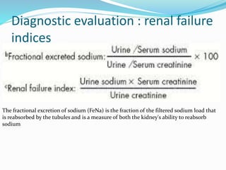


This document provides an overview of acute kidney injury (AKI), formerly known as acute renal failure. It discusses the definition and epidemiology of AKI and describes the main causes as pre-renal, intrinsic renal, and post-renal. Pre-renal AKI is the most common type and is caused by reduced renal blood flow. The document outlines the diagnostic evaluation, complications, treatment approaches including dialysis indications, and outcomes of AKI. It emphasizes the importance of identifying and eliminating nephrotoxic agents to optimize management of this condition.


