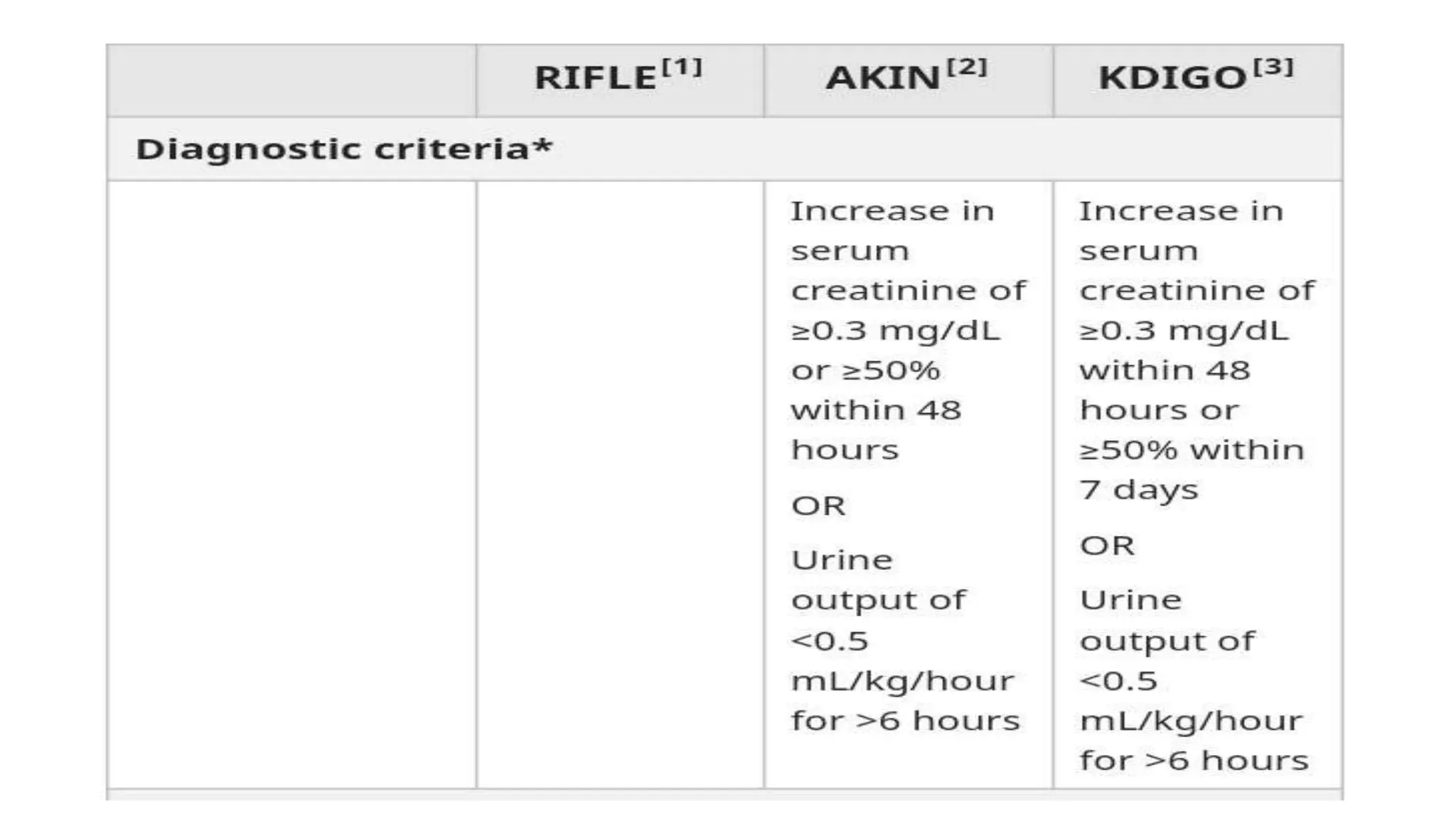
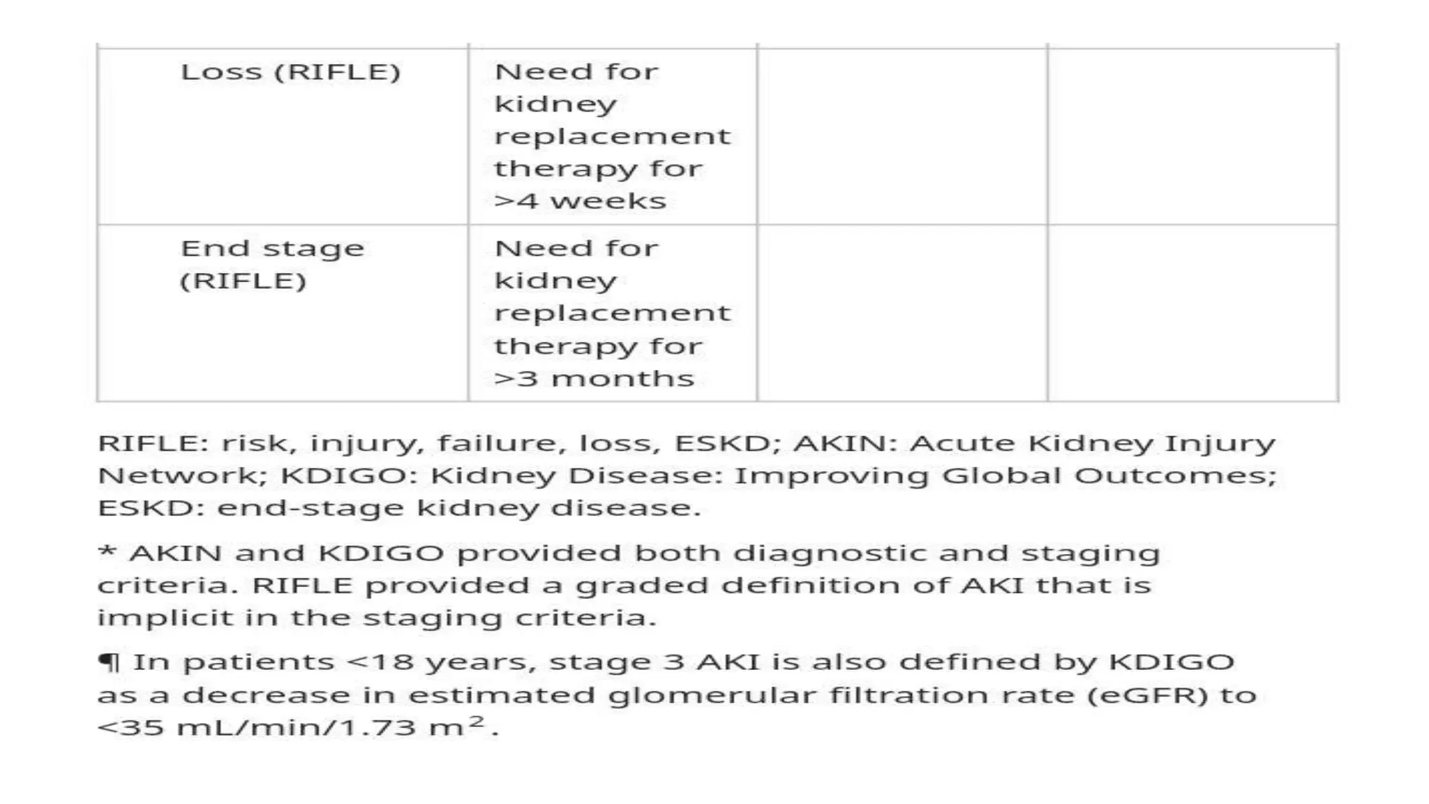
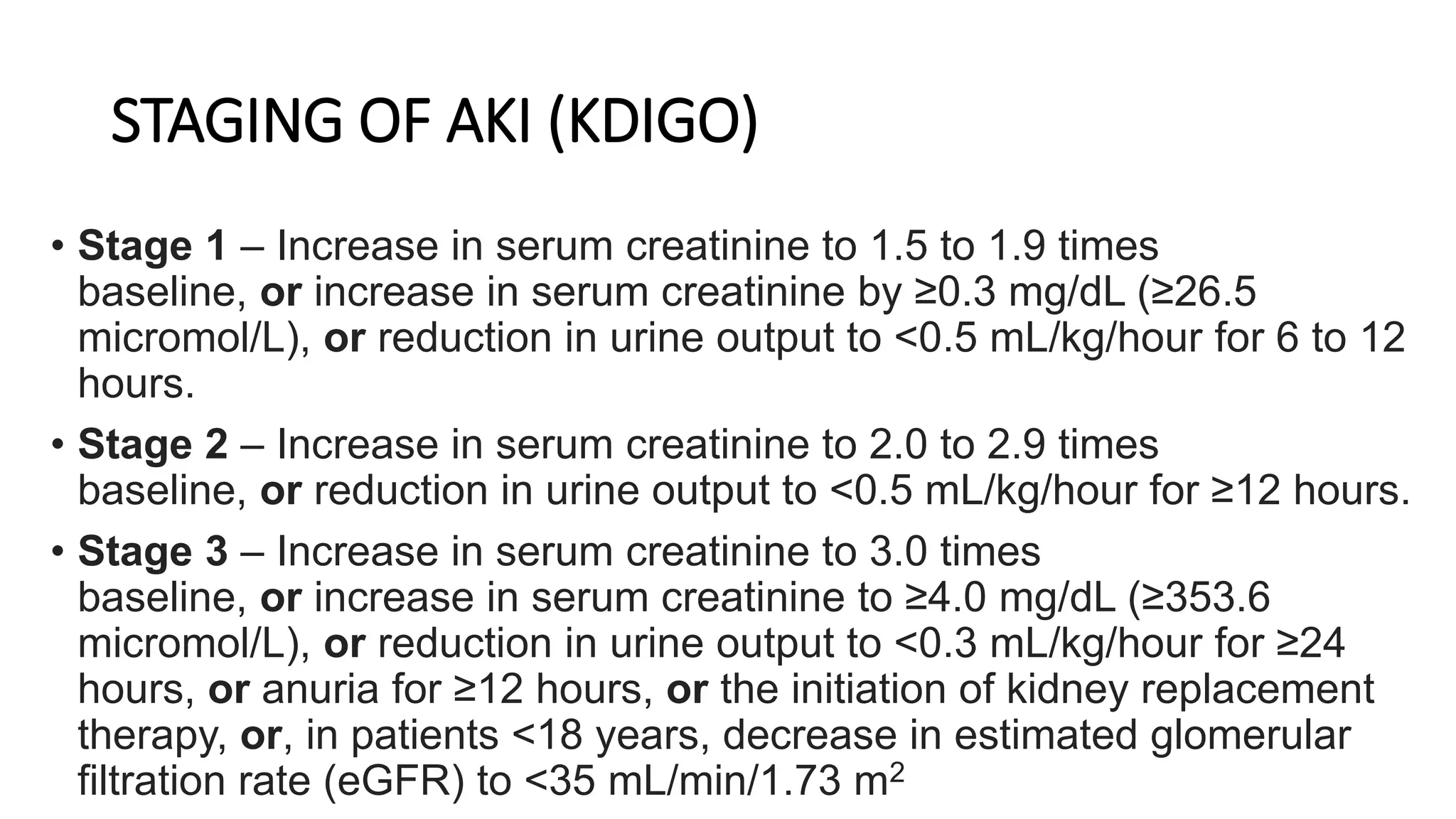
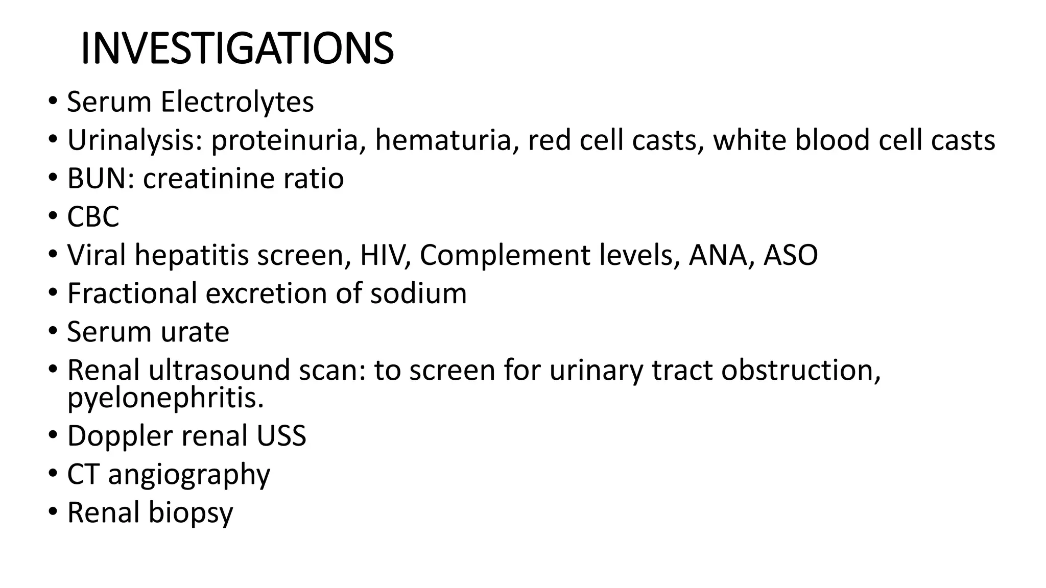
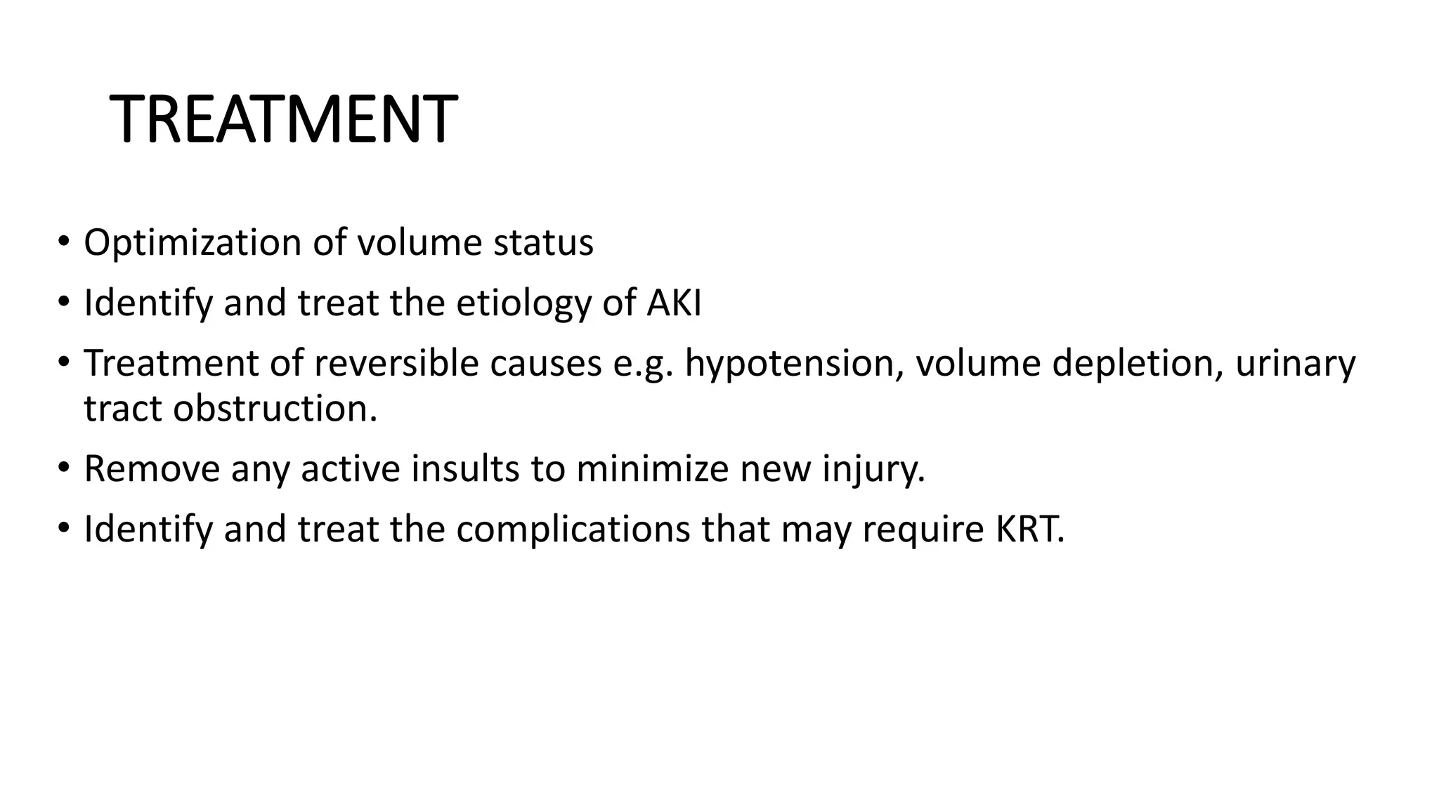
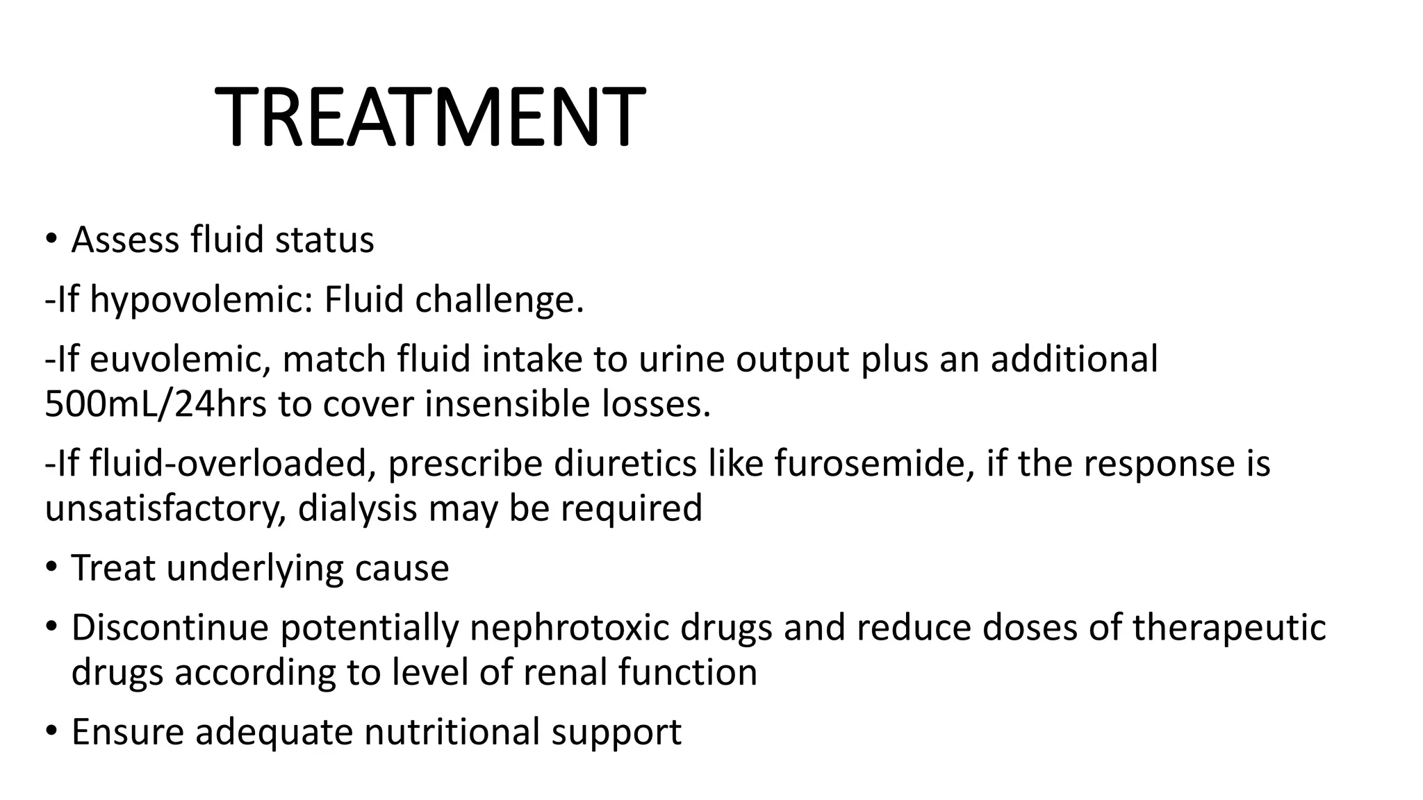
ACUTE KIDNEY INJURY
BY NAKATO SARAH
SUPERVISOR: DR.KIYINGI MICHEAL
OUTLINE
• Definition
• Epidemiology
• Causes
• Pathophysiology
• Clinical presentation
• Criteria and staging
• Investigations
• Management
• Complications
• Prognosis
• References
DEFINITION
• AKI refers to the sudden and often reversible loss of renal
function, which develops over days or weeks and is often
accompanied by a reduction in urine volume.
OR
• The abrupt decrease in kidney function, resulting in the
retention of urea and other nitrogenous waste products and in
the dysregulation of extracellular volume and electrolytes.
EPIDEMIOLOGY
•Estimates suggest that 13.3 million people worldwide are
affected by AKI annually, 85% of whom live in developing
countries.
•Further it is estimated that up to 1.7 million deaths occur
each year from AKI.
•Approximately 7% of all hospitalized patients and 20% of
acutely ill patients develop AKI.
CONTN
• In uncomplicated AKI, mortality is low, however when it is
associated with sepsis and multiple organ failure mortality is 50%–
70% and the outcome is usually determined by the severity of the
underlying disorder and other complications, rather than by kidney
injury itself
• Older patients are at higher risk of developing AKI and have a worse
outcome
• In Uganda, prevalence of AKI was 45.3% with 21.5% of children
having unresolved AKI at 24hours. AKI was more common in
Eastern Uganda (Ruth Namazzi et al, 2022)
PRE-RENAL CAUSES
1) volume depletion
• Renal losses - Diuretics, polyuria
• GI losses - Vomiting, diarrhea
• Cutaneous losses - Burns, Stevens-
Johnson syndrome
• Hemorrhage
• Pancreatitis
2) Decreased cardiac output
• Heart failure
• Pericardial effusion/tamponade
• Pulmonary embolus
• Acute myocardial infarction
• Severe valvular disease
3) Systemic vasodilation
• Sepsis
• Anaphylaxis
• Anesthetics
• Drug overdose
4) Afferent arteriolar
vasoconstriction
• Drugs - NSAIDs, amphotericin B,
calcineurin inhibitors,
norepinephrine, radiocontrast
agents
• Hepatorenal syndrome
INTRINSIC CAUSES
• ATN
• GN
• Interstitial nephritis
• Vascular causes
POST-RENAL CAUSES
• Ureteric obstruction - Stone disease, tumor, fibrosis, ligation
during pelvic surgery
• Bladder neck obstruction - Benign prostatic hyperplasia (BPH),
prostate cancer, neurogenic bladder, tricyclic antidepressants,
ganglion blockers, bladder tumor, stone disease,
hemorrhage/clot
• Urethral obstruction - Strictures, tumor, phimosis
CAUSES
PATHOPHYSIOLOGY
• Pre-renal; when perfusion to the kidney is reduced
• Renal; when the primary insult affects the kidney itself
• Post-renal; when there is obstruction to urine flow at any point from
the tubule to the urethra.
PRE-RENAL
• Pre-renal AKI results from a reduction in renal perfusion, typically due to a
reduction in systemic blood pressure.
• The drop in renal perfusion activates the renin–angiotensin–aldosterone
system, which promotes sodium retention in the kidney and systemic
vasoconstriction in order to restore blood pressure.
• Angiotensin also preferentially constricts the glomerular efferent arteriole,
while prostaglandins are released locally to vasodilate the afferent arteriole.
• The combined effect increases glomerular pressure to maintain GFR,
however if the blood pressure is very low, autoregulation fails and the GFR
falls.
• Importantly, in pre-renal AKI, the kidney is not damaged, therefore GFR can
improve rapidly if the renal perfusion is restored.
RENAL
• If the drop in renal perfusion is severe or sustained, pre-renal AKI may
progress to renal AKI as ischaemic injury causes ATN, the most extreme
example being with renal arterial or venous occlusion.
• Dead tubular cells may also be shed into the tubular lumen, leading to
tubular obstruction.
• While ischaemia is the most common cause of ATN in hospital, it may also
be caused by toxins and nephrotoxic drugs.
• Drugs can also cause allergic interstitial nephritis.
• The other common ‘renal’ cause of AKI is glomerulonephritis, in which there
is direct inflammatory damage to the glomeruli.
• COVID-19 infection is also associated with AKI in a significant proportion of
patients.
POST-RENAL
• Post-renal AKI occurs as the result of obstruction to the renal tract.
• This leads to elevation of intraluminal ureteral pressure transmitted to the
nephrons, with a subsequent fall in GFR.
• The obstruction needs to be bilateral to cause renal failure, therefore it is
unusual for renal stones to cause AKI, while obstruction of the bladder outlet
is a much more common cause.
• If the obstruction is not relieved, the low GFR is maintained by a drop in renal
perfusion via thromboxane A2 and angiotensin II.
• This leads to chronic renal injury over several weeks.
• If obstruction is reversed, the extent of recovery of renal function is
dependent on the duration of obstruction and the pre-morbid GFR
CLINICAL FEATURES
• Clinical features of AKI include short duration of symptoms, and an
absence of markers of CKD such as anemia, elevated PTH and small
kidneys observed on imaging.
PRE-RENAL
• Tachycardia
• Postural hypotension.
• Signs of poor peripheral perfusion, such as cold peripheries and delayed capillary
return.
• Weight loss
• Warm peripheries in the presence of hypotension may indicate sepsis
• Signs of dehydration
• Crush injuries, burns, abdominal surgeries
• Volume depletion (vomiting, diarrhea, burns, hemorrhage)
• Sepsis, Cardiac disease, Liver disease
• Drugs History (diuretics, ACE inhibitors, ARBs, NSAIDs, calcineurin inhibitors,
iodinated contrast)
RENAL
• Patients with glomerulonephritis demonstrate haematuria and proteinuria, and
may have clinical manifestations of an underlying disease, such as SLE or systemic
vasculitis.
• Drug history for example PPIs, NSAIDs and many antibiotics.
• Vascular disease is included here as diseases of the large and small renal vessels
typically present with hypertension and volume expansion, in contrast to the
volume depletion observed in pre-renal failure.
POST-RENAL
• Distended bladder
• DRE; Prostate
• Pelvic mass
CRITERIA AND STAGING
KDIGO ( KIDNEY DISEASE: IMPROVING GLOBAL OUTCOMES)
The KDIGO guidelines define AKI as follows:
• Increase in serum creatinine by ≥0.3 mg/dL (≥26.5 micromol/L) within
48 hours, or
• Increase in serum creatinine to ≥1.5 times baseline, which is known or
presumed to have occurred within the prior seven days, or
• Urine volume <0.5 mL/kg/hour for six hours
STAGING OF AKI (KDIGO)
• Stage 1 – Increase in serum creatinine to 1.5 to 1.9 times
baseline, or increase in serum creatinine by ≥0.3 mg/dL (≥26.5
micromol/L), or reduction in urine output to <0.5 mL/kg/hour for 6 to 12
hours.
• Stage 2 – Increase in serum creatinine to 2.0 to 2.9 times
baseline, or reduction in urine output to <0.5 mL/kg/hour for ≥12 hours.
• Stage 3 – Increase in serum creatinine to 3.0 times
baseline, or increase in serum creatinine to ≥4.0 mg/dL (≥353.6
micromol/L), or reduction in urine output to <0.3 mL/kg/hour for ≥24
hours, or anuria for ≥12 hours, or the initiation of kidney replacement
therapy, or, in patients <18 years, decrease in estimated glomerular
filtration rate (eGFR) to <35 mL/min/1.73 m2
INVESTIGATIONS
• Serum Electrolytes
• Urinalysis: proteinuria, hematuria, red cell casts, white blood cell casts
• BUN: creatinine ratio
• CBC
• Viral hepatitis screen, HIV, Complement levels, ANA, ASO
• Fractional excretion of sodium
• Serum urate
• Renal ultrasound scan: to screen for urinary tract obstruction,
pyelonephritis.
• Doppler renal USS
• CT angiography
• Renal biopsy
MANAGEMENT
• History taking
• Physical examination
• Investigations
• Treatment
Primary goals of treatment
• Maintenance of volume homeostasis
• Correction of biochemical abnormalities.
TREATMENT
• Optimization of volume status
• Identify and treat the etiology of AKI
• Treatment of reversible causes e.g. hypotension, volume depletion, urinary
tract obstruction.
• Remove any active insults to minimize new injury.
• Identify and treat the complications that may require KRT.
TREATMENT
• Assess fluid status
-If hypovolemic: Fluid challenge.
-If euvolemic, match fluid intake to urine output plus an additional
500mL/24hrs to cover insensible losses.
-If fluid-overloaded, prescribe diuretics like furosemide, if the response is
unsatisfactory, dialysis may be required
• Treat underlying cause
• Discontinue potentially nephrotoxic drugs and reduce doses of therapeutic
drugs according to level of renal function
• Ensure adequate nutritional support
CONTN
• If K+>6.5mmol/L and ECG changes of hyperkalemia are present:
Administer calcium gluconate to stabilize myocardium
Decrease intake of potassium in diet.
Lower potassium by oral potassium exchange resin to prevent potassium absorption
Administering intravenous glucose/insulin, sodium bicarbonate, and beta agonists to
move potassium intracellularly.
• These are holding measures until a definitive method of removing potassium is achieved
(restoration of renal function or dialysis).
• Screen for intercurrent infections and treat promptly if present
• Vasodilators??
INDICATIONS FOR URGENT KRT/RRT
• Fluid overload e.g. Pulmonary edema
• Hyperkalemia >6.5mEq/L or associated with signs and symptoms
e.g. cardiac, muscle weakness. Or hyperkalemia >5.5mEq/L if
there is ongoing tissue breakdown e.g. rhabdomyolysis.
• Signs of uremia e.g. pericarditis, mental status change.
• Severe metabolic acidosis pH>7.1 with hypervolemia.
• Acute poisoning e.g. methanol, ethylene glycol, lithium.
AKI IN OLD AGE
• Physiological change: Nephrons decline in number with age and average GFR
falls progressively, so many older patients will have established CKD and less
functional reserve. Small acute declines in renal function may therefore have a
significant impact.
• Creatinine: As muscle mass falls with age, less creatinine is produced each day.
Serum creatinine can therefore underestimate the severity of renal failure.
• Renal tubular function: Declines with age, leading to loss of urinary
• concentrating ability.
• Drugs: Increased drug prescription in older people (diuretics, ACE inhibitors
and NSAIDs) may contribute to the risk of AKI.
• Causes: infection, renal vascular disease, prostatic obstruction, myeloma and
severe cardiac dysfunction are common.
• Mortality: rises with age, primarily because of comorbid conditions.
COMPLICATIONS OF AKI
• Volume overload recognized by Pulmonary edema, HTN,
raised JVP
• Electrolyte disturbances
• Metabolic acidosis
• Neurological complications
• Chronic kidney disease
PROGNOSIS
REFERENCES
• Davidsons Principles and Practice of Medicine, 24th edition.
• UpToDate, 2023
• Medscape,2022

AKI.pptx

  • 1.
    ACUTE KIDNEY INJURY BYNAKATO SARAH SUPERVISOR: DR.KIYINGI MICHEAL
  • 2.
    OUTLINE • Definition • Epidemiology •Causes • Pathophysiology • Clinical presentation • Criteria and staging • Investigations • Management • Complications • Prognosis • References
  • 3.
    DEFINITION • AKI refersto the sudden and often reversible loss of renal function, which develops over days or weeks and is often accompanied by a reduction in urine volume. OR • The abrupt decrease in kidney function, resulting in the retention of urea and other nitrogenous waste products and in the dysregulation of extracellular volume and electrolytes.
  • 4.
    EPIDEMIOLOGY •Estimates suggest that13.3 million people worldwide are affected by AKI annually, 85% of whom live in developing countries. •Further it is estimated that up to 1.7 million deaths occur each year from AKI. •Approximately 7% of all hospitalized patients and 20% of acutely ill patients develop AKI.
  • 5.
    CONTN • In uncomplicatedAKI, mortality is low, however when it is associated with sepsis and multiple organ failure mortality is 50%– 70% and the outcome is usually determined by the severity of the underlying disorder and other complications, rather than by kidney injury itself • Older patients are at higher risk of developing AKI and have a worse outcome • In Uganda, prevalence of AKI was 45.3% with 21.5% of children having unresolved AKI at 24hours. AKI was more common in Eastern Uganda (Ruth Namazzi et al, 2022)
  • 6.
    PRE-RENAL CAUSES 1) volumedepletion • Renal losses - Diuretics, polyuria • GI losses - Vomiting, diarrhea • Cutaneous losses - Burns, Stevens- Johnson syndrome • Hemorrhage • Pancreatitis 2) Decreased cardiac output • Heart failure • Pericardial effusion/tamponade • Pulmonary embolus • Acute myocardial infarction • Severe valvular disease 3) Systemic vasodilation • Sepsis • Anaphylaxis • Anesthetics • Drug overdose 4) Afferent arteriolar vasoconstriction • Drugs - NSAIDs, amphotericin B, calcineurin inhibitors, norepinephrine, radiocontrast agents • Hepatorenal syndrome
  • 7.
    INTRINSIC CAUSES • ATN •GN • Interstitial nephritis • Vascular causes
  • 8.
    POST-RENAL CAUSES • Uretericobstruction - Stone disease, tumor, fibrosis, ligation during pelvic surgery • Bladder neck obstruction - Benign prostatic hyperplasia (BPH), prostate cancer, neurogenic bladder, tricyclic antidepressants, ganglion blockers, bladder tumor, stone disease, hemorrhage/clot • Urethral obstruction - Strictures, tumor, phimosis
  • 9.
  • 10.
    PATHOPHYSIOLOGY • Pre-renal; whenperfusion to the kidney is reduced • Renal; when the primary insult affects the kidney itself • Post-renal; when there is obstruction to urine flow at any point from the tubule to the urethra.
  • 11.
    PRE-RENAL • Pre-renal AKIresults from a reduction in renal perfusion, typically due to a reduction in systemic blood pressure. • The drop in renal perfusion activates the renin–angiotensin–aldosterone system, which promotes sodium retention in the kidney and systemic vasoconstriction in order to restore blood pressure. • Angiotensin also preferentially constricts the glomerular efferent arteriole, while prostaglandins are released locally to vasodilate the afferent arteriole. • The combined effect increases glomerular pressure to maintain GFR, however if the blood pressure is very low, autoregulation fails and the GFR falls. • Importantly, in pre-renal AKI, the kidney is not damaged, therefore GFR can improve rapidly if the renal perfusion is restored.
  • 13.
    RENAL • If thedrop in renal perfusion is severe or sustained, pre-renal AKI may progress to renal AKI as ischaemic injury causes ATN, the most extreme example being with renal arterial or venous occlusion. • Dead tubular cells may also be shed into the tubular lumen, leading to tubular obstruction. • While ischaemia is the most common cause of ATN in hospital, it may also be caused by toxins and nephrotoxic drugs. • Drugs can also cause allergic interstitial nephritis. • The other common ‘renal’ cause of AKI is glomerulonephritis, in which there is direct inflammatory damage to the glomeruli. • COVID-19 infection is also associated with AKI in a significant proportion of patients.
  • 14.
    POST-RENAL • Post-renal AKIoccurs as the result of obstruction to the renal tract. • This leads to elevation of intraluminal ureteral pressure transmitted to the nephrons, with a subsequent fall in GFR. • The obstruction needs to be bilateral to cause renal failure, therefore it is unusual for renal stones to cause AKI, while obstruction of the bladder outlet is a much more common cause. • If the obstruction is not relieved, the low GFR is maintained by a drop in renal perfusion via thromboxane A2 and angiotensin II. • This leads to chronic renal injury over several weeks. • If obstruction is reversed, the extent of recovery of renal function is dependent on the duration of obstruction and the pre-morbid GFR
  • 15.
    CLINICAL FEATURES • Clinicalfeatures of AKI include short duration of symptoms, and an absence of markers of CKD such as anemia, elevated PTH and small kidneys observed on imaging.
  • 16.
    PRE-RENAL • Tachycardia • Posturalhypotension. • Signs of poor peripheral perfusion, such as cold peripheries and delayed capillary return. • Weight loss • Warm peripheries in the presence of hypotension may indicate sepsis • Signs of dehydration • Crush injuries, burns, abdominal surgeries • Volume depletion (vomiting, diarrhea, burns, hemorrhage) • Sepsis, Cardiac disease, Liver disease • Drugs History (diuretics, ACE inhibitors, ARBs, NSAIDs, calcineurin inhibitors, iodinated contrast)
  • 17.
    RENAL • Patients withglomerulonephritis demonstrate haematuria and proteinuria, and may have clinical manifestations of an underlying disease, such as SLE or systemic vasculitis. • Drug history for example PPIs, NSAIDs and many antibiotics. • Vascular disease is included here as diseases of the large and small renal vessels typically present with hypertension and volume expansion, in contrast to the volume depletion observed in pre-renal failure. POST-RENAL • Distended bladder • DRE; Prostate • Pelvic mass
  • 18.
    CRITERIA AND STAGING KDIGO( KIDNEY DISEASE: IMPROVING GLOBAL OUTCOMES) The KDIGO guidelines define AKI as follows: • Increase in serum creatinine by ≥0.3 mg/dL (≥26.5 micromol/L) within 48 hours, or • Increase in serum creatinine to ≥1.5 times baseline, which is known or presumed to have occurred within the prior seven days, or • Urine volume <0.5 mL/kg/hour for six hours
  • 23.
    STAGING OF AKI(KDIGO) • Stage 1 – Increase in serum creatinine to 1.5 to 1.9 times baseline, or increase in serum creatinine by ≥0.3 mg/dL (≥26.5 micromol/L), or reduction in urine output to <0.5 mL/kg/hour for 6 to 12 hours. • Stage 2 – Increase in serum creatinine to 2.0 to 2.9 times baseline, or reduction in urine output to <0.5 mL/kg/hour for ≥12 hours. • Stage 3 – Increase in serum creatinine to 3.0 times baseline, or increase in serum creatinine to ≥4.0 mg/dL (≥353.6 micromol/L), or reduction in urine output to <0.3 mL/kg/hour for ≥24 hours, or anuria for ≥12 hours, or the initiation of kidney replacement therapy, or, in patients <18 years, decrease in estimated glomerular filtration rate (eGFR) to <35 mL/min/1.73 m2
  • 24.
    INVESTIGATIONS • Serum Electrolytes •Urinalysis: proteinuria, hematuria, red cell casts, white blood cell casts • BUN: creatinine ratio • CBC • Viral hepatitis screen, HIV, Complement levels, ANA, ASO • Fractional excretion of sodium • Serum urate • Renal ultrasound scan: to screen for urinary tract obstruction, pyelonephritis. • Doppler renal USS • CT angiography • Renal biopsy
  • 25.
    MANAGEMENT • History taking •Physical examination • Investigations • Treatment Primary goals of treatment • Maintenance of volume homeostasis • Correction of biochemical abnormalities.
  • 26.
    TREATMENT • Optimization ofvolume status • Identify and treat the etiology of AKI • Treatment of reversible causes e.g. hypotension, volume depletion, urinary tract obstruction. • Remove any active insults to minimize new injury. • Identify and treat the complications that may require KRT.
  • 27.
    TREATMENT • Assess fluidstatus -If hypovolemic: Fluid challenge. -If euvolemic, match fluid intake to urine output plus an additional 500mL/24hrs to cover insensible losses. -If fluid-overloaded, prescribe diuretics like furosemide, if the response is unsatisfactory, dialysis may be required • Treat underlying cause • Discontinue potentially nephrotoxic drugs and reduce doses of therapeutic drugs according to level of renal function • Ensure adequate nutritional support
  • 28.
    CONTN • If K+>6.5mmol/Land ECG changes of hyperkalemia are present: Administer calcium gluconate to stabilize myocardium Decrease intake of potassium in diet. Lower potassium by oral potassium exchange resin to prevent potassium absorption Administering intravenous glucose/insulin, sodium bicarbonate, and beta agonists to move potassium intracellularly. • These are holding measures until a definitive method of removing potassium is achieved (restoration of renal function or dialysis). • Screen for intercurrent infections and treat promptly if present • Vasodilators??
  • 29.
    INDICATIONS FOR URGENTKRT/RRT • Fluid overload e.g. Pulmonary edema • Hyperkalemia >6.5mEq/L or associated with signs and symptoms e.g. cardiac, muscle weakness. Or hyperkalemia >5.5mEq/L if there is ongoing tissue breakdown e.g. rhabdomyolysis. • Signs of uremia e.g. pericarditis, mental status change. • Severe metabolic acidosis pH>7.1 with hypervolemia. • Acute poisoning e.g. methanol, ethylene glycol, lithium.
  • 30.
    AKI IN OLDAGE • Physiological change: Nephrons decline in number with age and average GFR falls progressively, so many older patients will have established CKD and less functional reserve. Small acute declines in renal function may therefore have a significant impact. • Creatinine: As muscle mass falls with age, less creatinine is produced each day. Serum creatinine can therefore underestimate the severity of renal failure. • Renal tubular function: Declines with age, leading to loss of urinary • concentrating ability. • Drugs: Increased drug prescription in older people (diuretics, ACE inhibitors and NSAIDs) may contribute to the risk of AKI. • Causes: infection, renal vascular disease, prostatic obstruction, myeloma and severe cardiac dysfunction are common. • Mortality: rises with age, primarily because of comorbid conditions.
  • 31.
    COMPLICATIONS OF AKI •Volume overload recognized by Pulmonary edema, HTN, raised JVP • Electrolyte disturbances • Metabolic acidosis • Neurological complications • Chronic kidney disease
  • 32.
  • 33.
    REFERENCES • Davidsons Principlesand Practice of Medicine, 24th edition. • UpToDate, 2023 • Medscape,2022