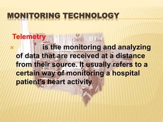
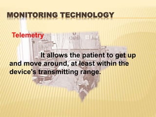
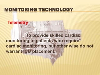
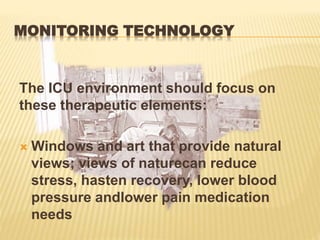
Telemetry allows for remote cardiac monitoring of patients who do not require intensive care unit placement but still need monitoring. It transmits data from a patient's heart to monitoring staff while allowing the patient mobility. An ideal intensive care unit environment focuses on reducing stress for patients through access to natural light, views, family involvement, and other therapeutic elements. Laboratory tests aim to be precise, accurate, sensitive and specific to reliably determine medical conditions and distinguish those with a condition from those without.
Telemetry monitoring allows cardiac patients to move freely while their heart is monitored. It is used for patients who need continuous EKG monitoring but do not require intensive care. A diagnostic information system can aggregate over 5,000 different patient test results into a standardized, easy to read format. This increases efficiency and accuracy of patient care. Mechanical ventilators deliver gas into a patient's airways to support breathing. Modes include time cycled, volume cycled and flow cycled ventilation. Intensive care units aim to reduce stress and promote recovery through factors like natural lighting, family involvement and reduced noise.
The document discusses various types of biomedical equipment used in medical settings. It defines medical equipment as devices designed to aid in diagnosis, monitoring, or treatment of medical conditions. It then proceeds to describe several pieces of equipment in 1-2 paragraphs each, including their purposes and basic functions. The equipment discussed includes cervical traction, syringe drives, pulse oximetry, electrosurgical units, laser microtomes, anemostats, cardiopulmonary bypass machines, aesthesiometers, home ultrasounds, nasogastric intubation, lung counters, dermatomes, Hirtz compasses, rhinoscopes, and plethysmographs.
Hybrid operating rooms (ORs) allow both minimally invasive surgeries and open procedures to be performed using imaging guidance in the same room. This enables more patients to undergo less invasive surgeries. Key considerations in designing a hybrid OR include determining the imaging system and equipment needed based on planned procedures, allocating sufficient space, and ensuring staff are properly trained on the new environment and equipment. Common hybrid ORs feature angiography, CT, or MRI systems to provide real-time imaging guidance during surgeries.
This document provides an overview of biomedical instrumentation. It discusses key topics such as:
- The development of biomedical instrumentation from early devices like the electrocardiograph to modern advances enabled by surplus electronics after WWII.
- Key considerations for designing medical instrumentation systems, including range, sensitivity, linearity, and frequency response.
- Components of the man-instrument system including the subject, stimuli, transducers, signal conditioning equipment, and displays.
- Objectives of instrumentation systems like information gathering, diagnosis, evaluation, monitoring and control.
- Biometrics as the measurement of physiological variables and parameters that biomedical instrumentation provides tools to measure.
This document provides an overview of biomedical instrumentation. It discusses how instrumentation is used to monitor and control process variables for measurement and control. Biomedical instrumentation specifically creates instruments to measure, record, and transmit data to and from the body. Some key types of biomedical instrumentation systems are direct/indirect, invasive/noninvasive, contact/remote for sensing and actuating in real-time or statically. Several important instruments are discussed in detail, including X-rays, electrocardiography, magnetic resonance imaging, ultrasound, and computed tomography. The document outlines the basic workings, advantages, and disadvantages of these key biomedical instruments.
The document discusses guidelines for organizing a critical care unit. Some key points covered are:
- A critical care unit should have 6-14 beds and be divided into pods of 10-15 beds each with dedicated staff.
- The unit should be located with easy access to emergency, operating rooms, and diagnostic departments. It should have sufficient space for patient transfer and equipment.
- Recommended floor space is 125-150 square feet per patient or 300 square feet for private rooms. Additional space is needed for staff areas.
- Essential equipment includes ventilators, monitors, infusion pumps, defibrillators, beds, and supplies for patient care and medication storage. Staff must be trained on equipment use and maintenance
Advanced Cardiovascular Surgery Hybrid Operating Room (Nuts & Bolts)Jacques Kpodonu,MD
1) The document discusses the key factors for building an advanced cardiovascular hybrid operating room, including clinical requirement assessment, planning, project management, and partner management.
2) Some of the main considerations for a hybrid OR layout are ensuring adequate space for imaging equipment, surgical equipment, anesthesia equipment, and radiation shielding. Workflow for medical staff and patients must also be considered.
3) Building a hybrid OR requires defining roles and responsibilities, deciding on equipment, and iterative planning and verification between all involved parties to ensure needs are met. Total investment for a hybrid OR project can be over $6 million.
The document provides an overview of medical equipment used at Columbia Asia Hospital in Kolkata, India. It describes equipment in various departments like radiology, cardiology, ICU, NICU, OT, cath lab, dialysis and gastro. For each major piece of equipment, it lists the manufacturer and some key features. It also details some major and minor breakdowns of equipment during the training period and how they were addressed.
Apollo Hospitals Group opens a new 400-bed multi-specialty hospital in Ahmedabad, Gujarat, strengthening healthcare infrastructure in the state. The hospital offers a wide range of diagnostic services and specialties, including cardiology, neurology, orthopedics, oncology and more. It aims to provide quality healthcare and raise standards in Gujarat through specialized treatment and state-of-the-art technology.
The document discusses computed tomography (CT) of the chest and protocols for performing chest CT scans. It provides details on how chest CT is used to examine abnormalities found on other imaging tests and help diagnose conditions causing chest symptoms. It describes the CT scanning process and equipment. Common uses of chest CT are outlined, along with lung disorders it can demonstrate and benefits compared to other imaging modalities. Specific protocols for routine chest CT, high-resolution CT, low-dose CT, airway CT, and aortic angiography CT are enumerated.
Ultrasound Guided Procedures
www.mskus.training
MSKUS Hands-on Scanning,
MSK US Training is a corporation specializing in Musculoskeletal Ultrasonography (MSK US) Imaging Contracted Clinical MSK US & Training, providing workshops at participants location. This is a unique modality that can enhance your patient care needs. It is technically demanding and is completely operator dependent. Extensive experience and proper training are essential to performing consistent, high-quality examinations. Focused MSK US hands-on scanning applications are important to the development of proficient MSK US diagnosis. Our highly organized & systematic hands-on scanning format of instruction, in conjunction with protocol scanning images, ensures the participants develop skills
1) The study evaluated the utility of the balloon-assisted technique (BAT) versus conventional techniques (CT) for trans catheter closure of very large atrial septal defects (ASDs) ≥35 mm.
2) BAT had a higher success rate of 90.3% compared to 16.6% for CT. Posterior malalignment of the septum was associated with failure of CT.
3) BAT helps facilitate controlled delivery and alignment of the closure device in very large ASDs with challenging anatomical features like posterior malalignment and is often useful when CT fails.
1) Procedural sedation is used for many medical procedures and aims to provide analgesia, amnesia, and reduce anxiety while maintaining airway reflexes and spontaneous breathing.
2) While pulse oximetry became standard in the 1980s, capnography has emerged as the new gold standard for monitoring procedural sedation as it can detect respiratory issues that oximetry may miss.
3) Overlake Hospital developed a new monitoring protocol using capnography based on evidence that it improves patient safety during procedural sedation. They saw reduced need for respiratory therapist intervention after implementing routine capnography use.
Echocardiographic guidance is critical for procedural success of paravalvular leak closure. Transesophageal echocardiography (TEE) and particularly three-dimensional echocardiography represent the gold standards. Fusion imaging provides real-time integration of three-dimensional echocardiography and X-ray fluoroscopy and can further facilitate spatial orientation, wire placement and device deployment. Intracardiac echocardiography (ICE) is a secondary approach possibly beneficial in selected cases.
Ambulatory Anesthesia and Non–Operating Room Anesthesia (NORA)Saeid Safari
GUIDELINES FOR AMBULATORY ANESTHESIA AND SURGERY (American Society of Anesthesiologists)
GUIDELINES FOR AMBULATORY ANESTHESIA AND SURGERY
Non–Operating Room Anesthesia (NORA)
Organization and guidelines for angloitalian meeting roma 010 on non operati...Claudio Melloni
This document provides guidelines and requirements for non-operating room anesthesia (NORA) and office-based anesthesia. It discusses considerations for patient characteristics, pre-anesthetic preparation, appropriate locations and equipment, monitoring during procedures, and specific requirements for accreditation of ambulatory surgery centers. Key points include the need for adherence to basic standards of care, special precautions for higher-risk patients, adequate facilities and equipment, interdepartmental cooperation, and understanding of procedural requirements.
Computers have become integral tools in the medical field, helping doctors discover, test, and apply new techniques while also providing an infrastructure to share medical knowledge globally. Computers are used extensively in areas like CT scans, heart rate monitoring, surgery, research, and spectroscopy, creating new opportunities to save lives and advance human health. In particular, technologies like CT scans and MRI machines use computer processing to generate detailed images of the inside of the body.
The LUCAS 2 is a mechanical chest compression device that provides consistent, high-quality CPR according to AHA guidelines. It can be used by first responders, medical professionals, and in pre-hospital and in-hospital settings for patients in cardiac arrest. The LUCAS 2 compresses the chest at a rate of 102 times per minute and a depth of 2.1 inches, allowing for full chest recoil between compressions. While it increases perfusion and standardizes compressions, there can be delays in application and it may not fit all patient sizes.
Monitoring is essential in any kind of medical practice. It is the observation of disease, condition and several other parameters over time. Usually a medical monitor is used for continuously measuring vital signs.
Computers play an important role in veterinary surgery by assisting with diagnosis, surgical planning and guidance, patient management, and other applications. They allow virtual simulations that can replace animal testing and provide educational opportunities. Computers also help manage patient data and records in veterinary hospitals. Advanced imaging technologies like CT scans, MRIs, ultrasounds, and digital radiography integrate computer processing and allow veterinarians to non-invasively visualize internal structures. New computer-assisted techniques like laparoscopic surgery, robotics, and natural orifice procedures further aid veterinary specialists.
Vibra Hospital of Southeastern Massachusetts has decreased the average time patients spend on ventilators by 29% by using volumetric capnography technology. Volumetric capnography continuously monitors exhaled carbon dioxide levels, allowing clinicians to optimize ventilator settings and assess patient status during weaning in real-time. This helps clinicians safely decrease ventilator dependency and differentiate between respiratory and other issues during weaning. The hospital also uses volumetric capnography to help COPD patients transition between ventilator, high-flow oxygen, and weaning.
This document discusses the digital operating room (DOR) and related technologies. It describes 5 levels of DOR maturity from 2005 to 2025, moving from vendor-specific integration to an intelligent infrastructure. A key goal is optimizing intra-operative processes through standards like DICOM and IHE profiles for surgery. Patient and process modeling aims to integrate multimodal data for personalized, evidence-based care. Standards and integrated models may facilitate knowledge discovery from surgical data, situations and workflows.
Anesthesia carries risks that can lead to patient death or injury. Several factors contribute to risks in the operating room including equipment issues, patient health factors, human performance errors, and system failures. Some key errors that can cause severe harm are airway issues, medication errors, and procedure mistakes. Maintaining vigilance, checklists, standards, training, and learning from adverse events can help improve safety. Thorough documentation and review of incidents is important for quality assurance.
Microelectromechanical systems (MEMS) have been used in the medical industry since the 1980s. They provide advantages over other technologies as they are small, reliable, inexpensive to manufacture, and stable. MEMS pressure sensors, accelerometers, and custom microstructures have a variety of applications including monitoring blood pressure, respiration, eye surgery equipment, hospital beds, dialysis machines, and drug delivery systems.
The document discusses critical care nursing. It defines critical care as care for extremely ill patients whose condition is unstable or potentially unstable. Critical care units provide comprehensive care for critically ill patients deemed recoverable. Critical care nursing involves specialized nursing care for patients with life-threatening conditions. The document outlines the design of critical care units, including bed size and spacing, environmental factors, and staffing needs. It also discusses the roles and responsibilities of critical care nurses in caring for critically ill patients.
This document discusses ambulatory and fast track anesthesia. It covers topics such as the benefits of ambulatory surgery including lower costs and greater efficiency. It describes different facility designs for ambulatory surgery and lists many common procedures that can be done on an outpatient basis. The document outlines considerations for patient selection and preoperative preparation including pharmacologic and non-pharmacologic techniques. It also discusses various anesthetic techniques for ambulatory surgery like general anesthesia, regional anesthesia, and monitored anesthesia care. Fast tracking approaches to minimize side effects like PONV are also summarized.
The document outlines the technology code of ethics for North Muskegon Public Schools. It states that use of technology is a privilege to enhance learning, but with access comes responsibility for equipment and information. The code gives users rights to use authorized hardware, software, and access outside resources to facilitate learning. It also lists responsibilities for appropriate use including maintaining privacy, seeking permission, and adhering to network etiquette. Violations will result in disciplinary actions such as losing computer privileges.
This document provides tips for creating effective PowerPoint presentations. It recommends brainstorming the audience and message before designing slides. Each slide should focus on a single idea to avoid confusion. Slides should be ordered logically and include photos to illustrate concepts. Appropriate animation, sounds, and fonts should be used to match the mood. Color also impacts mood - red can evoke emotion while yellow grabs attention and green creates comfort. Dark blue feels mature and white conveys sincerity. Black is not recommended as a background color. Proper use of these design elements can help relay the intended message to audiences.
Patient X, a 58-year-old grocery store manager, was experiencing abdominal pain several nights a week and occasional discomfort in the afternoon. An endoscopy revealed a peptic ulcer and infection with Helicobacter pylori bacteria. He was prescribed medication to reduce stomach acid and instructed to return for another endoscopy in 6 months. Peptic ulcer disease is common in the Philippines and a leading cause of death, especially among those with poor lifestyles. The duodenum is responsible for digesting food using enzymes secreted by the pancreas and bile from the liver and gallbladder.
An acute myocardial infarction (MI), commonly known as a heart attack, occurs when blood flow to the heart is blocked, injuring the heart muscle. Risk factors include previous cardiovascular disease, older age, smoking, high cholesterol, diabetes, high blood pressure, obesity, and chronic kidney disease. Treatment involves restoring blood flow through procedures like angioplasty or thrombolysis, along with medications like aspirin, nitroglycerin, and statins. Recovery involves cardiac rehabilitation with exercise training and lifestyle changes to manage risk factors and prevent future heart attacks.
This document provides information on cardiac failure or congestive heart failure (CHF). CHF occurs when the heart muscle is too weak or stiff to pump blood efficiently. As a result, blood moves through the heart and body more slowly and pressure in the heart increases. The heart cannot pump enough oxygen and nutrients to meet the body's needs. Risk factors include hypertension, diabetes, dyslipidemia, coronary artery disease, and sleep disorders. Diagnosis involves physical exam, blood tests, chest x-ray, echocardiogram, and other cardiac tests. Treatment focuses on managing symptoms through lifestyle changes, medications like ACE inhibitors, beta blockers, diuretics, and devices or procedures for severe cases. Nursing care addresses
The document outlines the technology code of ethics for North Muskegon Public Schools. It establishes that use of technology is a privilege to enhance learning, and with that privilege comes responsibility. It details rights and responsibilities for appropriate use of hardware, software, internet, and printers. Violations will result in disciplinary actions, with consequences increasing for repeated offenses such as loss of privileges. Users must sign a declaration agreeing to abide by the code of ethics.
The document outlines the technology code of ethics for North Muskegon Public Schools. It states that use of technology is a privilege to enhance learning, but with access comes responsibility. It details rights and responsibilities for users, including only using technology for educational purposes, properly caring for equipment, and adhering to network etiquette. Violations will result in disciplinary action, such as losing computer privileges. Users must sign a declaration agreeing to the code of ethics before using school technology resources.
The physical assessment examines the head, scalp, hair, face, eyelids, eyes, ears, mouth, neck, skin, nails, upper extremities, lower extremities, thoracic cavity, and abdomen. For each area, normal findings and actual findings are described and analyzed. The assessment found abnormalities in the skin, with black and blue areas on the lower back, and the abdomen, which was distended with tenderness on palpation. Otherwise, the physical exam was normal with no alterations noted.
Acute pulmonary failure occurs when abnormal gas exchange results from dysfunction of the respiratory system that threatens life. Risk factors include smoking, infections, lung disease, and neurological or muscular disorders. Symptoms include altered mental status, cyanosis, respiratory distress, and abnormal blood gases. Treatment focuses on stabilizing the patient, treating the underlying cause, and supporting respiratory function through oxygen, ventilation, suctioning, and positioning. Nursing care aims to maintain the airway, enhance gas exchange and nutrition, prevent complications, and provide education to the patient.
Acute renal failure occurs when the kidneys are unable to excrete waste products from the body, causing them to accumulate in the blood. It can be caused by conditions that decrease renal blood flow or damage the kidneys. Patients are classified as oliguric, excreting less than 500mL of urine per day, or nonoliguric, excreting more than 500mL daily. Risk factors include advanced age, diabetes, heart or liver disease. Diagnosis involves urine and blood tests to check kidney function and imaging tests in some cases. Treatment focuses on treating the underlying cause, managing fluid balance and electrolytes, and dialysis in severe cases.
GSM technology is used to monitor the different parameters of an ICU patient remotely and also control over medicine dosage is provided. Measurements of vital signs and behavioral patterns can be translated into accurate predictors of health risk ,even at an early stage and can be combined with alarm triggering systems in order to initiate the appropriate actions. The conventional methods including wet adhesive Ag/AgCl electrodes for HR and HRV, the capnograph device for respiratory status and pulse oximetry for oxyhemoglobin saturation provide excellent signals but are expensive, troublesome and inconvenient. A method to monitor physiological information based on GSM offers a new means for health monitoring. In this paper, we review the latest developments in monitoring and discuss the challenges and future directions for this field.
This document summarizes a remote health monitoring system using wearable body sensors to monitor cardiovascular disease patients. The system consists of three parts: 1) Wearable body sensors that collect physiological data from patients, 2) A personal server (PDA) that prioritizes and transfers data to 3) A medical server connected to the cloud where data can be accessed by medical staff. The system aims to efficiently respond to emergencies by prioritizing vital sign data and notifying medical staff of changes in a patient's heart health.
All medical imaging equipment manufactured today is supposed to conform to the DICOM standards. Viewing of the images thus produced cannot be done by ordinary imaging programs available on a regular PC. A special diagnostic medical imaging program is required, known as a DICOM workstation. For commercial use in medical diagnosis, such diagnostic medical imaging programs need to be FDA approved and need a special license. These measures ensure that any application developed for clinical purposes is capable of accurate depiction of high quality medical images.
Computers have revolutionized the field of medicine and patient treatment in several key ways. Computers now allow doctors to remotely monitor patients' vital signs, perform minimally invasive robotic surgeries, and communicate digitally with other medical experts. Hospitals also utilize electronic health records, clinical databases, simulations, and medical imaging controlled by computers to improve diagnosis, treatment outcomes, and overall patient care. Computers have become an essential tool for modern medicine.
LECTUREThe Impact of Technology on Clinical and IT SystemsIn.docxsmile790243
LECTURE
The Impact of Technology on Clinical and IT Systems
Introduction
One of the factors driving change in the health care delivery system is the rapidly evolving technology that emerges from research and development. Emerging technologies create rapid and profound change in the delivery system and may have drastic financial impacts. However, adapting new technologies without a clear understanding of what they can do for and to the system is never a good idea. They must be evaluated for their abilities to enhance the quality of care, along with their capacity to drive new revenue in a procedure-based delivery system. Finally, the cost of new technology is highly correlated with how new it is, and whether it is a stand-alone product with no competition. All of these factors combine to make it essential to do careful business and clinical analyses prior to committing to even the most appealing new technology.
In this module, we will examine two types of new technology: clinical applications and the electronic medical record (EMR).
Clinical Technology
In the realm of clinical technology, there are numerous subgroups. In selected subgroups, we will explore examples of new technology that is in the research and development pipeline.
Cardiovascular
The underlying theme of technology in cardiovascular care is the shift from significantly invasive approaches, such as open cardiac bypass surgery requiring a split sterna surgical approach and the use of a heart lung machine to maintain the patient during surgery, toward minimally invasive or noninvasive techniques. Ultra-wide band radar devices allow the measurement of cardiac output, heart rate, heart rhythm, and patterns of blood flow without any invasion of the body. The device is roughly the size of a deck of cards and can be worn in a shirt pocket without leads or monitor pads. The use of this type of radar-based approach allows noninvasive monitoring without pain or limitation of movement by patients.
Another cardiovascular application is the use of bio-absorbable, drug-eluting stents to open coronary arteries. The old technology required a surgical intervention that involved removing an artery from another part of the body and suturing it to the blocked coronary artery to provide a bridge for blood to flow past the blockage. This generally required hours in the operating room, with a patient on a heart bypass machine, and several days to a week in the intensive care unit after surgery. This has been largely replaced by placing stents or coils in the coronary arteries to hold them open. This is done in the cardiac catheterization lab under sedation or light anesthesia and is accomplished by threading a catheter through the arm or leg vein up to the heart and into the artery. However, historically these types of stents could block up again. The newest technology involves placing a bio-absorbable stent that eventually melts into the arterial wall, along with the drug-eluting aspect, which preven ...
The Impact of Technology on Clinical and IT SystemsIntroduction.docxoreo10
The Impact of Technology on Clinical and IT Systems
Introduction
One of the factors driving change in the health care delivery system is the rapidly evolving technology that emerges from research and development. Emerging technologies create rapid and profound change in the delivery system and may have drastic financial impacts. However, adapting new technologies without a clear understanding of what they can do for and to the system is never a good idea. They must be evaluated for their abilities to enhance the quality of care, along with their capacity to drive new revenue in a procedure-based delivery system. Finally, the cost of new technology is highly correlated with how new it is, and whether it is a stand-alone product with no competition. All of these factors combine to make it essential to do careful business and clinical analyses prior to committing to even the most appealing new technology.
In this module, we will examine two types of new technology: clinical applications and the electronic medical record (EMR).
Clinical Technology
In the realm of clinical technology, there are numerous subgroups. In selected subgroups, we will explore examples of new technology that is in the research and development pipeline.
Cardiovascular
The underlying theme of technology in cardiovascular care is the shift from significantly invasive approaches, such as open cardiac bypass surgery requiring a split sterna surgical approach and the use of a heart lung machine to maintain the patient during surgery, toward minimally invasive or noninvasive techniques. Ultra-wide band radar devices allow the measurement of cardiac output, heart rate, heart rhythm, and patterns of blood flow without any invasion of the body. The device is roughly the size of a deck of cards and can be worn in a shirt pocket without leads or monitor pads. The use of this type of radar-based approach allows noninvasive monitoring without pain or limitation of movement by patients.
Another cardiovascular application is the use of bio-absorbable, drug-eluting stents to open coronary arteries. The old technology required a surgical intervention that involved removing an artery from another part of the body and suturing it to the blocked coronary artery to provide a bridge for blood to flow past the blockage. This generally required hours in the operating room, with a patient on a heart bypass machine, and several days to a week in the intensive care unit after surgery. This has been largely replaced by placing stents or coils in the coronary arteries to hold them open. This is done in the cardiac catheterization lab under sedation or light anesthesia and is accomplished by threading a catheter through the arm or leg vein up to the heart and into the artery. However, historically these types of stents could block up again. The newest technology involves placing a bio-absorbable stent that eventually melts into the arterial wall, along with the drug-eluting aspect, which prevents clot ...
What is a Brain CT Imaging Perfusion Study?Carestream
Computed tomography perfusion (aka CTP) imaging shows which areas of the brain are supplied or perfused adequately with blood and provides detailed information on delivery of blood or blood flow to the brain. Here are 10 things you need to know about the procedure.
Application of instrumentation in medical worldkanhaiya jha
This document discusses various medical instrumentation technologies and their applications. It begins by discussing digital thermometers and how they use thermoresistors to precisely measure body temperature. It then describes magnetic resonance imaging (MRI) in detail, including its history, working procedure, required magnetic field strengths, diagnostic capabilities, advantages over CT scans, and potential disadvantages like noise and motion restrictions. The document also discusses glucometers, which allow home testing of blood glucose levels, and continuous glucose monitors which can track glucose trends. Overall, the document outlines how instrumentation has improved medical diagnostics and patient monitoring.
This presentation provides an overview of the principles, technology, and applications of ultrasound imaging in medical diagnostics, highlighting its benefits, limitations, and advancements in the field.
Computers are used extensively in healthcare. Doctors, nurses, pharmacists, and other healthcare workers use computers to record patient notes, transmit orders, view patient profiles, and manage overall hospital administration. Computers also play a key role in diagnostic imaging like CT scans, MRI scans, echocardiograms, and ultrasounds. They analyze medical test results, perform functions like EKGs, and guide minimally invasive surgeries. Computers further aid in pharmacy management, patient record keeping, and accessing medical information online. Overall, computers have become integral to patient care and healthcare delivery across multiple departments and specialties.
this Ppt will be able to tell you about
- Role of INFORMATION TECHNOLOGY IN HOSPITALS
- VARIOUS TRANSACTIONS
- SOFTWARES AVAILABLE
- LIVE EXAMPLES OF HOSPITALS USING SOFTWARES
sustainable capacity building Utilization of Advancements in Medical Technolo...biodun olusesi
This document discusses the role of resident doctors in utilizing and staying immersed in new advances in medical technology. It notes that medical technology is continuously evolving and affecting all areas of medicine. The role of resident doctors is to both utilize current technology and stay informed about future advances in order to provide the best possible care for patients.
This document provides an introduction to interventional radiology techniques for veterinary critical care specialists. It discusses how interventional radiology has evolved in human medicine from a purely diagnostic tool to a subspecialty with many therapeutic applications. The document suggests interventional radiology and endoscopy techniques may replace more invasive surgeries for veterinary patients. It emphasizes the importance of critical care specialists becoming familiar with these new techniques and their applications, as they are often the first point of contact for clients. The document aims to introduce some of the basic equipment used in interventional radiology, such as fluoroscopy machines and contrast agents, and provide examples of current clinical applications.
A gsm based intelligent wireless mobile patient monitoring systemeSAT Journals
Abstract Monitoring one’s heart rate and body temperature continuously from a remote area is impossible for a medical expert by using typical monitoring devices. To overcome this problem we can implement a GSM based system using microcontroller and LM35 sensor which is low-cost and use-friendly. Here, a heart beat sensor is used to detect the heart rate and an LM35 sensor to sense the body temperature. These signals are processed by a PIC microcontroller. Then, an SMS alert will be sent to the medical expert by using a GSM module. Thus, doctors can monitor the health condition of a patient continuously from a remote place and can suggest the patient about taking an immediate remedy. As a result, we can save many lives by providing them a quick service using this system. Keywords: Telemedicine, Remote monitoring system, Heart Beat Rate, Body Temperature, Photoplethysmograph, LM35, Microcontroller, GSM Modem etc…
Respiration Pathway_Dräger Ventilation Story (1).pptxAthulRaj169213
This document discusses ventilation strategies and technologies from a medical device company. It focuses on improving acute care in intensive care through solutions that assist in achieving treatment goals, comprehensive data services, and care-centered workplaces. Some key technologies and strategies discussed include non-invasive ventilation, automated leak compensation, anti-air shower, oxygen therapy, integrated CO2 monitoring, recruitment trends, APRV, smart pulmonary view, AutoFlow, VC-MMV, PC-AC, and PulmoVista 500. The overall aim is to optimize ventilation settings, reduce complications, facilitate weaning, and improve patient outcomes.
Intelligent Healthcare Monitoring in IoTIJAEMSJORNAL
The developing of IoT-based health care systems must ensure and increase the safety of the patients, their quality of life and other health care activities. We may not be aware of the health condition of the patient during the sleeping hours. To overcome this problem. This paper proposes an intelligent healthcare monitoring system which monitors and maintains the patient health condition at regular intervals. The heart rate sensor and temperature sensor would help us analyze the patients’ current health condition. In case of major fluctuations in consecutive intervals a buzzer is run in order to notify the hospital staff and doctors. The monitored details are stored in the cloud "ThingSpeak". The doctor can view the patient health condition using Virtuino simulator. This system would help in reducing the random risks of tracing a patient medical highly. Arduino UNO is used to implement this intelligent healthcare monitoring system.
The document describes several healthcare IT systems implemented in Tamil Nadu, India:
1. A disease surveillance system for HIV/AIDS that automates data gathering, reporting, and provides mapping and analytics at the village level.
2. An electronic medical record system for ART patients with biometric identification and access to records across centers.
3. A patient management system using digital pen technology for physicians to record exam notes and prescriptions.
4. Remote patient monitoring services for ICU, radiology and cardiology using devices like ECG monitors and mobile transmission of data.
5. An ambulatory patient monitoring system called SmartChair that measures vital signs and transmits data via SMS or to a central
1) The study compares radiation doses received by patients undergoing fluoroscopy exams using conventional versus digital fluoroscopy equipment over a 5 year period across two hospitals.
2) Measurements of dose-area product showed radiation doses were reduced by up to 60% with the use of digital equipment compared to conventional equipment.
3) While digital equipment allows for more images to be taken, this did not necessarily increase total patient radiation dose due to the lower dose contribution of digital imaging compared to conventional imaging.
Health Monitoring System in Emergency Using IoTIRJET Journal
This document describes a health monitoring system using IoT that measures ECG, blood pressure, heart rate, and body temperature. Sensors are connected to a DE1-SoC board and measure these vital signs with high accuracy (over 98% on average). The data is transmitted via Ethernet in under 2 milliseconds to an IBM Bluemix cloud platform. A web interface displays the medical measurements and recommendations to users and doctors for remote patient monitoring. Testing with volunteers found the system accurately measures vital signs and has potential to benefit patients by allowing health checks anywhere without costly hospital visits.
This document discusses using ZigBee technology for patient location, identification, and tracking in hospitals. It describes the key requirements for such a system in a healthcare environment, including precision, real-time access, secure identification, and tracking capabilities. It then provides a brief overview of other indoor location technologies like RFID and Wi-Fi and explains why ZigBee is the most appropriate solution, allowing for precision tracking at the room or area level in real time while meeting other usability and security requirements. The ZigBee system would improve patient safety, clinical processes, and resource optimization.
This document provides information about strokes, including types, symptoms, risk factors, diagnosis, and treatment. It discusses the two main types of strokes: ischemic, caused by blockage of blood vessels in the brain, and hemorrhagic, caused by bleeding in the brain. Symptoms of stroke can include weakness, numbness, vision problems, dizziness, and confusion. Risk factors include age, high blood pressure, atrial fibrillation, diabetes, smoking, obesity, and family history. Diagnosis involves physical exams, imaging tests like CT scans and MRIs, and blood tests. Treatment focuses on preventing future strokes through medications like blood thinners, statins, and blood pressure medications as well as rehabilitation therapies.
1. Congestive heart failure occurs when the heart muscle is unable to pump blood efficiently, often due to conditions that stiffen or weaken the heart such as high blood pressure or coronary artery disease.
2. As the heart pumps less effectively, blood moves more slowly through the body and the heart has to work harder. The heart chambers may enlarge and fluid can build up in the lungs and other organs, causing congestion.
3. Treatment focuses on managing fluid levels, improving heart function, and treating the underlying cause. Medications target the renin-angiotensin-aldosterone system and sympathetic nervous system, while diuretics help remove excess fluid.
This document provides information on acute kidney failure (ARF), including its definition, risk factors, pathophysiology, diagnosis, and nursing care considerations. ARF occurs when the kidneys are unable to excrete waste from the body due to high levels of toxins. It is characterized by three phases: onset, maintenance, and recovery. Nursing interventions focus on monitoring fluid balance, electrolytes, output, diet, and preventing infections to support the patient's recovery.
The document defines acute respiratory failure as abnormal gas exchange resulting from dysfunction of the respiratory system that is potentially life-threatening. It can be caused by issues in the lungs, chest wall, or central nervous system control of breathing. Signs include altered mental status, cyanosis, increased work of breathing, and abnormal blood gases. Diagnostic tests may include blood gases, chest x-ray, and pulmonary function tests. Risk factors include smoking, age, infection, and chronic lung disease. Treatment involves addressing the underlying cause, supplemental oxygen, bronchodilators, diuretics, and vasodilators. Nursing care focuses on monitoring respiratory status, clearing secretions, managing oxygen needs, and patient education.
An acute myocardial infarction (MI), commonly known as a heart attack, occurs when blood flow to the heart is blocked causing damage to heart muscle. Diagnosis involves ECGs, blood tests of cardiac markers, and symptoms like chest pain. Treatment focuses on restoring blood flow through medications and preventing further complications. Nursing interventions for an MI aim to support cardiac output and tissue perfusion, manage pain and activity levels, and provide education on lifestyle changes and medication management upon discharge.
A 58-year-old male was admitted to the hospital on June 22, 2014 with abdominal pain and was placed under the care of Dr. Mark Cacho and Dr. Brandt Lojo. Over the next three days, the doctors ordered various tests, medications, and procedures for the patient, including a complete blood count, plain saline solution, antibiotics, and an endoscopy with biopsy, which was performed on the third day. The patient was advised to rest after the endoscopy procedure and follow a diet of small, frequent meals while avoiding acidic foods.
The document outlines a treatment plan for ulcers, including medications to take (ranitidine, omeprazole, amoxicillin), avoiding certain exercises and foods, quitting smoking, following up with a doctor, and seeking immediate care for severe symptoms like abdominal pain or bloody vomit. Exercise is recommended but to avoid straining the abdomen. The patient should reduce stress, not stop medications without consulting their doctor, and contact their doctor if symptoms worsen or new ones arise.
Smoking and aging are major risk factors for duodenal ulcers. Smoking decreases the secretion of bicarbonate from the pancreas and increases the secretion of hydrochloric acid and pepsin into the stomach and duodenum. This increases the concentration and activity of acid and pepsin in the digestive tract. The duodenal mucosa cannot withstand the digestive action of hydrochloric acid and pepsin, damaging the mucosa and preventing it from secreting enough mucus to act as a protective barrier against hydrochloric acid. This decreases the resistance of the mucosa and destroys blood vessels, leading to further erosion of the mucosa and pain and bleeding in the abdomen.
The document contains multiple sections from nursing notes on different patients. It includes assessments of patients' symptoms and concerns, nursing diagnoses, objectives for interventions, details of interventions provided and their rationales, and evaluations of outcomes. Key information includes patients presenting with anxiety about their health, pain, knowledge deficits, and weight gain related to changes in diet. Nurses addressed these issues through monitoring, education, and lifestyle counseling aimed at reducing anxiety and pain levels, increasing knowledge, and identifying unhealthy eating habits within 8 hours of interventions.
This document summarizes four drugs used in combination to treat Helicobacter pylori infection and duodenal ulcers: clarithromycin, omeprazole, amoxicillin, and bismuth subsalicylate. It provides the classification, indication, contraindications, side effects, and important nursing considerations for each drug. The drugs are commonly administered together to eradicate H. pylori bacteria and treat associated gastritis and duodenal ulcers. Nurses should monitor patients for anticipated responses and side effects and educate them about proper administration and potential symptoms like black stools.
Patient X, a 58-year-old man, has been hospitalized since June 27th. During his hospitalization, he has reminded his daughters to focus on their education and advised his son to live a healthy lifestyle without drinking or smoking. As a person in middle adulthood, Patient X is experiencing the developmental task of generativity versus stagnation, which involves establishing stability and transmitting cultural values to younger generations. An endoscopy found ulceration in Patient X's duodenal area and tested positive for H. pylori infection and damage to the stomach lining.
3. MONITORING TECHNOLOGY
Telemetry
is the monitoring and analyzing
of data that are received at a distance
from their source. It usually refers to a
certain way of monitoring a hospital
patient's heart activity
6. MONITORING TECHNOLOGY
The ICU environment should focus on
these therapeutic elements:
Windows and art that provide natural
views; views of naturecan reduce
stress, hasten recovery, lower blood
pressure andlower pain medication
needs
7. MONITORING TECHNOLOGY
3. Post cardiothoracic surgery.
4. Drug toxicities limited to potential
arrhythmogenic compounds such as digitalis
and tricyclics.
5. New onset supraventricular arrhythmias
(atrial fibrillation or flutter, PSVT, etc.)
6. Suspected pacemaker malfunction.
7. Observation post pacemaker or defibrillator
placement or post ablation.
8. MONITORING TECHNOLOGY
8. Syncope when an arrhythmia or other cardiac disorder is
a suspected etiology.
9. Suspected defibrillator discharge.
10. Unstable angina of low or intermediate grade (new
onset angina without EKG or enzymatic changes,
crescendo angina in patient with history of coronary
disease).
11. Study drug protocols requiring cardiac monitoring as a
part of the protocol.
9. MONITORING TECHNOLOGY
Prior to discharge from the Telemetry Unit,
patients shall meet the following criteria:
1. Physician shall order the discontinuation of
Telemetry monitoring.
2. Patient shall be free of chest pain for 24 hours
prior to discharge.
3. Patient's vital signs shall be stable
10. MONITORING TECHNOLOGY
4. Patient does not require intravenous
vasoactive medications.
5. Patient shall be able to perform minimal
ADLs without shortness of breath.
6. Patient shall have normal sinus rhythm,
stable rhythm, or controlled arrhythmia for
thepast 24 hours
11. DIAGNOSTIC INFORMATION
SYSTEM
Today's physicians and other
caregivers have access to more than
5,000 different patient tests to help them
evaluate, diagnose, and treat patients.
The problem is that physicians receive
these test results from a wide range of
disparate sources that use vastly
different reporting formats
12. DIAGNOSTIC INFORMATION
SYSTEM
Displaying test results in fragmented,
variable and incomplete formats
prevents physicians fromefficiently
reviewing and analyzing vital patient
information. This can lead to:
Clinicians spending far too much time
(an estimated 30%) gathering and
organizing data
13. DIAGNOSTIC INFORMATION
SYSTEM
Critical information being missed which
can cause medical errors
Redundant testing (an estimated 14%
to 30% of the 30 billion diagnostic tests
done annually in the US are non-
contributory and avoidable)
14. DIAGNOSTIC INFORMATION
SYSTEM
The inability to share or exchange vital
clinical information among providers
Underutilization of EHRs and other
health IT applications and an
unnecessary reliance on paper,
telephones, and fax machines
15. In order to provide cost-
effective, high quality care, physicians
and other caregivers need immediate
access to accurate, timely, and
comprehensive clinical information.
16. THE PATENTED DIS SOLUTION
The patented DIS solution
provides rapid access to cumulative
patient information in an easy to read and
analyze format that increases the accuracy
and efficiency of patient care and improves
the productivity of physicians, nurses and
others at all points of care.
17. THE PATENTED DIS SOLUTION
DIS can aggregate and collate
more than 5,000 different patient test
results and display them all in one place in
the same user-friendly format. Its ease of
access and use adds immediate value to
the health care delivery process:
Converts raw patient test results data into
organized, meaningful information for
easy access,viewing, and analysis
18. THE PATENTED DIS SOLUTION
Provides a standardized reporting
format that unifies test results from all
data sources for usersat all points of
care3.
Integrates test results data on all levels
(data, application, system and network)
increasing efficiency, accuracy and
productivity
19. THE PATENTED DIS SOLUTION
Reduces the number of pages and
screens on which results are displayed
by an average of 80percent5.
Saves time and helps physicians
minimize duplicate and non-
contributory testing
20. THE PATENTED DIS SOLUTION
The DIS technology solution was
specifically designed for immediate use by
all test results data users in physician
offices, hospital inpatient facilities and
outpatient clinics and community health
centers. Its implementation will depend on
collaborative innovation among a number
of interdependent stakeholders.
21. THE PATENTED DIS SOLUTION
These include all the test
results data providers in hospital and
community-based clinical laboratories
and in imaging and other testing
facilities and the vendors of EHRs and
PHRs, as well as the vendors of HIE data
integration and exchange platforms and
the HIOs/HIEs that they serve
22. MECHANICAL VENTILATOR
Is a machine that generates a
controlled flow of gas into a patients
airways. Oxygen and air are received from
cylinders or wall outlets, the gas is
pressure reduced and blended according
to the prescribed inspired oxygen tension
(FiO2), accumulated in a receptacle within
the machine, and delivered to the patient
using one of many available modes of
ventilation.
23. MECHANICAL VENTILATOR
The mechanics of inspiratory support
are more complex than previously
considered. It has been established that
cyclical inflation and deflation injures
lung parenchyma and worsens outcome:
(1). Large tidal volume ventilation, to
normalize blood gases has been shown
to worsen outcome in lung injury
24. MECHANICAL VENTILATOR
(2), presumably due to excessive
pressure induced stretch injury of the
parenchyma. Modernventilation strategy
involves attempting to achieve an
adequate minute volume with the lowest
possibleairway pressure (as this relates
to the degree of alveolar distension).
The pressure that we areinterested in
minimizing is at the level of the alveolus,
the plateau pressure.
25. MECHANICAL VENTILATOR
Ventilator cycling refers to the
mechanism by which the phase of the
breath switches from inspiration to
expiration. Modes of ventilation are time
cycled, volume cycled or flow cycled. Time
cycling refers to the application of a set
controlled breath rate. In controlled
ventilation a number of mandatory breaths
are delivered to the patient at a
predetermined interval.
26. MECHANICAL VENTILATOR
The respiratory rate may be
controlled by the operator or the patient.
The patient may breathe spontaneously,
and with modern ventilators these
breaths are supported either by
delivering facsimiles of the controlled
breaths synchronously with the patient’s
effort or by allowing the patient more
subjective control.
27. MECHANICAL VENTILATOR
Pressure support is a form of
flow cycled ventilation in which the
patient triggers the ventilator and a
pressure limited flow of gas is delivered.
The patient determines the duration of
the breath and the tidal volume, which
may vary from breath to breath
28. CT SCAN
CT scans are a specialized type
of x-ray. The patient lies down on a
couch which slides into a large circular
opening. The x-ray tube rotates
around the patient and a computer
collects the results. These results are
translated into images that look like a
"slice" of the person.
29. CT SCAN
Sometimes a radiologist will
decide that contrast agents should be
used. Contrast agents are iodine based
and are absorbed by abnormal
tissues. They make it easier for the
doctor to see tumors within the brain
tissue. There are some (rare) risks
associated with contrast agents and you
should make sure that you discuss this
with the doctor before arriving for the
30. CT SCAN
CT is very good for imaging
bone structures. In fact, it's usually the
imaging mode of choice when looking at
the inner ears. It can easily detect
tumors within the auditory canals and
can demonstrate the entire cochlea on
most patients.
31. MRI
MRI is a completely different
animal! Unlike CT it uses magnets and
radio waves to create the images. No x-
rays are used in an MRI scanner.
32. MRI
The patient lies on a couch that
looks very similar the ones used for
CT. They are then placed in a very long
cylinder and asked to remain perfectly
still. The machine will produce a lot of
noise and examinations typically run
about 30 minutes.
33. MRI
The cylinder that you are lying in
is actually a very large magnet. The
computer will send radio waves through
your body and collect the signal that is
emitted from the hydrogen atoms in your
cells. This information is collected by an
antenna and fed into a sophisticated
computer that produces the images. These
images look similar to a CAT scan but they
have much higher detail in the soft tissues.
Unfortunately, MRI does not do a very good
job with bones.
34. MRI
One of the great advantages of
MRI is the ability to change the contrast
of the images. Small changes in the
radio waves and the magnetic fields can
completely change the contrast of the
image. Different contrast settings will
highlight different types of tissue.
35. MRI
Another advantage of MRI is the
ability to change the imaging plane
without moving the patient. If you look at
the images to the left you should notice
that they look very different. The top two
images are what we call axial
images. This is what you would see if
you cut the patient in half and looked at
them from the top.
36. MRI
The image on the bottom is a
coronal image. This slices the patient
from front to back. Most MRI machines
can produce images in any plane. CT
can not do this.
37. MRI
Contrast agents are also used
in MRI but they are not made of iodine.
There are fewer documented cases of
reactions to MRI contrast and it is
considered to be safer than x-ray dye.
Once again, you should discuss
contrast agents with your physician
before you arrive for the examination.
38. ICU
The ICU environment should focus on
the set herapeutic elements:
Windows and art that provide natural
views; views of nature can reduce
stress, hasten recovery, lower blood
pressure and lower pain medication
needs
39. ICU
Family participation, including
facilities for overnight stay and
comfortable waiting rooms
Providing familiarity in the ICU
environment through personalization,
warmer colors, natural materials, and
artwork
40. ICU
Providing a measure of privacy and
personal control through adjustable
lighting, adjustable curtains and blinds,
accessible bed controls, and TV, VCR
and CD players
Noise reduction through computerized
pagers and silent alarms
Medical team continuity that allows one
team to follow the patient through his or
her entire stay
41. ICU
A tele-ICU system involves a
command center with the technological
capability to remotely monitor patients in
off-site intensive care units. The command
center is staffed with intensivist physicians
and critical care nurses who use two-way
audio-visual monitoring systems to provide
support and guidance to the bedside staff
in those ICUs.
42. ICU
And critical care nurses play a
major role in the success of a tele-ICU.
They have a great deal of potential when
it comes to bridging distance barriers,
influencing patient safety and even
mentoring bedside nurses, noted Karen
Harvey, MSN, RN, certification programs
specialist for the corporation
43. LABORATORY TESTING
What are the indicators of test reliability?
Four indicators are most
commonly used to determine the reliability
of a clinical laboratory test. Two of these,
accuracy and precision, reflect how well the
test method performs day to day in a
laboratory. The other two, sensitivity and
specificity, deal with how well the test is
able to distinguish disease from absence of
disease.
44. LABORATORY TESTING
The accuracy and precision of each
test method are established and are
frequently monitored by the professional
laboratory personnel. Sensitivity and
specificity data are determined by
research studies and are generally
found in medical literature.
45. LABORATORY TESTING
Although each test has its own
performance measures and appropriate
uses, laboratory tests are designed to be
as precise, accurate, specific, and
sensitive as possible. These basic
concepts are the corner stonesof
reliability of your test results and
provide the confidence your health care
provider has in using the clinical
laboratory.
46. LABORATORY TESTING
Accuracy and Precision
Statistical measurements
of accuracy and precision reveal a lab
test's basic reliability. These terms,
which describe sources of variability, are
not interchangeable. A test method can
be precise (reliable reproducibility)
without being accurate (measuring what
it is supposed to measure and its true
value) or vice versa.
47. LABORATORY TESTING
Precision (Repeatability)
A test method is said to be precise
when repeated analyses on the same
sample give similar results. When a test
method is precise, the amount of random
variation is small. The test method can be
trusted because results are reliably
reproduced time after time.
48. LABORATORY TESTING
Accuracy (Trueness)
A test method is said to be accurate
when the test value approaches the
absolute ³true´ value of the
substance(analyte) being measured.
Results from every test performed are
compared to known "control specimens"
that have undergone multiple evaluations
and compared to the "gold" standard for
that assay, thus analyzed to the best testing
standards available
49. LABORATORY TESTING
Accuracy (Trueness)
Although a test that is 100% accurate and
100% precise is ideal, in practice, test
methodology, instrumentation, and laboratory
operations all contribute to small but
measurable variations in results. The small
amount of variability that typically occurs does
not usually detract from the test ¶s value and
statistically is insignificant. The level of
precision and accuracy that can be obtained is
specific to each test method but is constantly
monitored for reliability through comprehensive
quality control and quality assurance
procedures.
50. LABORATORY TESTING
Accuracy (Trueness)
Therefore, when your blood is
tested more than once by the same
laboratory, your test results should not
change much unless your condition has
changed. There may be some differences
between laboratories in precision and
accuracy due to different analytical
instrumentation or methodologies,
however, the test results are reported with
standardized reference intervals specific
for that laboratory.
51. LABORATORY TESTING
Sensitivity and Specificity
The tests that a provider chooses
in order to diagnose or monitor a medical
condition are based on their in
herentability to distinguish whether you
have the condition or do not have the
condition. Depending on the symptoms
and medical history, a provider will order
tests to confirm a condition (tests with high
sensitivity) or tests to rule out the
condition (tests with high specificity).
52. LABORATORY TESTING
Sensitivity
Sensitivity is the ability of a test to
correctly identify individuals who have a given
disease or condition.
For example, ascertain test may have proven to
be 90% sensitive. If 100 people are known to
have a certain disease, the test that identifies
that disease will correctly do so for 90 of those
100 cases (90%). The other 10 people (10%)
tested will not show the expected result for this
test.
For that 10%, the finding of a "normal" result can
be misleading and is termed false-negative
53. LABORATORY TESTING
Sensitivity
A test's sensitivity becomes
particularly important when you are
seeking to exclude a dangerous disease,
such astesting for the presence of the
HIV antibody. Screening for HIV antibody
often utilizes an ELISA test method,
whichhas sensitivity over 99%. However,
a person may get a false-negative if
tested too soon after the initial infection
(lessthan 6 weeks).
54. LABORATORY TESTING
Sensitivity
Thus, the result of a false-
negative gives a person the sense of
being disease-free when in fact theyare
not. The more sensitive a test, the fewer
false-negative results will be produced.
55. LABORATORY TESTING
Specificity
Specificity is the ability of a test
to correctly exclude individuals who do
not have a given disease or condition.
For example, a certain test may have
proven to be 90% specific. If 100 healthy
individuals are tested with that
method,only 90 of those 100 healthy
people (90%) will be found "normal"
(disease-free) by the test.
56. LABORATORY TESTING
Specificity
The other 10 people (who do
not have the disease) will appear to be
positive for that test.
For that 10%, their "abnormal" findings
are a misleading false-positive result.
When it is necessary to confirm a
diagnosis that requires dangerous
therapy, a test's specificity is one of the
crucial indicators.
57. LABORATORY TESTING
Specificity
A patient who has been told
that he is positive for a specific test yet
truly does not have that disease may be
subjected to potentially painful or
dangerous treatment, additional expense,
and unwarranted anxiety. The more
specific a test, the fewer false-positive
results it produces.
58. LABORATORY TESTING
The FDA requires that developers
and manufacturers of a new test provide
target values for test results and provide
evidence for the expected ranges as well
as information on test limitations and other
factors that could generate false results.
Thus it is critical for the health care
provider to correlate the laboratory results
with an individual's clinical condition to
determine if repeat testing would be
needed.