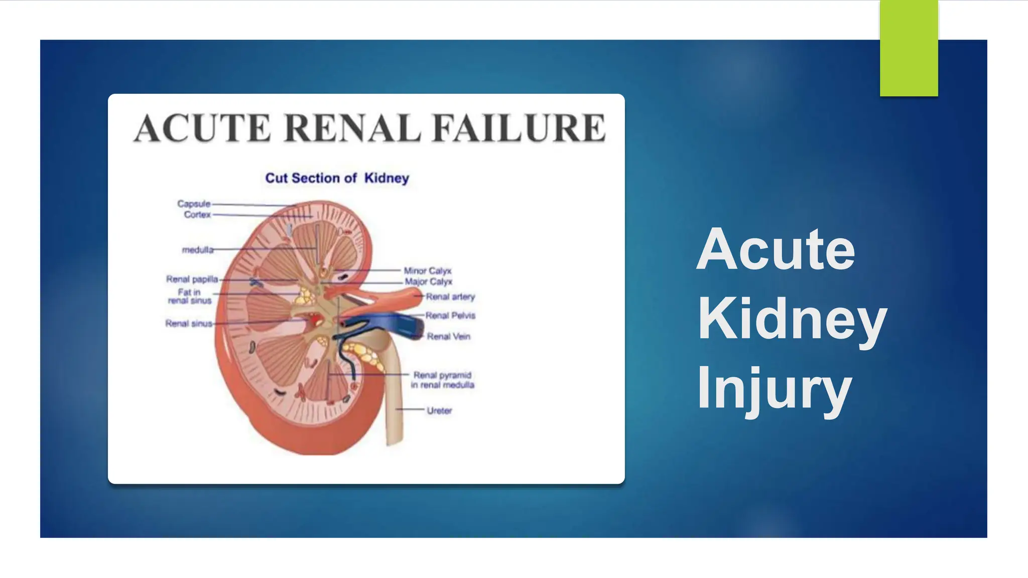
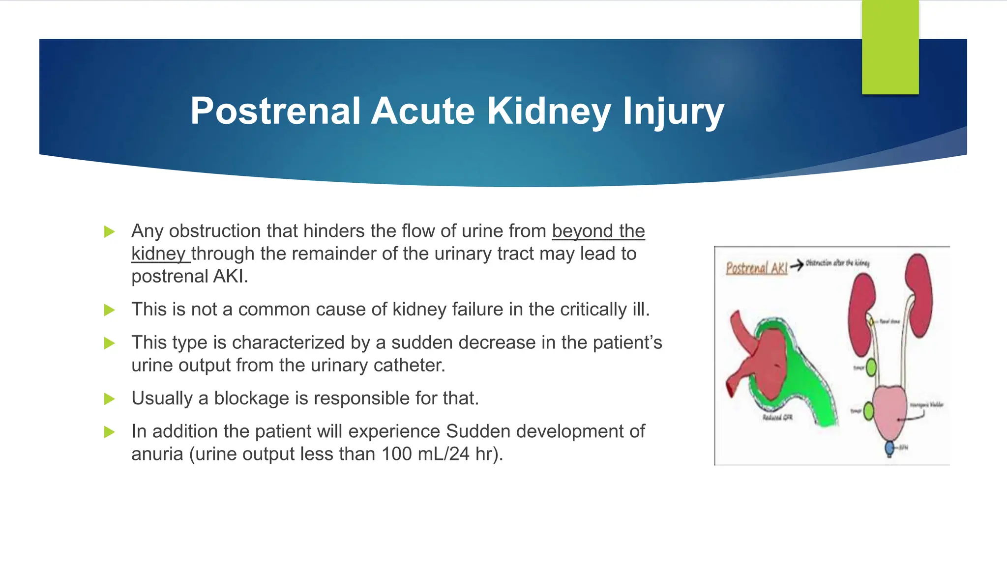
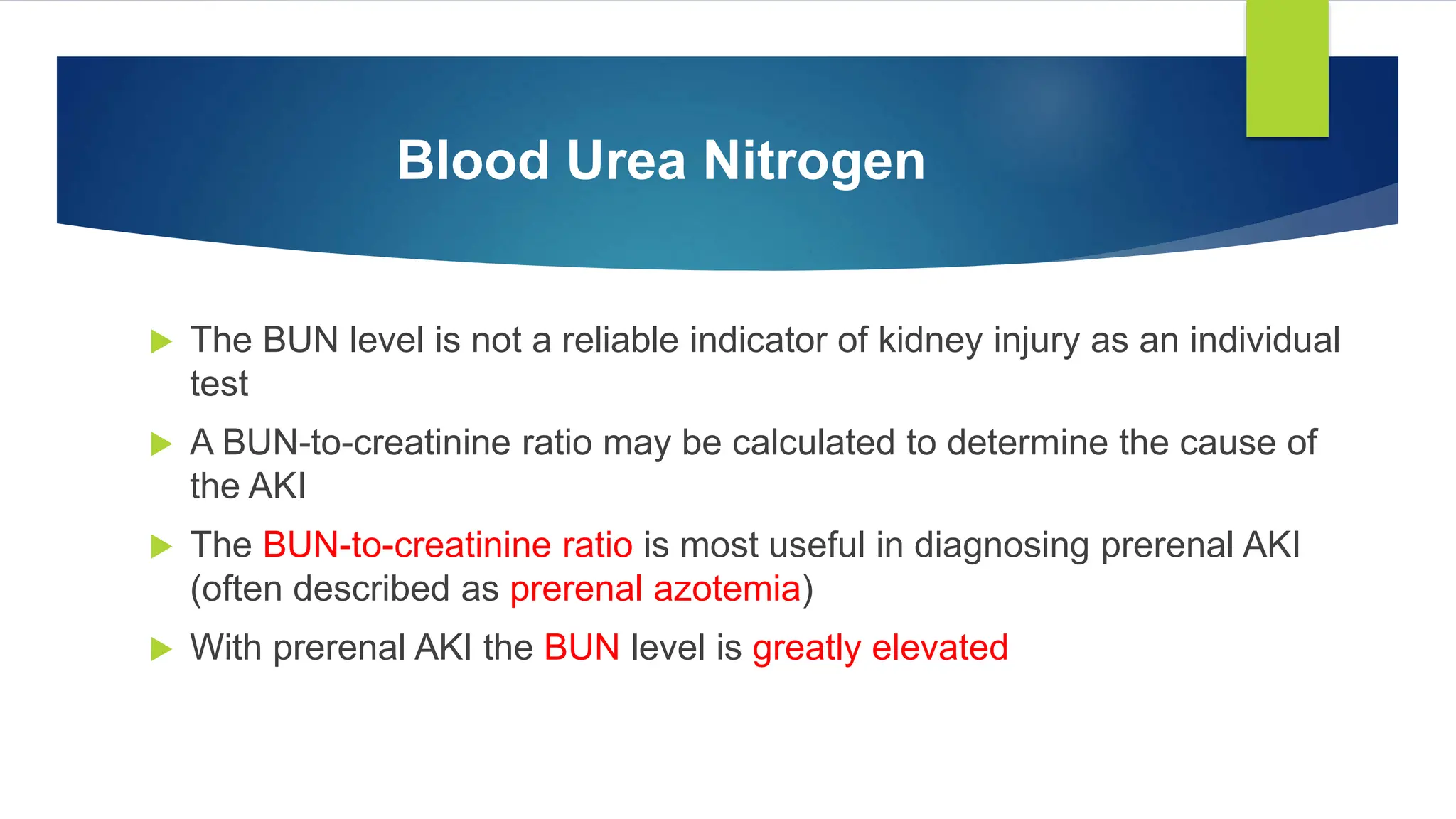
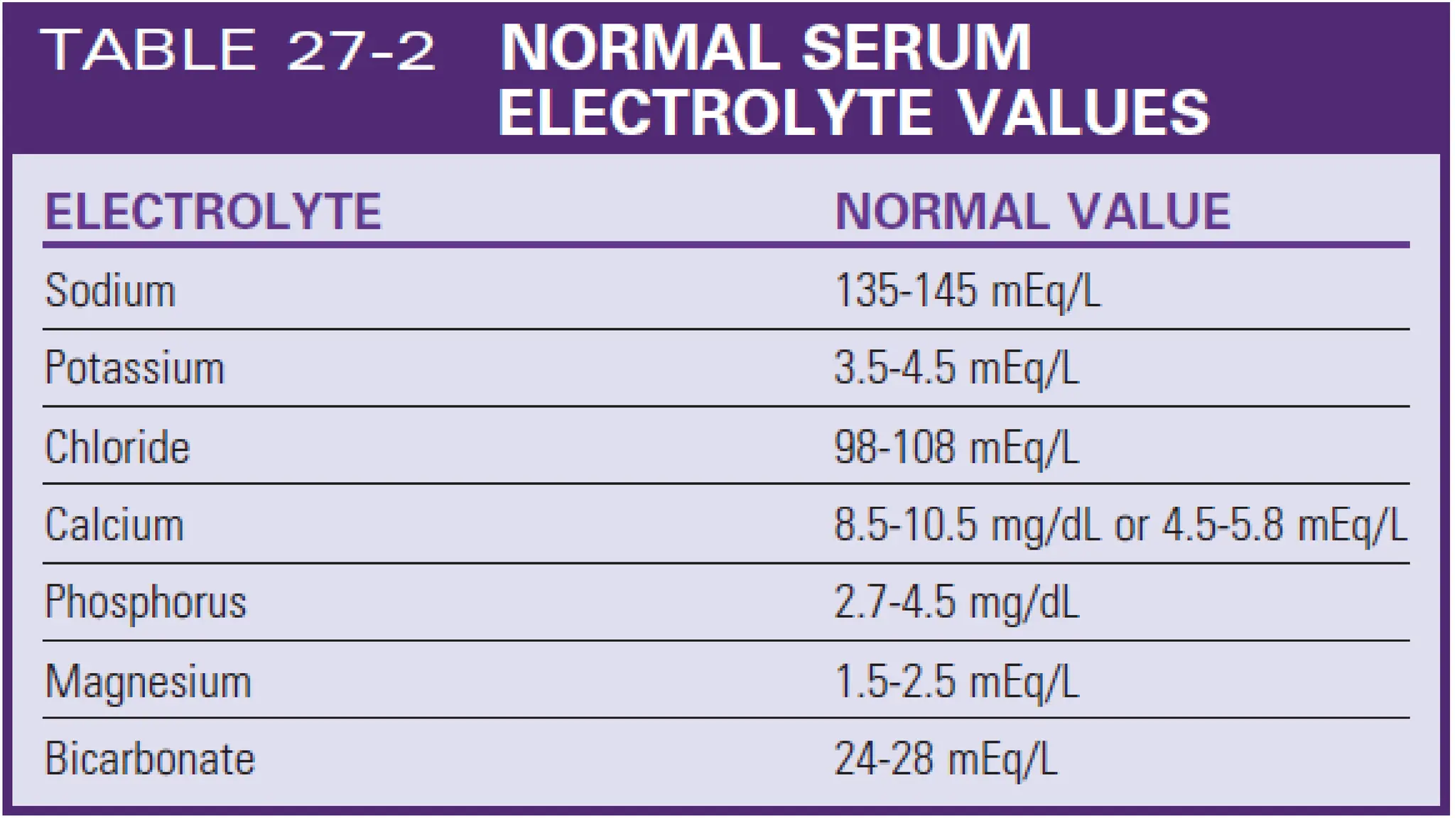
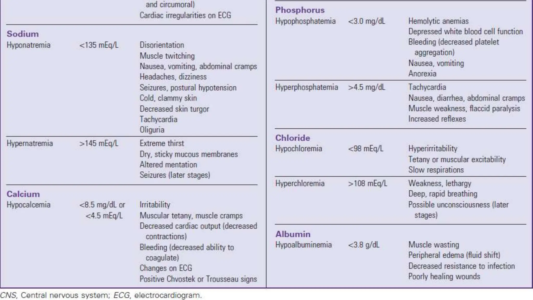
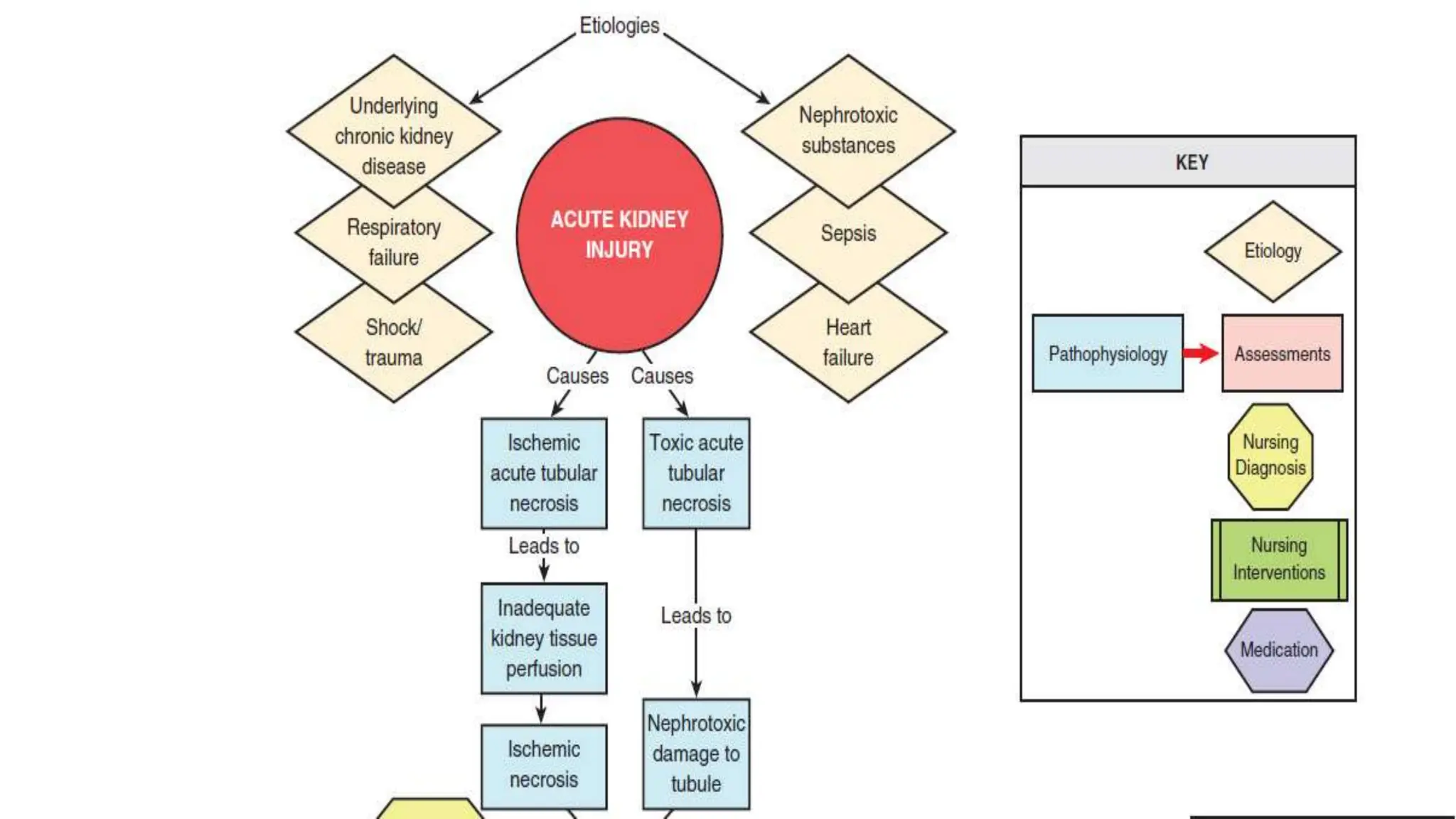
Acute
Kidney
Injury
OBJECTIVES
 Define Acute Kidney Injury (AKI) Explain the causes of acute kidney injury (AKI)
 Understand the pathophysiology of AKI
 Differentiate between types of AKI
 Understand common laboratory changes associated with AKI
 Identify the signs and symptoms of AKI and contributing factors
 Discuss common medical problem associated with AKI
 Discuss the management of AKI
 Discuss the nursing management for patients with AKI
 Develop a nursing care plan for patients with AKI
1
Introduction
 Acute kidney injury (AKI) is a relatively new term used to describe the spectrum
of acute-onset kidney disorders that can range from mild impairment of kidney
function through acute kidney failure that requires renal replacement therapy
(dialysis).
 Severe AKI is characterized by a sudden decline in glomerular filtration rate
(GFR), with subsequent retention of products in the blood that are normally
excreted by the kidneys; this disrupts electrolyte balance, acid–base
homeostasis, and fluid volume equilibrium
1
Critical Illness and Acute Kidney Injury
 Critical care patients with AKI have a longer length of hospital stay, more
complications and higher mortality
 One of the reasons for poor survival is that critical care patients often have coexisting
non kidney conditions that increase their susceptibility to the development of AKI
 High-risk diagnoses include heart failure, shock, respiratory failure, and sepsis A
patient is not admitted to the critical care unit with a diagnosis of AKI alone; there is
always coexisting hemodynamic, cardiac, pulmonary, or neurologic compromise
1
Definitions of Acute Kidney Injury
 Measurement of kidney function is indirect, and the diagnosis of AKI is
predominantly derived from changes in urine output and elevation of serum
creatinine level, with the understanding that changes in these values reflect a
decline in the GFR.
 Urine output is sometimes a problematic measure to use because diuretics
artificially increase the urine output but may not alter the course of kidney
failure
1
Acute Kidney Injury Network Criteria
Types of Acute Kidney Injury
Pre-renal Acute Kidney Injury
 Any condition that decreases blood flow, blood pressure, or
kidney perfusion before arterial blood reaches the renal
artery that supplies the kidney may be anatomically
described as prerenal AKI.
 When arterial hypoperfusion due to low cardiac output,
hemorrhage, vasodilation, thrombosis, or other cause
reduces the blood flow to the kidney, glomerular filtration
decreases, and consequently, urine output decreases.
 If normal perfusion and cardiac output are restored quickly, the kidney will
recover and not suffer permanent injury.
 However, if the prerenal insult is not corrected, the GFR will decline, the blood
urea nitrogen (BUN) concentration will rise (prerenal azotemia),1 and the patient
will develop oliguria and be at risk for significant kidney damage
 The term azotemia is used to describe an acute rise in the BUN level often
associated with prerenal AKI.
 Uremia is another term used to describe an elevated BUN value.
Intrarenal Acute Kidney Injury
 Any condition that produces an ischemic or toxic insult
directly at parenchymal nephron tissue places the patient
at risk for development of intrarenal AKI.
 Ischemic damage may be caused by prolonged
hypotension or low cardiac output.
 Toxic injury reaction may occur in response to substances
that damage the kidney tubular endothelium, such as some
antimicrobial medications and the contrast dye used in
radiologic diagnostic studies.
Postrenal Acute Kidney Injury
 Any obstruction that hinders the flow of urine from beyond the
kidney through the remainder of the urinary tract may lead to
postrenal AKI.
 This is not a common cause of kidney failure in the critically ill.
 This type is characterized by a sudden decrease in the patient’s
urine output from the urinary catheter.
 Usually a blockage is responsible for that.
 In addition the patient will experience Sudden development of
anuria (urine output less than 100 mL/24 hr).
OBJECTIVES
 1. Explain the causes of acute kidney injury (AKI).
 2. Describe urine production during the nonoliguric, oliguric, and diuretic phases of acute tubular necrosis
 3. Differentiate between the three types of AKI based on history and physical examination, laboratory
values, and diagnostic tests
1
ASSESSMENT AND
DIAGNOSIS
Acidosis
 Acidosis (pH less than 7.35) is one of the trademarks of
severe acute kidney insult.
 Metabolic acidosis occurs as a result of the accumulation
of un-excreted waste products
 Low serum albumin concentration, which often occurs in
AKI, has a slight alkalinizing effect, but it is not enough to
correct the metabolic acidosis
 Even respiratory compensation and mechanical ventilatory
support are rarely sufficient to reverse the metabolic
acidosis.
Blood Urea Nitrogen
 The BUN level is not a reliable indicator of kidney injury as an individual
test
 A BUN-to-creatinine ratio may be calculated to determine the cause of
the AKI
 The BUN-to-creatinine ratio is most useful in diagnosing prerenal AKI
(often described as prerenal azotemia)
 With prerenal AKI the BUN level is greatly elevated
Serum Creatinine
 Creatinine is a byproduct of muscle metabolism that is formed non-enzymatically from
creatinine in muscles
 Creatinine is completely excreted when kidney function is normal
 When the kidneys are not working, the serum creatinine level rises
 Even small increases in serum creatinine represent a significant decrease in GFR
 Serum creatinine level is assessed daily to follow the trend of kidney function and to determine
whether it is stable, getting better, or getting worse
Creatinine Clearance
 If the patient is making sufficient urine, the urinary creatinine clearance can be
measured
 A normal urinary creatinine clearance rate is 120 mL/min, but this value decreases
with kidney failure
 Critical care patients with severe AKI will manifest elevated serum creatinine and may
be oliguric
 Consequently, the urinary creatinine clearance rate is rarely measured during critical
illness
OBJECTIVES
 1. Explain the causes of acute kidney injury (AKI).
 2. Describe urine production during the nonoliguric, oliguric, and diuretic phases of acute tubular necrosis
 3. Differentiate between the three types of AKI based on history and physical examination, laboratory
values, and diagnostic tests
1
Physical Assessment
Signs and symptoms
1. Thirst
2. Decreased skin turgor,
3. Fatigue
4. Pulmonary congestion
5. Increasing heart failure
6. Rising blood pressure
7. Edema
Contributing factors
 Fluid retention caused by inadequate
urine output.
 Low serum albumin levels create a lower
oncotic pressure in the vasculature, and
more fluid seeps out into the interstitial
spaces to cause peripheral edema.
 Inflammation associated with AKI or a
coexisting nonrenal disease increases
vascular permeability
Heart Failure and Acute Kidney Injury
 There is a strong association between kidney failure and
heart failure.
 In studies of critically ill patients with AKI, 54%3 have
acute kidney failure and heart failure.
 Several of the risk factors for atherosclerotic
cardiovascular disease are also detrimental to the kidney
over the long term, notably hypertension and diabetes.
 Maintenance of a BP below 130/80 mm Hg and a blood
glucose within the normal range decrease the risk of both
developing CKD and the atherosclerotic cardiac diseases
such as coronary artery disease or peripheral-arterial
disease.
Respiratory Failure and Acute Kidney
Injury
 There is a significant association between
respiratory failure and kidney failure.
 In studies of critically ill patients with kidney failure,
over 50% have respiratory failure.
 Mechanical ventilation can alter kidney function.
 Positive-pressure ventilation reduces blood flow to
the kidney, lowers the GFR, and decreases urine
output.
 These effects are worsen with the addition of positive end-expiratory
pressure (PEEP).
 AKI increases inflammation, causes the lung vasculature to become
more permeable, and contributes to the development of acute respiratory
distress syndrome (ARDS).
 Patients with AKI are more likely to require prolonged mechanical
ventilatory support
Sepsis and Acute Kidney Injury
 Sepsis causes almost half of the cases of AKI in the critically ill
 Sepsis and septic shock create hemodynamic instability and reduce perfusion to the
kidney.
 Immunologic, toxic, and inflammatory factors may alter the function of the kidney
microvasculature and tubular cells.
 Clinical guidelines for hemodynamic support in sepsis emphasize the need for adequate
fluid resuscitation, because reversal of hypotension and restoration of hemodynamic
stability can often be achieved with fluids alone.
 Unfortunately, in severely septic patients, inflammation increases vascular permeability,
and much of this fluid may move into the third space (interstitial space).
 If the blood pressure remains low, the use of vasopressors is recommended to
 raise refractory low blood pressure after volume resuscitation.
 Vasopressors raise blood pressure and increase systemic vascular resistance
(SVR), but they also may raise the vascular resistance within the kidney
microvasculature.
 Other practices aimed at reversing the deleterious effects of sepsis include
maintaining the patient’s hemoglobin level at 7 to 9 g/dL and blood glucose level
below 150 mg/dL and ensuring optimal hydration as evidenced by a central
venous pressure above 8 mm Hg
Trauma and Acute Kidney Injury
 Trauma patients with major crush injuries have an elevated risk of kidney failure
because of the release of creatine and myoglobin from damaged muscle cells, a
condition called rhabdomyolysis.
 Myoglobin in large quantities is toxic to the kidney.
 A major goal of treatment is to prevent rhabdomyolysis-induced AKI.
 Mortality rate is low, provided adequate crystalloid volume is administered early in
the course of treatment.
 It is important to measure the serum potassium levels.
 Life-threatening hyperkalemia can occur as cell lysis permits intracellular
potassium to be released into the bloodstream
Trauma and Acute Kidney Injury
 The level of creatine kinase (CK), a marker of systemic muscle damage, increases in patients with
rhabdomyolysis.
 Crystalloid volume resuscitation is the primary treatment for preservation of adequate kidney function
and prevention of
 AKI.
 In many hospitals, the intravenous (IV) fluids are alkalinized by the addition of sodium bicarbonate,
and the urine output is increased by IV administration of the diuretic mannitol.
 A bicarbonate and mannitol regimen is instituted to prevent acidosis and hyperkalemia,
because both are frequent complications of rhabdomyolysis.
 Close attention is paid to hourly urine output that can be dark brown or tea-colored CK levels,
serum creatinine levels, serum potassium levels, and any signs of compartment syndrome in all
patients admitted with this diagnosis.
Management
 Promote hydration and avoid dehydration
 Hemodynamic monitoring including surveillance of the central venous pressure, pulmonary artery
occlusion pressure, cardiac output, and cardiac index.
 The daily weight, combined with accurate intake and output monitoring, is a powerful indicator of
fluid gains or losses over 24 hours
 A 1-kg weight gain over 24 hours represents 1000 mL (1 liter) of additional fluid retention.
 Electrolyte levels require frequent observation
 Sodium bicarbonate
 Calcium Replacement
 Dietary–Phosphorus-Binding Medications- to maintain the calcium levels in the blood
Management
 If hyperkalemia is identified, all potassium supplements are stopped.
 If the patient is producing urine, IV diuretics can be administered.
 Acute hyperkalemia can be treated temporarily by IV administration of insulin and glucose.
 An infusion of 50 mL of 50% dextrose accompanied by 10 units of regular insulin forces
potassium out of the serum and into the cells.
 Fluid Restriction to prevent the development of interstitial edema
 Renal replacement therapy
 Diuretics-helpful for symptoms such as pulmonary edema
Management
 Low-dose dopamine (2 to 3 mcg/kg/min) is frequently infused to stimulate blood
flow to the kidney
 Dopamine is effective in increasing urine output in the short term,
 Dietary-Phosphorus Binders
Nutrition
 Diet or nutritional supplementation for the patient with AKI in the critical care unit is designed to
account for the diminished excretory capacity of the kidney.
 The recommended energy intake is between 20 and 30 kilocalories/kg per day, with 1.2 to 1.5
grams/kg of protein per day to control azotemia (increased
 Oral nutrition is preferred, and if the patient cannot eat, enteral nutrition is recommended over
parenteral (intravenous) nutrition.
 Fluids are limited, and monitoring of blood glucose levels is recommended
 The electrolytes potassium, sodium, and phosphorus are strictly limited.
2
Nursing Diagnoses For the Patient with
Acute Kidney Injury
3
Nursing Diagnoses For the Patient with
Acute Kidney Injury
 Excess Fluid Volume related to decreased kidney function
 Decreased Cardiac Output related to fluid volume excess, disturbances in renin–angiotensin
system
 Imbalanced Nutrition: Less Than Body Requirements related to anorexia, nausea and vomiting,
dietary restrictions, and altered oral mucous membranes
 Risk for Skin Integrity related to poor nutritional status, immobility, and edema
3
 Anxiety related to unexpected serious illness and uncertain prognosis, unfamiliar environment,
and current symptoms
 Activity Intolerance related to shortness of breath, fatigue, anemia, uremia, and dialysis procedure
 Disturbed Sleep Pattern related to fragmented sleep in hospital environment• Risk for Infection
related to decreased functioning of immune system
 Deficient Knowledge related to pathophysiology and etiology of acute episode, dietary restrictions,
medications, complications, prognosis, and follow-up care
Nursing Diagnoses For the Patient with
Acute Kidney Injury
3
3
3
3
3
3
Acute Kidney Injury.pptx

Acute Kidney Injury.pptx

  • 1.
  • 2.
    OBJECTIVES  Define AcuteKidney Injury (AKI) Explain the causes of acute kidney injury (AKI)  Understand the pathophysiology of AKI  Differentiate between types of AKI  Understand common laboratory changes associated with AKI  Identify the signs and symptoms of AKI and contributing factors  Discuss common medical problem associated with AKI  Discuss the management of AKI  Discuss the nursing management for patients with AKI  Develop a nursing care plan for patients with AKI 1
  • 3.
    Introduction  Acute kidneyinjury (AKI) is a relatively new term used to describe the spectrum of acute-onset kidney disorders that can range from mild impairment of kidney function through acute kidney failure that requires renal replacement therapy (dialysis).  Severe AKI is characterized by a sudden decline in glomerular filtration rate (GFR), with subsequent retention of products in the blood that are normally excreted by the kidneys; this disrupts electrolyte balance, acid–base homeostasis, and fluid volume equilibrium 1
  • 4.
    Critical Illness andAcute Kidney Injury  Critical care patients with AKI have a longer length of hospital stay, more complications and higher mortality  One of the reasons for poor survival is that critical care patients often have coexisting non kidney conditions that increase their susceptibility to the development of AKI  High-risk diagnoses include heart failure, shock, respiratory failure, and sepsis A patient is not admitted to the critical care unit with a diagnosis of AKI alone; there is always coexisting hemodynamic, cardiac, pulmonary, or neurologic compromise 1
  • 5.
    Definitions of AcuteKidney Injury  Measurement of kidney function is indirect, and the diagnosis of AKI is predominantly derived from changes in urine output and elevation of serum creatinine level, with the understanding that changes in these values reflect a decline in the GFR.  Urine output is sometimes a problematic measure to use because diuretics artificially increase the urine output but may not alter the course of kidney failure 1
  • 6.
    Acute Kidney InjuryNetwork Criteria
  • 7.
    Types of AcuteKidney Injury Pre-renal Acute Kidney Injury  Any condition that decreases blood flow, blood pressure, or kidney perfusion before arterial blood reaches the renal artery that supplies the kidney may be anatomically described as prerenal AKI.  When arterial hypoperfusion due to low cardiac output, hemorrhage, vasodilation, thrombosis, or other cause reduces the blood flow to the kidney, glomerular filtration decreases, and consequently, urine output decreases.
  • 8.
     If normalperfusion and cardiac output are restored quickly, the kidney will recover and not suffer permanent injury.  However, if the prerenal insult is not corrected, the GFR will decline, the blood urea nitrogen (BUN) concentration will rise (prerenal azotemia),1 and the patient will develop oliguria and be at risk for significant kidney damage  The term azotemia is used to describe an acute rise in the BUN level often associated with prerenal AKI.  Uremia is another term used to describe an elevated BUN value.
  • 9.
    Intrarenal Acute KidneyInjury  Any condition that produces an ischemic or toxic insult directly at parenchymal nephron tissue places the patient at risk for development of intrarenal AKI.  Ischemic damage may be caused by prolonged hypotension or low cardiac output.  Toxic injury reaction may occur in response to substances that damage the kidney tubular endothelium, such as some antimicrobial medications and the contrast dye used in radiologic diagnostic studies.
  • 10.
    Postrenal Acute KidneyInjury  Any obstruction that hinders the flow of urine from beyond the kidney through the remainder of the urinary tract may lead to postrenal AKI.  This is not a common cause of kidney failure in the critically ill.  This type is characterized by a sudden decrease in the patient’s urine output from the urinary catheter.  Usually a blockage is responsible for that.  In addition the patient will experience Sudden development of anuria (urine output less than 100 mL/24 hr).
  • 11.
    OBJECTIVES  1. Explainthe causes of acute kidney injury (AKI).  2. Describe urine production during the nonoliguric, oliguric, and diuretic phases of acute tubular necrosis  3. Differentiate between the three types of AKI based on history and physical examination, laboratory values, and diagnostic tests 1
  • 12.
    ASSESSMENT AND DIAGNOSIS Acidosis  Acidosis(pH less than 7.35) is one of the trademarks of severe acute kidney insult.  Metabolic acidosis occurs as a result of the accumulation of un-excreted waste products  Low serum albumin concentration, which often occurs in AKI, has a slight alkalinizing effect, but it is not enough to correct the metabolic acidosis  Even respiratory compensation and mechanical ventilatory support are rarely sufficient to reverse the metabolic acidosis.
  • 13.
    Blood Urea Nitrogen The BUN level is not a reliable indicator of kidney injury as an individual test  A BUN-to-creatinine ratio may be calculated to determine the cause of the AKI  The BUN-to-creatinine ratio is most useful in diagnosing prerenal AKI (often described as prerenal azotemia)  With prerenal AKI the BUN level is greatly elevated
  • 14.
    Serum Creatinine  Creatinineis a byproduct of muscle metabolism that is formed non-enzymatically from creatinine in muscles  Creatinine is completely excreted when kidney function is normal  When the kidneys are not working, the serum creatinine level rises  Even small increases in serum creatinine represent a significant decrease in GFR  Serum creatinine level is assessed daily to follow the trend of kidney function and to determine whether it is stable, getting better, or getting worse
  • 15.
    Creatinine Clearance  Ifthe patient is making sufficient urine, the urinary creatinine clearance can be measured  A normal urinary creatinine clearance rate is 120 mL/min, but this value decreases with kidney failure  Critical care patients with severe AKI will manifest elevated serum creatinine and may be oliguric  Consequently, the urinary creatinine clearance rate is rarely measured during critical illness
  • 16.
    OBJECTIVES  1. Explainthe causes of acute kidney injury (AKI).  2. Describe urine production during the nonoliguric, oliguric, and diuretic phases of acute tubular necrosis  3. Differentiate between the three types of AKI based on history and physical examination, laboratory values, and diagnostic tests 1
  • 17.
    OBJECTIVES  1. Explainthe causes of acute kidney injury (AKI).  2. Describe urine production during the nonoliguric, oliguric, and diuretic phases of acute tubular necrosis  3. Differentiate between the three types of AKI based on history and physical examination, laboratory values, and diagnostic tests 1
  • 18.
    OBJECTIVES  1. Explainthe causes of acute kidney injury (AKI).  2. Describe urine production during the nonoliguric, oliguric, and diuretic phases of acute tubular necrosis  3. Differentiate between the three types of AKI based on history and physical examination, laboratory values, and diagnostic tests 1
  • 19.
    OBJECTIVES  1. Explainthe causes of acute kidney injury (AKI).  2. Describe urine production during the nonoliguric, oliguric, and diuretic phases of acute tubular necrosis  3. Differentiate between the three types of AKI based on history and physical examination, laboratory values, and diagnostic tests 1
  • 20.
    OBJECTIVES  1. Explainthe causes of acute kidney injury (AKI).  2. Describe urine production during the nonoliguric, oliguric, and diuretic phases of acute tubular necrosis  3. Differentiate between the three types of AKI based on history and physical examination, laboratory values, and diagnostic tests 1
  • 21.
    OBJECTIVES  1. Explainthe causes of acute kidney injury (AKI).  2. Describe urine production during the nonoliguric, oliguric, and diuretic phases of acute tubular necrosis  3. Differentiate between the three types of AKI based on history and physical examination, laboratory values, and diagnostic tests 1
  • 22.
    OBJECTIVES  1. Explainthe causes of acute kidney injury (AKI).  2. Describe urine production during the nonoliguric, oliguric, and diuretic phases of acute tubular necrosis  3. Differentiate between the three types of AKI based on history and physical examination, laboratory values, and diagnostic tests 1
  • 23.
    Physical Assessment Signs andsymptoms 1. Thirst 2. Decreased skin turgor, 3. Fatigue 4. Pulmonary congestion 5. Increasing heart failure 6. Rising blood pressure 7. Edema
  • 24.
    Contributing factors  Fluidretention caused by inadequate urine output.  Low serum albumin levels create a lower oncotic pressure in the vasculature, and more fluid seeps out into the interstitial spaces to cause peripheral edema.  Inflammation associated with AKI or a coexisting nonrenal disease increases vascular permeability
  • 25.
    Heart Failure andAcute Kidney Injury  There is a strong association between kidney failure and heart failure.  In studies of critically ill patients with AKI, 54%3 have acute kidney failure and heart failure.  Several of the risk factors for atherosclerotic cardiovascular disease are also detrimental to the kidney over the long term, notably hypertension and diabetes.  Maintenance of a BP below 130/80 mm Hg and a blood glucose within the normal range decrease the risk of both developing CKD and the atherosclerotic cardiac diseases such as coronary artery disease or peripheral-arterial disease.
  • 26.
    Respiratory Failure andAcute Kidney Injury  There is a significant association between respiratory failure and kidney failure.  In studies of critically ill patients with kidney failure, over 50% have respiratory failure.  Mechanical ventilation can alter kidney function.  Positive-pressure ventilation reduces blood flow to the kidney, lowers the GFR, and decreases urine output.
  • 27.
     These effectsare worsen with the addition of positive end-expiratory pressure (PEEP).  AKI increases inflammation, causes the lung vasculature to become more permeable, and contributes to the development of acute respiratory distress syndrome (ARDS).  Patients with AKI are more likely to require prolonged mechanical ventilatory support
  • 29.
    Sepsis and AcuteKidney Injury  Sepsis causes almost half of the cases of AKI in the critically ill  Sepsis and septic shock create hemodynamic instability and reduce perfusion to the kidney.  Immunologic, toxic, and inflammatory factors may alter the function of the kidney microvasculature and tubular cells.  Clinical guidelines for hemodynamic support in sepsis emphasize the need for adequate fluid resuscitation, because reversal of hypotension and restoration of hemodynamic stability can often be achieved with fluids alone.  Unfortunately, in severely septic patients, inflammation increases vascular permeability, and much of this fluid may move into the third space (interstitial space).
  • 31.
     If theblood pressure remains low, the use of vasopressors is recommended to  raise refractory low blood pressure after volume resuscitation.  Vasopressors raise blood pressure and increase systemic vascular resistance (SVR), but they also may raise the vascular resistance within the kidney microvasculature.  Other practices aimed at reversing the deleterious effects of sepsis include maintaining the patient’s hemoglobin level at 7 to 9 g/dL and blood glucose level below 150 mg/dL and ensuring optimal hydration as evidenced by a central venous pressure above 8 mm Hg
  • 32.
    Trauma and AcuteKidney Injury  Trauma patients with major crush injuries have an elevated risk of kidney failure because of the release of creatine and myoglobin from damaged muscle cells, a condition called rhabdomyolysis.  Myoglobin in large quantities is toxic to the kidney.  A major goal of treatment is to prevent rhabdomyolysis-induced AKI.  Mortality rate is low, provided adequate crystalloid volume is administered early in the course of treatment.  It is important to measure the serum potassium levels.  Life-threatening hyperkalemia can occur as cell lysis permits intracellular potassium to be released into the bloodstream
  • 33.
    Trauma and AcuteKidney Injury  The level of creatine kinase (CK), a marker of systemic muscle damage, increases in patients with rhabdomyolysis.  Crystalloid volume resuscitation is the primary treatment for preservation of adequate kidney function and prevention of  AKI.  In many hospitals, the intravenous (IV) fluids are alkalinized by the addition of sodium bicarbonate, and the urine output is increased by IV administration of the diuretic mannitol.
  • 34.
     A bicarbonateand mannitol regimen is instituted to prevent acidosis and hyperkalemia, because both are frequent complications of rhabdomyolysis.  Close attention is paid to hourly urine output that can be dark brown or tea-colored CK levels, serum creatinine levels, serum potassium levels, and any signs of compartment syndrome in all patients admitted with this diagnosis.
  • 35.
    Management  Promote hydrationand avoid dehydration  Hemodynamic monitoring including surveillance of the central venous pressure, pulmonary artery occlusion pressure, cardiac output, and cardiac index.  The daily weight, combined with accurate intake and output monitoring, is a powerful indicator of fluid gains or losses over 24 hours  A 1-kg weight gain over 24 hours represents 1000 mL (1 liter) of additional fluid retention.  Electrolyte levels require frequent observation  Sodium bicarbonate  Calcium Replacement  Dietary–Phosphorus-Binding Medications- to maintain the calcium levels in the blood
  • 36.
    Management  If hyperkalemiais identified, all potassium supplements are stopped.  If the patient is producing urine, IV diuretics can be administered.  Acute hyperkalemia can be treated temporarily by IV administration of insulin and glucose.  An infusion of 50 mL of 50% dextrose accompanied by 10 units of regular insulin forces potassium out of the serum and into the cells.  Fluid Restriction to prevent the development of interstitial edema  Renal replacement therapy  Diuretics-helpful for symptoms such as pulmonary edema
  • 37.
    Management  Low-dose dopamine(2 to 3 mcg/kg/min) is frequently infused to stimulate blood flow to the kidney  Dopamine is effective in increasing urine output in the short term,  Dietary-Phosphorus Binders
  • 38.
    Nutrition  Diet ornutritional supplementation for the patient with AKI in the critical care unit is designed to account for the diminished excretory capacity of the kidney.  The recommended energy intake is between 20 and 30 kilocalories/kg per day, with 1.2 to 1.5 grams/kg of protein per day to control azotemia (increased  Oral nutrition is preferred, and if the patient cannot eat, enteral nutrition is recommended over parenteral (intravenous) nutrition.  Fluids are limited, and monitoring of blood glucose levels is recommended  The electrolytes potassium, sodium, and phosphorus are strictly limited.
  • 39.
  • 40.
    Nursing Diagnoses Forthe Patient with Acute Kidney Injury 3
  • 41.
    Nursing Diagnoses Forthe Patient with Acute Kidney Injury  Excess Fluid Volume related to decreased kidney function  Decreased Cardiac Output related to fluid volume excess, disturbances in renin–angiotensin system  Imbalanced Nutrition: Less Than Body Requirements related to anorexia, nausea and vomiting, dietary restrictions, and altered oral mucous membranes  Risk for Skin Integrity related to poor nutritional status, immobility, and edema 3
  • 42.
     Anxiety relatedto unexpected serious illness and uncertain prognosis, unfamiliar environment, and current symptoms  Activity Intolerance related to shortness of breath, fatigue, anemia, uremia, and dialysis procedure  Disturbed Sleep Pattern related to fragmented sleep in hospital environment• Risk for Infection related to decreased functioning of immune system  Deficient Knowledge related to pathophysiology and etiology of acute episode, dietary restrictions, medications, complications, prognosis, and follow-up care
  • 43.
    Nursing Diagnoses Forthe Patient with Acute Kidney Injury 3
  • 45.
  • 46.
  • 47.
  • 48.
  • 49.