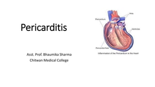
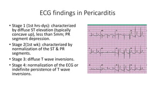
Pericarditis
Asst. Prof. Bhaumika Sharma
Chitwan Medical College
Pericarditis
• The pericardium is a fibroelastic sac made up of visceral and
parietal layers separated by a (potential) space, the
pericardial cavity.
• In healthy individuals, the pericardial cavity contains 15 to 50
mL of an ultrafiltrate of plasma.
• Constrictive pericarditis is a condition in which a chronic
inflammatory thickening of the pericardium compresses the heart so
it is unable to fill normally during diastole.
Diseases of Pericardium
• Diseases of the pericardium present clinically in one of four ways:
1. Acute or recurrent pericarditis
2. Pericardial effusion without major hemodynamic compromise
3. Cardiac tamponade
4. Constrictive pericarditis
Note: Acute Pericarditis (AP) refers to inflammation of the pericardial
sac. The term myopericarditis, or perimyocarditis, is used for case fo AP
that also demonstrate myocardial inflammation.
Etiology
Idiopathic
Infections Viral, tubercular
Neoplasm A. Metastatic
B. Primary: rhabdomyosarcoma, fibroma, angioma, lipoma
C. Paraneoplastic
Cardiac A. Early infarction pericarditis
B. Late postcardiac injury syndrome (Dressler’s syndrome)
C. Myocarditis
D. Dissecting aortic aneurysm
Autoimmune A. Rheumatic disease: including lupus, RA, vasculitis etc.
Drugs Procainamide, isoniazid, hydralazine, phenytoin, penicillin, doxorubicin etc.
Metabolic A. hypothyroidism: primarily pericardial effusion
B. Uremia
C. Ovarian hyperstimulation syndrome
Trauma A. Blunt, Penetrating
Aortic Aneurysm
Infection
A. viral-coxsackievirus, echovirus, adenovirus, EBV,
CMV, influenza, varicella, rubella, HIV, hepatitis B,
mumps etc
B. Bacterial-staphylococcus, streptococcus,
pneumococcus, Haemophilus, Neiseria
(gonorrhoeae or meningitidis), tuberculosis,
salmonella etc
C. Mycoplasma
D. Fungal
E. Parasitic-echinococcus, amebiasis, toxoplasmosis
F. Infective endocarditis
Pericarditis
• Acute pericarditis refers to inflammation of pericardial sac.
• Acute pericarditis is the most common disorder involving the
pericardium.
Classification
Acute Subacute Chronic
< 6 weeks 6 weeks – 6 months > 6 months
1. Effusive
2. Fibrous
1. Effusive –constrictive
2. Constrictive
1. Effusive
2. Adhesive
3. Constrictive
Clinical Manifestation
1. Pain (anterior chest)
2. Pericardial friction rub
3. Dyspnea
4. Fever, sweating, child
5. Dysrhythmias
Chest Pain Characteristics
• Sudden in onset
• Retrosternal in location
• Pleuritic and sharp in nature
• Exacerbated by inspiration and coughing
• Worsens when supine and improves upon sitting upright or leaning
forward
• Can often radiate to the neck, arms, or left shoulder, trapezius muscle
• Duration: hours to days
• Response to NTG: no
Pericardial Friction Rub
• Presents in 85%
• High-pitched scratchy or squeaky sound best heard at the LSB with
patient leaning forward.
Diagnostic Evaluation
• Echocardiogram: most sensitive method
• Chest X-ray: may show heart enlargement
• ECG: to evaluate for MI
• WBC and Differential elevations
• Antinuclear antibody (ANA) for lupus erythematosus
• Purified protein derivative (PPD) test positive in TB, ASO titre elevated if
rheumatic fever present
• Pericardiocentesis: examination and diagnosis
• BUN or Urea, creatinine
• Elevated ESR
• Troponin level elevated, returns to normal within 1-2 weeks
ECG findings in Pericarditis
• Stage 1 (1st hrs-dys): characterized
by diffuse ST elevation (typically
concave up), less than 5mm; PR
segment depression.
• Stage 2(1st wk): characterized by
normalization of the ST & PR
segments.
• Stage 3: diffuse T wave inversions.
• Stage 4: normalization of the ECG or
indefinite persistence of T wave
inversions.
Management
• To determine etiology, administer pharmacologic therapy, alert for possible
complication of cardiac tamponade.
1. Bacterial pericarditis: penicillin or antimicrobial agents
2. Rheumatic fever: penicillin G and other antimicrobial agents
3. Tuberculosis: ATT
4. Fungal pericarditis: amphotericin B and fluconazole
5. Systemic lupus erythematosus: corticosteroids
6. Renal pericarditis: dialysis, biochemical control of ESRD
7. Neoplastic pericarditis: intrapericardial instillation of chemotherapy; radiotherapy
8. Post-MI syndrome: best rest, aspirin, prednisone
9. Post-pericardiotomy syndrome (after open heart surgery): treat symptomatically
10. Emergency pericardiocentesis if cardiac tamponade develops
11. Partial pericardiectomy or total pericardiectomy for recurrent constrictive
pericarditis
Complications
1. Cardiac tamponade
2. Heart failure
3. Hemopericardium (esp. post-MI receiving anticoagulants)
Nursing Management
1. Evaluate complaint of chest pain.
a. Ask for pain characteristics
b. Elevate head of bed; position pillow on over the bed table for leaning
c. Assess if this relieves pain.
2. Auscultate heart sounds
a. Listen to friction rub
b. Listen to the heart with patient in different positions.
3. Evaluate history for precipitating factors
Nursing Diagnoses
• Acute Pain related to pericardial inflammation
• Decreased Cardiac Output related to impaired ventricular
expansion
Nursing Interventions
Reducing Discomfort
1. Provide prescribed drug for pain and symptom relief
2. Relieve anxiety of patient and family by explaining the difference
between pain of pericarditis and pain of recurrent MI.
3. Explain to patient and family that pericarditis does not indicate
further heart damage.
4. Encourage patient to remain on bed rest when chest pain, fever and
friction rub occur.
5. Assist patient to position of comfort.
Maintaining Cardiac Output
• Assess heart rate, rhythm, BP, respirations at least hourly in
the acute phase; continuously if hemodynamically
unstable.
• Assess for signs of cardiac tamponade increased heart rate,
decreased BP, presence of paradoxical pulse, distended
neck veins, restlessness, muffled heart sounds.
• Prepare for emergency pericardiocentesis or surgery. Keep
pericardiocentesis tray at bedside.
• Assess for signs of heart failure.
• Monitor closely for the development of dysrhythmias.
Patient Education and Health Maintenance
• Teach patient the etiology of pericarditis.
• Instruct patient about s/s of pericarditis and the need
for long-term medication therapy to help relieve
symptoms.
• Review all medications with the patient purpose,
adverse effects, dosage, and special precautions.
Evaluation: Expected Outcomes
• Verbalizes relief of pain
• Pulse and heart rate stable, no dysrhythmias, no
friction rub

Pericarditis for student

  • 1.
    Pericarditis Asst. Prof. BhaumikaSharma Chitwan Medical College
  • 11.
    Pericarditis • The pericardiumis a fibroelastic sac made up of visceral and parietal layers separated by a (potential) space, the pericardial cavity. • In healthy individuals, the pericardial cavity contains 15 to 50 mL of an ultrafiltrate of plasma.
  • 12.
    • Constrictive pericarditisis a condition in which a chronic inflammatory thickening of the pericardium compresses the heart so it is unable to fill normally during diastole.
  • 14.
    Diseases of Pericardium •Diseases of the pericardium present clinically in one of four ways: 1. Acute or recurrent pericarditis 2. Pericardial effusion without major hemodynamic compromise 3. Cardiac tamponade 4. Constrictive pericarditis Note: Acute Pericarditis (AP) refers to inflammation of the pericardial sac. The term myopericarditis, or perimyocarditis, is used for case fo AP that also demonstrate myocardial inflammation.
  • 15.
    Etiology Idiopathic Infections Viral, tubercular NeoplasmA. Metastatic B. Primary: rhabdomyosarcoma, fibroma, angioma, lipoma C. Paraneoplastic Cardiac A. Early infarction pericarditis B. Late postcardiac injury syndrome (Dressler’s syndrome) C. Myocarditis D. Dissecting aortic aneurysm Autoimmune A. Rheumatic disease: including lupus, RA, vasculitis etc. Drugs Procainamide, isoniazid, hydralazine, phenytoin, penicillin, doxorubicin etc. Metabolic A. hypothyroidism: primarily pericardial effusion B. Uremia C. Ovarian hyperstimulation syndrome Trauma A. Blunt, Penetrating
  • 16.
  • 17.
    Infection A. viral-coxsackievirus, echovirus,adenovirus, EBV, CMV, influenza, varicella, rubella, HIV, hepatitis B, mumps etc B. Bacterial-staphylococcus, streptococcus, pneumococcus, Haemophilus, Neiseria (gonorrhoeae or meningitidis), tuberculosis, salmonella etc C. Mycoplasma D. Fungal E. Parasitic-echinococcus, amebiasis, toxoplasmosis F. Infective endocarditis
  • 18.
    Pericarditis • Acute pericarditisrefers to inflammation of pericardial sac. • Acute pericarditis is the most common disorder involving the pericardium.
  • 19.
    Classification Acute Subacute Chronic <6 weeks 6 weeks – 6 months > 6 months 1. Effusive 2. Fibrous 1. Effusive –constrictive 2. Constrictive 1. Effusive 2. Adhesive 3. Constrictive
  • 20.
    Clinical Manifestation 1. Pain(anterior chest) 2. Pericardial friction rub 3. Dyspnea 4. Fever, sweating, child 5. Dysrhythmias
  • 21.
    Chest Pain Characteristics •Sudden in onset • Retrosternal in location • Pleuritic and sharp in nature • Exacerbated by inspiration and coughing • Worsens when supine and improves upon sitting upright or leaning forward • Can often radiate to the neck, arms, or left shoulder, trapezius muscle • Duration: hours to days • Response to NTG: no
  • 22.
    Pericardial Friction Rub •Presents in 85% • High-pitched scratchy or squeaky sound best heard at the LSB with patient leaning forward.
  • 23.
    Diagnostic Evaluation • Echocardiogram:most sensitive method • Chest X-ray: may show heart enlargement • ECG: to evaluate for MI • WBC and Differential elevations • Antinuclear antibody (ANA) for lupus erythematosus • Purified protein derivative (PPD) test positive in TB, ASO titre elevated if rheumatic fever present • Pericardiocentesis: examination and diagnosis • BUN or Urea, creatinine • Elevated ESR • Troponin level elevated, returns to normal within 1-2 weeks
  • 24.
    ECG findings inPericarditis • Stage 1 (1st hrs-dys): characterized by diffuse ST elevation (typically concave up), less than 5mm; PR segment depression. • Stage 2(1st wk): characterized by normalization of the ST & PR segments. • Stage 3: diffuse T wave inversions. • Stage 4: normalization of the ECG or indefinite persistence of T wave inversions.
  • 25.
    Management • To determineetiology, administer pharmacologic therapy, alert for possible complication of cardiac tamponade. 1. Bacterial pericarditis: penicillin or antimicrobial agents 2. Rheumatic fever: penicillin G and other antimicrobial agents 3. Tuberculosis: ATT 4. Fungal pericarditis: amphotericin B and fluconazole 5. Systemic lupus erythematosus: corticosteroids 6. Renal pericarditis: dialysis, biochemical control of ESRD 7. Neoplastic pericarditis: intrapericardial instillation of chemotherapy; radiotherapy 8. Post-MI syndrome: best rest, aspirin, prednisone 9. Post-pericardiotomy syndrome (after open heart surgery): treat symptomatically 10. Emergency pericardiocentesis if cardiac tamponade develops 11. Partial pericardiectomy or total pericardiectomy for recurrent constrictive pericarditis
  • 26.
    Complications 1. Cardiac tamponade 2.Heart failure 3. Hemopericardium (esp. post-MI receiving anticoagulants)
  • 27.
    Nursing Management 1. Evaluatecomplaint of chest pain. a. Ask for pain characteristics b. Elevate head of bed; position pillow on over the bed table for leaning c. Assess if this relieves pain. 2. Auscultate heart sounds a. Listen to friction rub b. Listen to the heart with patient in different positions. 3. Evaluate history for precipitating factors
  • 28.
    Nursing Diagnoses • AcutePain related to pericardial inflammation • Decreased Cardiac Output related to impaired ventricular expansion
  • 29.
    Nursing Interventions Reducing Discomfort 1.Provide prescribed drug for pain and symptom relief 2. Relieve anxiety of patient and family by explaining the difference between pain of pericarditis and pain of recurrent MI. 3. Explain to patient and family that pericarditis does not indicate further heart damage. 4. Encourage patient to remain on bed rest when chest pain, fever and friction rub occur. 5. Assist patient to position of comfort.
  • 30.
    Maintaining Cardiac Output •Assess heart rate, rhythm, BP, respirations at least hourly in the acute phase; continuously if hemodynamically unstable. • Assess for signs of cardiac tamponade increased heart rate, decreased BP, presence of paradoxical pulse, distended neck veins, restlessness, muffled heart sounds. • Prepare for emergency pericardiocentesis or surgery. Keep pericardiocentesis tray at bedside. • Assess for signs of heart failure. • Monitor closely for the development of dysrhythmias.
  • 31.
    Patient Education andHealth Maintenance • Teach patient the etiology of pericarditis. • Instruct patient about s/s of pericarditis and the need for long-term medication therapy to help relieve symptoms. • Review all medications with the patient purpose, adverse effects, dosage, and special precautions.
  • 32.
    Evaluation: Expected Outcomes •Verbalizes relief of pain • Pulse and heart rate stable, no dysrhythmias, no friction rub