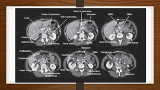
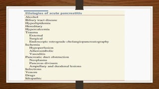
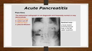
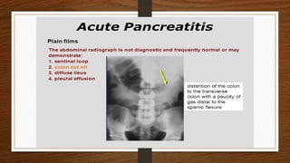
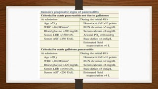
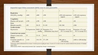
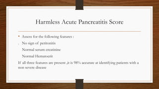
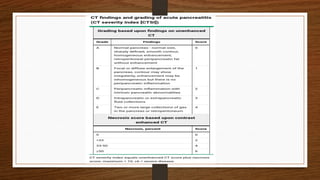
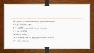
Acute pancreatitis
Mentor : Dr Mahipendra Tiwari
Prepared by : Resident Shikhar Shrestha,Surgery
Objectives
To understand the anatomy and physiology of the pancreas
To define and classify acute pancreatitis
To know epidemiology and etiology of acute pancreatitis
To understand pathophysiology of acute pancreatitis
To discuss on presentation and diagnosis of acute appendicitis
To know the treatment and prognosis
• Pancreas is perhaps most unforgiving organ in the human body
• Leading most surgeons to avoid even palpating it , unless necessary
• Minor trauma to pancreas can result in release of pancreatic enzymes
• And cause life threating pancreatitis
physiology
Definition
• Defined as acute condition ,
• Presenting with abdominal pain
• A 3 folds or greater rise in the serum level of pancreatic enzymes amylase
and lipase
• And characteristics findings of inflammation on CECT
Bailey and love 27th
edition
Classification
• Two classification system have been proposed : on the basis of severity
1. Three grades (mild, moderately severe and severe ) of Revised Atlanta
classification
2. Four catagories(mild, moderate severe and critical) of Determinant based
classification
Schwartz 11th
ed
Epidemiology
• In context of Nepal, incidence of AP ranges from 10 to 50 per 100,000 per
annum
• Overall mortality of approximately 4-6% and increases to 17-39 %in severe
cases
• In Nepal exact data are yet to be discovered
• But study claims alcoholic pancreatitis as the leading cause,accounting for 66
% of all causes
Etiology
• Gallstones
• Alcoholism
• Post ERCP
• Abdominal trauma
• Following biliary, uppe
• Drugs (corticosteroids, azathioprine,propofol valproic acid, thiazides, oestrogens)
• r gastrointestinal or cardiothoracic surgery
• Hyperparathyroidism , Hypercalcaemia
• Pancreas divisum
• Autoimmune pancreatitis
• Hereditary pancreatitis
• Viral infections (mumps, coxsackie B)
• Malnutrition
• Scorpion bite
• Idiopathic
• Gallstones: Are the most common cause (worldwide)
• Accounts for 40 to 70% of cases
• However ,only 3 -7 % of patients with gallstones develop pancreatitis
1
• Three possibilities have been suggested
1. Common channel hypothesis : Reflux of bile into the pancreatic duct due
to transient obstruction of ampulla during passage of stone
2. Edema resulting from the passage of a stone
3. Obstruction of pancreatic duct and leading to ductal hypertension
• Small gallstones are associated with increase risk of pancreatitis
• Risk of developing acute pancreatitis in patients with gallstone is greater in
men
• However incidence of gallstone pancreatitis is higher in women due to a
higher prevalence of gall stones
• Alcohol : responsible for approximately 25 to 35 % of cases of acute
pancreatitis ,worldwide
1.Act by increasing the synthesis of enzymes by pancreatic acinar cells to
synthesize the digestive and lysosomal enzymes
2.Sensitization of acinar cells to cholecystokinin induced premature activation
of zymogens
3.Ethanol induces ductal permeability which cause prematurely activated
enzyme to cause damage to the pancreatic parenchyma
4.Decreases the level of trypsin inhibitor concentration
5. Also induces sensitization of the Pancreas to the toxic effects of coxsackie
virus
• Hyper lipidemia :Lipase liberates toxic fatty acid into pancreatic micro
circulation
• Leads to impairment of pancreatic microcirculation and ischemia
• Hereditary pancreatitis : Autosomal dominant disease related to mutation of
trypsinogenic gene
• Drugs : Thaizides diuretics, frusemide, Estrogen replacement therapy,steroid
therapy , propofol
Pathophysiology
• Occurs in various degree of severity,
• Causes of which are multifactorial
• Pancreatitis begins with :
Premature activation of pancreatic enzymes within pancreas
Activation of digestive zymogen inside acinar cells
Causes injury to acinar cells
• Events subsequent to acinar cell injury :
Inflammatory cell activation and recruitment
Generation and release of cytokines and other chemical mediators
That causes systemic inflammation and multiple organ dysfunction
Diagnosis
• Modalities of diagnosis
1.Detailed history
2.Thorough examination
3.Appropirate investigations
Detailed History
• Clinical Presentation
Pain is the cardinal symptom
Characteristically develops quickly, reaching maximum intensity within
minutes
Pain is severe , constant and refractory to the usual dose of analgesics
Usually experienced in the epigastrium,but may be localized to the upper
quadrant or diffused throughout abdomen
• Radiates to back in 50% of the patient
• In some ,relieved by leaning forward
• Nausea ,repeated vomiting and retching are marked
• Retching may persists even if stomach is kept empty by nasogastric
aspiration
Thorough Examination
Appearance can be ambiguous
Can be fair looking or at the extreme ,gravely ill look with profound shock
toxicity and confusion
Tachypnea and tachycardia is usual
Hypotension can be present
Body temperature is often normal, but rises as inflammation develops
Mild icteric due to biliary obstruction in gall stone pancreatitis
Bleeding into fascial plane can produce bluish discoloration
Flanks (Grey turner’s sign)or umbilicus(cullen’s sign)
Abdominal distension due to ileus or, more rarely ascites
Usually muscle guarding in upper abdomen
Pleural effusion present in 10-20% of patients
Another rare sign is tetany due to hypocalcemia
Appropriate Investigations
• Investigations of acute pancreatitis must be based on :
1. Is a diagnosis of acute pancreatitis correct ?
2. How severe is the attack ?
3. What is the etiology ?
laboratory
• Serum amylase or lipase > 3 times upper limit of normal
• Serum amylase concentrates rises immediately with onset of disease
• Peaks within several hours and remains elevated for 3-5 days
• No correlation between extent of serum amylase elevation and severity
• Serum lipase if available,is more sensitive and specific then amylase
Imaging
• Non specific sign on abdominal radiographs :
Colon cut off sign
Generalised or localized illeus (sentinel loop)
Renal halo sign
Chest radiograph may reveal a pleural effusion
• USG doesnot establish the diagnosis of acute pancreatitis
• Should be performed :
To detect gallstone,as a potential cause
To rule out acute cholecystitis
• CECT is indicated in following :
1. If there is diagnostic uncertainty.
2. In patients with severe acute pancreatitis, to distinguish interstitial from
necrotising pancreatitis
3. Severity of pancreatitis detected on CT be staged a/c to balthazar score
4. In patients with organ failure,signs of sepsis and clinical deterioration
5. When localized collection is suspected,fluid collection,psuedocyst
• MRI and MRCP ,are used increasingly to diagnose and assess severity
• MRI appears to be comparable to CT :
As effective as CT in demonstrating presence and extent of pancreatic necrosis
and fluid collection
Regarding the severity of the disease
Probably superior for indicating the suitability of collections for non surgical
drainage
Severity scoring system
• Ranson’s criteria
• Glass gow score
• SOFA score
• CT severity index(balthazar score)
• Apache II score
• Systemic inflammatory response syndrome score
• BISAP score
• Harmless acute pancreatitis score
Harmless Acute Pancreatitis Score
• Assess for the following features :
. No sign of peritonitis
Normal serum creatinine
Normal Hematocrit
If all three features are present ,it is 98% accurate at identifying patients with a
non severe disease
Treatment
• After initial assessment if a patient is considered to have mild acute pancreatitis
A conservative approach is indicated with IV fluids and frequent observation
Brief period of fasting for patient who is nauseated
Prolonged Nil by mouth has got little physiological justification
Antibiotics are not indicated
Apart from analgesic and anti emetics ,No drugs and intervention are warranted
Early Management of Severe acute Pancreatitis
• Admission to HDU/ICU
• Analgesia
• Aggressive fluid rehydration
• Supplemental oxygen
• Invasive monitoring of vital signs, central venous pressure, urine output,
blood gases
• Frequent monitoring of haematological and biochemicalparameters
(including liver and renal function, clotting, serum calcium, blood glucose)
Nasogastric drainage (only initially)
Antibiotics if cholangitis suspected; prophylactic antibiotics can be considered
CT scan essential if organ failure, clinical deterioration or signs of sepsis develop
ERCP within 72 hours for severe gallstone pancreatitis or signs of cholangitis
Supportive therapy for organ failure if it develops (inotropes, ventilatory support,
haemofiltration,)
If nutritional support is required, consider enteral (nasogastric) feeding
Local complications
Acute(< 4 weeks),No defined walls
Acute pancreatic fluid collection (Infected and Non infected)
Acute necrotic collection (Infected and Non infected)
Chronic( > 4 weeks) Defined walls
Pancreatic pseudocyst (Infected and Non infected)
Walled of necrosis (Infected and Non infected)
Systemic complications
1.Cardiovascular :
Shock
Arrhythmias
2. Pulmonary
ARDS ,Pulmonary Effusion
3.Renal failure
4.Haematological
5. Metabolic
Hypocalcaemia
Hyperglycaemia
Hyperlipidaemia
6. Gastrointestinal
Ileus ,GI hemmorrhage
7. Neurological
Visual disturbances
Confusion, irritability
Encephalopathy
8. Miscellaneous
Subcutaneous fat necrosis, Arthralgia
Prognosis
Ranson’s criteria :
using 11 component score,mortality is 0 to 3 %,score <3
11 to 15 % when the score is >=3
40 % when the score is >=6
The apachae II score :
Study suggests mortality is less than 4 % with a score <8
11 to 18 % with a score of >8
SIRS score :In one validation study, mortality rates were
25 % for persistent SIRS
8 % for SIRS at admission but not persistent
0 % for No SIRS
CT severity index :
23 % mortality with any degree of pancreatic necrosis
0 % with no necrosis
References
1.Bailey and love 27 th ed
2..Sabiston text book of surgery 21th ed
3..Schwartz 11th
edition
4.AGA institute technical review on acute pancreatitis.AGA institute technical review on acute
pancreatitis
5.Etiology incidence and survival of acute pancreatitis
6.Gallstone pancreatitis and the effect of cholecystectomy;a population based cohort study
7.The etiology of acute hemmorhagic pancreatitis
8.Pancreatic duct obstruction triggers acute necrotizing pancreatitis
9.Practice guidelines in acute pancreatitis
10.Acute pancreatitis :value of CT in establishing prognosis
11.Influence of duration of symptoms over perioperative outcomes during emergency laproscopic
cholecystectomy

Acute pancreatitis [Autosaved].pptx surgery

  • 1.
    Acute pancreatitis Mentor :Dr Mahipendra Tiwari Prepared by : Resident Shikhar Shrestha,Surgery
  • 2.
    Objectives To understand theanatomy and physiology of the pancreas To define and classify acute pancreatitis To know epidemiology and etiology of acute pancreatitis To understand pathophysiology of acute pancreatitis To discuss on presentation and diagnosis of acute appendicitis To know the treatment and prognosis
  • 3.
    • Pancreas isperhaps most unforgiving organ in the human body • Leading most surgeons to avoid even palpating it , unless necessary • Minor trauma to pancreas can result in release of pancreatic enzymes • And cause life threating pancreatitis
  • 10.
  • 13.
    Definition • Defined asacute condition , • Presenting with abdominal pain • A 3 folds or greater rise in the serum level of pancreatic enzymes amylase and lipase • And characteristics findings of inflammation on CECT Bailey and love 27th edition
  • 14.
    Classification • Two classificationsystem have been proposed : on the basis of severity 1. Three grades (mild, moderately severe and severe ) of Revised Atlanta classification 2. Four catagories(mild, moderate severe and critical) of Determinant based classification Schwartz 11th ed
  • 16.
    Epidemiology • In contextof Nepal, incidence of AP ranges from 10 to 50 per 100,000 per annum • Overall mortality of approximately 4-6% and increases to 17-39 %in severe cases • In Nepal exact data are yet to be discovered • But study claims alcoholic pancreatitis as the leading cause,accounting for 66 % of all causes
  • 17.
    Etiology • Gallstones • Alcoholism •Post ERCP • Abdominal trauma • Following biliary, uppe • Drugs (corticosteroids, azathioprine,propofol valproic acid, thiazides, oestrogens) • r gastrointestinal or cardiothoracic surgery
  • 18.
    • Hyperparathyroidism ,Hypercalcaemia • Pancreas divisum • Autoimmune pancreatitis • Hereditary pancreatitis • Viral infections (mumps, coxsackie B) • Malnutrition • Scorpion bite • Idiopathic
  • 19.
    • Gallstones: Arethe most common cause (worldwide) • Accounts for 40 to 70% of cases • However ,only 3 -7 % of patients with gallstones develop pancreatitis 1
  • 20.
    • Three possibilitieshave been suggested 1. Common channel hypothesis : Reflux of bile into the pancreatic duct due to transient obstruction of ampulla during passage of stone 2. Edema resulting from the passage of a stone 3. Obstruction of pancreatic duct and leading to ductal hypertension • Small gallstones are associated with increase risk of pancreatitis
  • 21.
    • Risk ofdeveloping acute pancreatitis in patients with gallstone is greater in men • However incidence of gallstone pancreatitis is higher in women due to a higher prevalence of gall stones
  • 22.
    • Alcohol :responsible for approximately 25 to 35 % of cases of acute pancreatitis ,worldwide 1.Act by increasing the synthesis of enzymes by pancreatic acinar cells to synthesize the digestive and lysosomal enzymes 2.Sensitization of acinar cells to cholecystokinin induced premature activation of zymogens 3.Ethanol induces ductal permeability which cause prematurely activated enzyme to cause damage to the pancreatic parenchyma 4.Decreases the level of trypsin inhibitor concentration 5. Also induces sensitization of the Pancreas to the toxic effects of coxsackie virus
  • 23.
    • Hyper lipidemia:Lipase liberates toxic fatty acid into pancreatic micro circulation • Leads to impairment of pancreatic microcirculation and ischemia
  • 24.
    • Hereditary pancreatitis: Autosomal dominant disease related to mutation of trypsinogenic gene • Drugs : Thaizides diuretics, frusemide, Estrogen replacement therapy,steroid therapy , propofol
  • 26.
    Pathophysiology • Occurs invarious degree of severity, • Causes of which are multifactorial • Pancreatitis begins with : Premature activation of pancreatic enzymes within pancreas Activation of digestive zymogen inside acinar cells Causes injury to acinar cells
  • 27.
    • Events subsequentto acinar cell injury : Inflammatory cell activation and recruitment Generation and release of cytokines and other chemical mediators That causes systemic inflammation and multiple organ dysfunction
  • 30.
    Diagnosis • Modalities ofdiagnosis 1.Detailed history 2.Thorough examination 3.Appropirate investigations
  • 31.
    Detailed History • ClinicalPresentation Pain is the cardinal symptom Characteristically develops quickly, reaching maximum intensity within minutes Pain is severe , constant and refractory to the usual dose of analgesics Usually experienced in the epigastrium,but may be localized to the upper quadrant or diffused throughout abdomen
  • 32.
    • Radiates toback in 50% of the patient • In some ,relieved by leaning forward • Nausea ,repeated vomiting and retching are marked • Retching may persists even if stomach is kept empty by nasogastric aspiration
  • 34.
    Thorough Examination Appearance canbe ambiguous Can be fair looking or at the extreme ,gravely ill look with profound shock toxicity and confusion Tachypnea and tachycardia is usual Hypotension can be present Body temperature is often normal, but rises as inflammation develops
  • 35.
    Mild icteric dueto biliary obstruction in gall stone pancreatitis Bleeding into fascial plane can produce bluish discoloration Flanks (Grey turner’s sign)or umbilicus(cullen’s sign) Abdominal distension due to ileus or, more rarely ascites Usually muscle guarding in upper abdomen Pleural effusion present in 10-20% of patients Another rare sign is tetany due to hypocalcemia
  • 36.
    Appropriate Investigations • Investigationsof acute pancreatitis must be based on : 1. Is a diagnosis of acute pancreatitis correct ? 2. How severe is the attack ? 3. What is the etiology ?
  • 37.
    laboratory • Serum amylaseor lipase > 3 times upper limit of normal • Serum amylase concentrates rises immediately with onset of disease • Peaks within several hours and remains elevated for 3-5 days • No correlation between extent of serum amylase elevation and severity • Serum lipase if available,is more sensitive and specific then amylase
  • 38.
    Imaging • Non specificsign on abdominal radiographs : Colon cut off sign Generalised or localized illeus (sentinel loop) Renal halo sign Chest radiograph may reveal a pleural effusion
  • 41.
    • USG doesnotestablish the diagnosis of acute pancreatitis • Should be performed : To detect gallstone,as a potential cause To rule out acute cholecystitis
  • 42.
    • CECT isindicated in following : 1. If there is diagnostic uncertainty. 2. In patients with severe acute pancreatitis, to distinguish interstitial from necrotising pancreatitis 3. Severity of pancreatitis detected on CT be staged a/c to balthazar score 4. In patients with organ failure,signs of sepsis and clinical deterioration 5. When localized collection is suspected,fluid collection,psuedocyst
  • 44.
    • MRI andMRCP ,are used increasingly to diagnose and assess severity • MRI appears to be comparable to CT : As effective as CT in demonstrating presence and extent of pancreatic necrosis and fluid collection Regarding the severity of the disease Probably superior for indicating the suitability of collections for non surgical drainage
  • 46.
    Severity scoring system •Ranson’s criteria • Glass gow score • SOFA score • CT severity index(balthazar score) • Apache II score • Systemic inflammatory response syndrome score • BISAP score • Harmless acute pancreatitis score
  • 50.
    Harmless Acute PancreatitisScore • Assess for the following features : . No sign of peritonitis Normal serum creatinine Normal Hematocrit If all three features are present ,it is 98% accurate at identifying patients with a non severe disease
  • 52.
    Treatment • After initialassessment if a patient is considered to have mild acute pancreatitis A conservative approach is indicated with IV fluids and frequent observation Brief period of fasting for patient who is nauseated Prolonged Nil by mouth has got little physiological justification Antibiotics are not indicated Apart from analgesic and anti emetics ,No drugs and intervention are warranted
  • 53.
    Early Management ofSevere acute Pancreatitis • Admission to HDU/ICU • Analgesia • Aggressive fluid rehydration • Supplemental oxygen • Invasive monitoring of vital signs, central venous pressure, urine output, blood gases • Frequent monitoring of haematological and biochemicalparameters (including liver and renal function, clotting, serum calcium, blood glucose)
  • 54.
    Nasogastric drainage (onlyinitially) Antibiotics if cholangitis suspected; prophylactic antibiotics can be considered CT scan essential if organ failure, clinical deterioration or signs of sepsis develop ERCP within 72 hours for severe gallstone pancreatitis or signs of cholangitis Supportive therapy for organ failure if it develops (inotropes, ventilatory support, haemofiltration,) If nutritional support is required, consider enteral (nasogastric) feeding
  • 56.
    Local complications Acute(< 4weeks),No defined walls Acute pancreatic fluid collection (Infected and Non infected) Acute necrotic collection (Infected and Non infected) Chronic( > 4 weeks) Defined walls Pancreatic pseudocyst (Infected and Non infected) Walled of necrosis (Infected and Non infected)
  • 58.
    Systemic complications 1.Cardiovascular : Shock Arrhythmias 2.Pulmonary ARDS ,Pulmonary Effusion 3.Renal failure 4.Haematological
  • 59.
    5. Metabolic Hypocalcaemia Hyperglycaemia Hyperlipidaemia 6. Gastrointestinal Ileus,GI hemmorrhage 7. Neurological Visual disturbances Confusion, irritability Encephalopathy 8. Miscellaneous Subcutaneous fat necrosis, Arthralgia
  • 60.
    Prognosis Ranson’s criteria : using11 component score,mortality is 0 to 3 %,score <3 11 to 15 % when the score is >=3 40 % when the score is >=6 The apachae II score : Study suggests mortality is less than 4 % with a score <8 11 to 18 % with a score of >8
  • 61.
    SIRS score :Inone validation study, mortality rates were 25 % for persistent SIRS 8 % for SIRS at admission but not persistent 0 % for No SIRS CT severity index : 23 % mortality with any degree of pancreatic necrosis 0 % with no necrosis
  • 62.
    References 1.Bailey and love27 th ed 2..Sabiston text book of surgery 21th ed 3..Schwartz 11th edition 4.AGA institute technical review on acute pancreatitis.AGA institute technical review on acute pancreatitis 5.Etiology incidence and survival of acute pancreatitis 6.Gallstone pancreatitis and the effect of cholecystectomy;a population based cohort study 7.The etiology of acute hemmorhagic pancreatitis 8.Pancreatic duct obstruction triggers acute necrotizing pancreatitis 9.Practice guidelines in acute pancreatitis 10.Acute pancreatitis :value of CT in establishing prognosis 11.Influence of duration of symptoms over perioperative outcomes during emergency laproscopic cholecystectomy