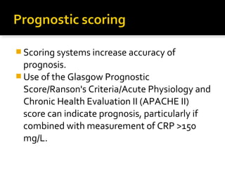
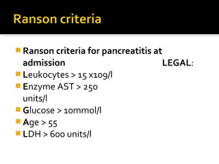
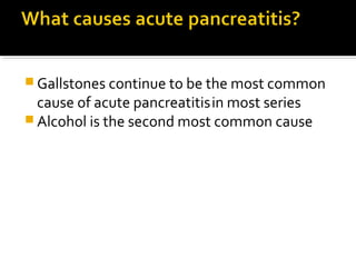
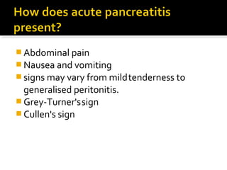
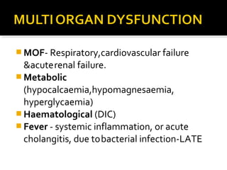
The document discusses acute pancreatitis, including causes, clinical features, diagnosis, severity grading, management, and prognosis. Gallstones and alcohol are the most common causes. Scoring systems like Ranson criteria and APACHE II can help indicate severity and prognosis. Management involves treatment of the underlying cause, supportive care, and monitoring for complications like pancreatic necrosis which may require intervention.
Acute Pancreatitis (According to American College of Gastroenterology 2013 gu...Jibran Mohsin
This Presentation focuses on definition, new classification, different scoring systems for severity, rationale for radiological signs and new management recommendations as per 2013 American College of Gastroenterology guidelines
Acute pancreatitis means inflammation of the pancreas that develops quickly. The main symptom is tummy (abdominal) pain. It usually settles in a few days but sometimes it becomes severe and very serious. The most common causes of acute pancreatitis are gallstones and drinking a lot of alcohol.
Revised Atlanta classification of Acute PancreatitisDr M Venkatesh
The most important change in Atlanta classification is the categorization of the various pancreatic collections.
In acute IEP, collections that do not have an enhancing capsule are called APFCs; after development of a capsule, they are referred to as
pseudocysts
In necrotizing pancreatitis,a collection without an enhancing capsule is called an ANC (usually in the first 4 weeks) and thereafter a WON, which has an enhancing capsule.
The most important distinction between collections in necrotizing pancreatitis and those associated with acute IEP is the presence of nonliquefied material in collections due to necrotizing pancreatitis.
Presentation describes the pathophysiology of Acute pancreatitis & its management in detail. Information is useful in practice although acute pancreatitis is quite rare
Approach to Management of Upper Gastrointestinal (GI) BleedingArun Vasireddy
Upper gastrointestinal bleeding is gastrointestinal bleeding in the upper gastrointestinal tract, commonly defined as bleeding arising from the esophagus, stomach, or duodenum. Blood may be observed in vomit (hematemesis) or in altered form in the stool (melena). Depending on the severity of the blood loss, there may be symptoms of insufficient circulating blood volume and shock. As a result, upper gastrointestinal bleeding is considered a medical emergency and typically requires hospital care for urgent diagnosis and treatment. Upper gastrointestinal bleeding can be caused by peptic ulcers, gastric erosions, esophageal varices, and some rarer causes such as gastric cancer.
The initial assessment includes measurement of the blood pressure and heart rate, as well as blood tests to determine hemoglobin concentration. In significant bleeding, fluid replacement is often required, as well as blood transfusion, before the source of bleeding can be determined by endoscopy of the upper digestive tract with an esophagogastroduodenoscopy. Depending on the source, endoscopic therapy can be applied to reduce rebleeding risk. Specific medical treatments (such as proton pump inhibitors for peptic ulcer disease) or procedures (such as TIPS for variceal hemorrhage) may be used. Recurrent or refractory bleeding may lead to need for surgery, although this has become uncommon as a result of improved endoscopic and medical treatment.
Abdominal pain is one of common problems
encountered by doctors, either in primary or
secondary health care (specialists). It may be
mild, but it may also a life-threatening sign. It
has been estimated that almost 50% adults have
experienced abdominal pain. In general, abdominal pain is categorized
based on the onset as acute or chronic pain.
Sudden onset of abdominal pain that lasts for less
than 24 hours is considered as acute abdominal
pain.
The problems of a surgeon
If 'I' operate 'and 'the' problem 'is' not 'surgical, Pt
exposed 'to' unnecessary 'risk ,'anesthetic,'etc.'
Risks 'greater' with 'concomitant 'illness,'older 'age'
If 'I' do 'not' operate 'and' problem 'is' surgical, 'patient 'at'
risk 'because' of 'wrong' therapy.'
Again 'the' older 'patient 'is' under 'greater' burden.'
this presentation includes anatomy physiology function of peritoneum ,also includes cause of peritonitis its severity ,various scoring system investigation and treatment.It includes the recent advancement and latest articles from latest books of surgery.
This is a lecture note for 5th-semester MBBS students. Lecture notes on hepatology, liver disease, and liver abscess. Introduction to a liver abscess, pyogenic liver abscess, causes, approach and management of liver abscess.
This is based on approach to a patient presenting to emergency department complaining of right hypochondriac pain. It includes anatomy, pathophysiology, epidemiology, clinical assessment, investigation, management, complication and disposition of a biliary infection.
The prostate is an exocrine gland of the male mammalian reproductive system
It is a walnut-sized gland that forms part of the male reproductive system and is located in front of the rectum and just below the urinary bladder
Function is to store and secrete a clear, slightly alkaline fluid that constitutes 10-30% of the volume of the seminal fluid that along with the spermatozoa, constitutes semen
A healthy human prostate measures (4cm-vertical, by 3cm-horizontal, 2cm ant-post ).
It surrounds the urethra just below the urinary bladder. It has anterior, median, posterior and two lateral lobes
It’s work is regulated by androgens which are responsible for male sex characteristics
Generalised disease of the prostate due to hormonal derangement which leads to non malignant enlargement of the gland (increase in the number of epithelial cells and stromal tissue)to cause compression of the urethra leading to symptoms (LUTS
Tom Selleck Health: A Comprehensive Look at the Iconic Actor’s Wellness Journeygreendigital
Tom Selleck, an enduring figure in Hollywood. has captivated audiences for decades with his rugged charm, iconic moustache. and memorable roles in television and film. From his breakout role as Thomas Magnum in Magnum P.I. to his current portrayal of Frank Reagan in Blue Bloods. Selleck's career has spanned over 50 years. But beyond his professional achievements. fans have often been curious about Tom Selleck Health. especially as he has aged in the public eye.
Follow us on: Pinterest
Introduction
Many have been interested in Tom Selleck health. not only because of his enduring presence on screen but also because of the challenges. and lifestyle choices he has faced and made over the years. This article delves into the various aspects of Tom Selleck health. exploring his fitness regimen, diet, mental health. and the challenges he has encountered as he ages. We'll look at how he maintains his well-being. the health issues he has faced, and his approach to ageing .
Early Life and Career
Childhood and Athletic Beginnings
Tom Selleck was born on January 29, 1945, in Detroit, Michigan, and grew up in Sherman Oaks, California. From an early age, he was involved in sports, particularly basketball. which played a significant role in his physical development. His athletic pursuits continued into college. where he attended the University of Southern California (USC) on a basketball scholarship. This early involvement in sports laid a strong foundation for his physical health and disciplined lifestyle.
Transition to Acting
Selleck's transition from an athlete to an actor came with its physical demands. His first significant role in "Magnum P.I." required him to perform various stunts and maintain a fit appearance. This role, which he played from 1980 to 1988. necessitated a rigorous fitness routine to meet the show's demands. setting the stage for his long-term commitment to health and wellness.
Fitness Regimen
Workout Routine
Tom Selleck health and fitness regimen has evolved. adapting to his changing roles and age. During his "Magnum, P.I." days. Selleck's workouts were intense and focused on building and maintaining muscle mass. His routine included weightlifting, cardiovascular exercises. and specific training for the stunts he performed on the show.
Selleck adjusted his fitness routine as he aged to suit his body's needs. Today, his workouts focus on maintaining flexibility, strength, and cardiovascular health. He incorporates low-impact exercises such as swimming, walking, and light weightlifting. This balanced approach helps him stay fit without putting undue strain on his joints and muscles.
Importance of Flexibility and Mobility
In recent years, Selleck has emphasized the importance of flexibility and mobility in his fitness regimen. Understanding the natural decline in muscle mass and joint flexibility with age. he includes stretching and yoga in his routine. These practices help prevent injuries, improve posture, and maintain mobilit
Ozempic: Preoperative Management of Patients on GLP-1 Receptor Agonists Saeid Safari
Preoperative Management of Patients on GLP-1 Receptor Agonists like Ozempic and Semiglutide
ASA GUIDELINE
NYSORA Guideline
2 Case Reports of Gastric Ultrasound
TEST BANK for Operations Management, 14th Edition by William J. Stevenson, Ve...kevinkariuki227
TEST BANK for Operations Management, 14th Edition by William J. Stevenson, Verified Chapters 1 - 19, Complete Newest Version.pdf
Anti ulcer drugs and their Advance pharmacology ||
Anti-ulcer drugs are medications used to prevent and treat ulcers in the stomach and upper part of the small intestine (duodenal ulcers). These ulcers are often caused by an imbalance between stomach acid and the mucosal lining, which protects the stomach lining.
||Scope: Overview of various classes of anti-ulcer drugs, their mechanisms of action, indications, side effects, and clinical considerations.
Title: Sense of Taste
Presenter: Dr. Faiza, Assistant Professor of Physiology
Qualifications:
MBBS (Best Graduate, AIMC Lahore)
FCPS Physiology
ICMT, CHPE, DHPE (STMU)
MPH (GC University, Faisalabad)
MBA (Virtual University of Pakistan)
Learning Objectives:
Describe the structure and function of taste buds.
Describe the relationship between the taste threshold and taste index of common substances.
Explain the chemical basis and signal transduction of taste perception for each type of primary taste sensation.
Recognize different abnormalities of taste perception and their causes.
Key Topics:
Significance of Taste Sensation:
Differentiation between pleasant and harmful food
Influence on behavior
Selection of food based on metabolic needs
Receptors of Taste:
Taste buds on the tongue
Influence of sense of smell, texture of food, and pain stimulation (e.g., by pepper)
Primary and Secondary Taste Sensations:
Primary taste sensations: Sweet, Sour, Salty, Bitter, Umami
Chemical basis and signal transduction mechanisms for each taste
Taste Threshold and Index:
Taste threshold values for Sweet (sucrose), Salty (NaCl), Sour (HCl), and Bitter (Quinine)
Taste index relationship: Inversely proportional to taste threshold
Taste Blindness:
Inability to taste certain substances, particularly thiourea compounds
Example: Phenylthiocarbamide
Structure and Function of Taste Buds:
Composition: Epithelial cells, Sustentacular/Supporting cells, Taste cells, Basal cells
Features: Taste pores, Taste hairs/microvilli, and Taste nerve fibers
Location of Taste Buds:
Found in papillae of the tongue (Fungiform, Circumvallate, Foliate)
Also present on the palate, tonsillar pillars, epiglottis, and proximal esophagus
Mechanism of Taste Stimulation:
Interaction of taste substances with receptors on microvilli
Signal transduction pathways for Umami, Sweet, Bitter, Sour, and Salty tastes
Taste Sensitivity and Adaptation:
Decrease in sensitivity with age
Rapid adaptation of taste sensation
Role of Saliva in Taste:
Dissolution of tastants to reach receptors
Washing away the stimulus
Taste Preferences and Aversions:
Mechanisms behind taste preference and aversion
Influence of receptors and neural pathways
Impact of Sensory Nerve Damage:
Degeneration of taste buds if the sensory nerve fiber is cut
Abnormalities of Taste Detection:
Conditions: Ageusia, Hypogeusia, Dysgeusia (parageusia)
Causes: Nerve damage, neurological disorders, infections, poor oral hygiene, adverse drug effects, deficiencies, aging, tobacco use, altered neurotransmitter levels
Neurotransmitters and Taste Threshold:
Effects of serotonin (5-HT) and norepinephrine (NE) on taste sensitivity
Supertasters:
25% of the population with heightened sensitivity to taste, especially bitterness
Increased number of fungiform papillae
Title: Sense of Smell
Presenter: Dr. Faiza, Assistant Professor of Physiology
Qualifications:
MBBS (Best Graduate, AIMC Lahore)
FCPS Physiology
ICMT, CHPE, DHPE (STMU)
MPH (GC University, Faisalabad)
MBA (Virtual University of Pakistan)
Learning Objectives:
Describe the primary categories of smells and the concept of odor blindness.
Explain the structure and location of the olfactory membrane and mucosa, including the types and roles of cells involved in olfaction.
Describe the pathway and mechanisms of olfactory signal transmission from the olfactory receptors to the brain.
Illustrate the biochemical cascade triggered by odorant binding to olfactory receptors, including the role of G-proteins and second messengers in generating an action potential.
Identify different types of olfactory disorders such as anosmia, hyposmia, hyperosmia, and dysosmia, including their potential causes.
Key Topics:
Olfactory Genes:
3% of the human genome accounts for olfactory genes.
400 genes for odorant receptors.
Olfactory Membrane:
Located in the superior part of the nasal cavity.
Medially: Folds downward along the superior septum.
Laterally: Folds over the superior turbinate and upper surface of the middle turbinate.
Total surface area: 5-10 square centimeters.
Olfactory Mucosa:
Olfactory Cells: Bipolar nerve cells derived from the CNS (100 million), with 4-25 olfactory cilia per cell.
Sustentacular Cells: Produce mucus and maintain ionic and molecular environment.
Basal Cells: Replace worn-out olfactory cells with an average lifespan of 1-2 months.
Bowman’s Gland: Secretes mucus.
Stimulation of Olfactory Cells:
Odorant dissolves in mucus and attaches to receptors on olfactory cilia.
Involves a cascade effect through G-proteins and second messengers, leading to depolarization and action potential generation in the olfactory nerve.
Quality of a Good Odorant:
Small (3-20 Carbon atoms), volatile, water-soluble, and lipid-soluble.
Facilitated by odorant-binding proteins in mucus.
Membrane Potential and Action Potential:
Resting membrane potential: -55mV.
Action potential frequency in the olfactory nerve increases with odorant strength.
Adaptation Towards the Sense of Smell:
Rapid adaptation within the first second, with further slow adaptation.
Psychological adaptation greater than receptor adaptation, involving feedback inhibition from the central nervous system.
Primary Sensations of Smell:
Camphoraceous, Musky, Floral, Pepperminty, Ethereal, Pungent, Putrid.
Odor Detection Threshold:
Examples: Hydrogen sulfide (0.0005 ppm), Methyl-mercaptan (0.002 ppm).
Some toxic substances are odorless at lethal concentrations.
Characteristics of Smell:
Odor blindness for single substances due to lack of appropriate receptor protein.
Behavioral and emotional influences of smell.
Transmission of Olfactory Signals:
From olfactory cells to glomeruli in the olfactory bulb, involving lateral inhibition.
Primitive, less old, and new olfactory systems with different path
Flu Vaccine Alert in Bangalore Karnatakaaddon Scans
As flu season approaches, health officials in Bangalore, Karnataka, are urging residents to get their flu vaccinations. The seasonal flu, while common, can lead to severe health complications, particularly for vulnerable populations such as young children, the elderly, and those with underlying health conditions.
Dr. Vidisha Kumari, a leading epidemiologist in Bangalore, emphasizes the importance of getting vaccinated. "The flu vaccine is our best defense against the influenza virus. It not only protects individuals but also helps prevent the spread of the virus in our communities," he says.
This year, the flu season is expected to coincide with a potential increase in other respiratory illnesses. The Karnataka Health Department has launched an awareness campaign highlighting the significance of flu vaccinations. They have set up multiple vaccination centers across Bangalore, making it convenient for residents to receive their shots.
To encourage widespread vaccination, the government is also collaborating with local schools, workplaces, and community centers to facilitate vaccination drives. Special attention is being given to ensuring that the vaccine is accessible to all, including marginalized communities who may have limited access to healthcare.
Residents are reminded that the flu vaccine is safe and effective. Common side effects are mild and may include soreness at the injection site, mild fever, or muscle aches. These side effects are generally short-lived and far less severe than the flu itself.
Healthcare providers are also stressing the importance of continuing COVID-19 precautions. Wearing masks, practicing good hand hygiene, and maintaining social distancing are still crucial, especially in crowded places.
Protect yourself and your loved ones by getting vaccinated. Together, we can help keep Bangalore healthy and safe this flu season. For more information on vaccination centers and schedules, residents can visit the Karnataka Health Department’s official website or follow their social media pages.
Stay informed, stay safe, and get your flu shot today!
Pulmonary Thromboembolism - etilogy, types, medical- Surgical and nursing man...VarunMahajani
Disruption of blood supply to lung alveoli due to blockage of one or more pulmonary blood vessels is called as Pulmonary thromboembolism. In this presentation we will discuss its causes, types and its management in depth.
MANAGEMENT OF ATRIOVENTRICULAR CONDUCTION BLOCK.pdfJim Jacob Roy
Cardiac conduction defects can occur due to various causes.
Atrioventricular conduction blocks ( AV blocks ) are classified into 3 types.
This document describes the acute management of AV block.
Couples presenting to the infertility clinic- Do they really have infertility...Sujoy Dasgupta
Dr Sujoy Dasgupta presented the study on "Couples presenting to the infertility clinic- Do they really have infertility? – The unexplored stories of non-consummation" in the 13th Congress of the Asia Pacific Initiative on Reproduction (ASPIRE 2024) at Manila on 24 May, 2024.
Prix Galien International 2024 Forum ProgramLevi Shapiro
June 20, 2024, Prix Galien International and Jerusalem Ethics Forum in ROME. Detailed agenda including panels:
- ADVANCES IN CARDIOLOGY: A NEW PARADIGM IS COMING
- WOMEN’S HEALTH: FERTILITY PRESERVATION
- WHAT’S NEW IN THE TREATMENT OF INFECTIOUS,
ONCOLOGICAL AND INFLAMMATORY SKIN DISEASES?
- ARTIFICIAL INTELLIGENCE AND ETHICS
- GENE THERAPY
- BEYOND BORDERS: GLOBAL INITIATIVES FOR DEMOCRATIZING LIFE SCIENCE TECHNOLOGIES AND PROMOTING ACCESS TO HEALTHCARE
- ETHICAL CHALLENGES IN LIFE SCIENCES
- Prix Galien International Awards Ceremony
Lung Cancer: Artificial Intelligence, Synergetics, Complex System Analysis, S...Oleg Kshivets
RESULTS: Overall life span (LS) was 2252.1±1742.5 days and cumulative 5-year survival (5YS) reached 73.2%, 10 years – 64.8%, 20 years – 42.5%. 513 LCP lived more than 5 years (LS=3124.6±1525.6 days), 148 LCP – more than 10 years (LS=5054.4±1504.1 days).199 LCP died because of LC (LS=562.7±374.5 days). 5YS of LCP after bi/lobectomies was significantly superior in comparison with LCP after pneumonectomies (78.1% vs.63.7%, P=0.00001 by log-rank test). AT significantly improved 5YS (66.3% vs. 34.8%) (P=0.00000 by log-rank test) only for LCP with N1-2. Cox modeling displayed that 5YS of LCP significantly depended on: phase transition (PT) early-invasive LC in terms of synergetics, PT N0—N12, cell ratio factors (ratio between cancer cells- CC and blood cells subpopulations), G1-3, histology, glucose, AT, blood cell circuit, prothrombin index, heparin tolerance, recalcification time (P=0.000-0.038). Neural networks, genetic algorithm selection and bootstrap simulation revealed relationships between 5YS and PT early-invasive LC (rank=1), PT N0—N12 (rank=2), thrombocytes/CC (3), erythrocytes/CC (4), eosinophils/CC (5), healthy cells/CC (6), lymphocytes/CC (7), segmented neutrophils/CC (8), stick neutrophils/CC (9), monocytes/CC (10); leucocytes/CC (11). Correct prediction of 5YS was 100% by neural networks computing (area under ROC curve=1.0; error=0.0).
CONCLUSIONS: 5YS of LCP after radical procedures significantly depended on: 1) PT early-invasive cancer; 2) PT N0--N12; 3) cell ratio factors; 4) blood cell circuit; 5) biochemical factors; 6) hemostasis system; 7) AT; 8) LC characteristics; 9) LC cell dynamics; 10) surgery type: lobectomy/pneumonectomy; 11) anthropometric data. Optimal diagnosis and treatment strategies for LC are: 1) screening and early detection of LC; 2) availability of experienced thoracic surgeons because of complexity of radical procedures; 3) aggressive en block surgery and adequate lymph node dissection for completeness; 4) precise prediction; 5) adjuvant chemoimmunoradiotherapy for LCP with unfavorable prognosis.
New Directions in Targeted Therapeutic Approaches for Older Adults With Mantl...i3 Health
i3 Health is pleased to make the speaker slides from this activity available for use as a non-accredited self-study or teaching resource.
This slide deck presented by Dr. Kami Maddocks, Professor-Clinical in the Division of Hematology and
Associate Division Director for Ambulatory Operations
The Ohio State University Comprehensive Cancer Center, will provide insight into new directions in targeted therapeutic approaches for older adults with mantle cell lymphoma.
STATEMENT OF NEED
Mantle cell lymphoma (MCL) is a rare, aggressive B-cell non-Hodgkin lymphoma (NHL) accounting for 5% to 7% of all lymphomas. Its prognosis ranges from indolent disease that does not require treatment for years to very aggressive disease, which is associated with poor survival (Silkenstedt et al, 2021). Typically, MCL is diagnosed at advanced stage and in older patients who cannot tolerate intensive therapy (NCCN, 2022). Although recent advances have slightly increased remission rates, recurrence and relapse remain very common, leading to a median overall survival between 3 and 6 years (LLS, 2021). Though there are several effective options, progress is still needed towards establishing an accepted frontline approach for MCL (Castellino et al, 2022). Treatment selection and management of MCL are complicated by the heterogeneity of prognosis, advanced age and comorbidities of patients, and lack of an established standard approach for treatment, making it vital that clinicians be familiar with the latest research and advances in this area. In this activity chaired by Michael Wang, MD, Professor in the Department of Lymphoma & Myeloma at MD Anderson Cancer Center, expert faculty will discuss prognostic factors informing treatment, the promising results of recent trials in new therapeutic approaches, and the implications of treatment resistance in therapeutic selection for MCL.
Target Audience
Hematology/oncology fellows, attending faculty, and other health care professionals involved in the treatment of patients with mantle cell lymphoma (MCL).
Learning Objectives
1.) Identify clinical and biological prognostic factors that can guide treatment decision making for older adults with MCL
2.) Evaluate emerging data on targeted therapeutic approaches for treatment-naive and relapsed/refractory MCL and their applicability to older adults
3.) Assess mechanisms of resistance to targeted therapies for MCL and their implications for treatment selection
5. Abdominal pain
Nausea and vomiting
signs may vary from mild tenderness to
generalised peritonitis.
Grey-Turner's sign
Cullen's sign
6. MOF- Respiratory,cardiovascular failure
´ renal failure.
Metabolic
(hypocalcaemia,hypomagnesaemia,
hyperglycaemia)
Haematological (DIC)
Fever - systemic inflammation, or acute
cholangitis, due to bacterial infection-LATE
7. a recognised entity
occurs in cases of shock of unknown origin,
during the postoperative period,
in renal transplant
peritoneal dialysis patients,
and in diabetic ketoacidosis.
8.
9. Typical clinical features
+ a high plasma concentration of pancreatic
enzymes
serum amylase concentrations decline quickly
over two to three days
Relate it to onset of abdominal pain
10. several non-pancreatic diseases (visceral
perforation, small bowel obstruction and
ischaemia, leaking aortic aneurysm, ectopic
pregnancy),
tumours also secrete amylase
11. superior sensitivity and specificity
preferable to serum amylase for the
diagnosis of acute pancreatitis
12. History
physical examination,
liver function tests,
and biliary ultrasonography will indicate the
correct cause in most cases.
If not, follow-up investigations, should
include fasting plasma lipids and calcium, viral
antibody titres, and repeat biliary
ultrasonography.
13.
14. detect free air in the abdomen,
colon cut-off sign, a sentinel loop, or an ileus.
calcifications within the pancreas - chronic
pancreatitis.
15. Plain radiographs clues
alternative abdominal emergency,
detect and stage complications of acute
severe pancreatitis, especially pancreatic
necrosis
16. pancreatic necrosis cannot be appreciated
until at least three days after the onset of
symptoms.
Patients with persisting organ failure,
signs of sepsis,
clinical deterioration occurring after an initial
improvement
Follow-up scans
17. also provide prognostic information based on
the following grading scale developed by
Balthazar:
A - Normal
B - Enlargement
C - Peripancreatic inflammation
D - Single fluid collection
E - Multiple fluid collections
18. The chances of infection and death are
virtually nil in grades A and B
steadily increase in grades C through E.
Patients with grade E pancreatitis have a 50%
chance of developing an infection and a 15%
chance of dying.
19. only be used in the following situations:
severe acute pancreatitis secondary to stones
biliary pancreatitis - worsening jaundice and
clinical deterioration despite maximal supportive
therapy.
with sphincterotomy and stone extraction, may
reduce the length of hospital stay, the
complication rate, and, possibly, the mortality
rate.
in the setting of suspected SOD (sphincter of
oddi dysfunction)
20. PRSS1 genetic testing is recommended in symptomatic
patients with any of the following features
n
Recurrent attacks of acute pancreatitis for which no cause
has been found
Idiopathic chronic pancreatitis
A family history of pancreatitis in a first or second degree
relative
Unexplained pancreatitis occurring in a child
21.
22. Supplemental oxygen
adequate fluid resuscitation
A urinary catheter
Central venous monitoring
All patients with severe acute pancreatitis
should be managed in a high dependency unit or
intensive therapy unit.
opiate analgesia.
A nasogastric tube is not useful routinely but
may be helpful if protracted vomiting occurs in
the presence of a radiologically demonstrated
ileus.
23. All patients with severe acute pancreatitis
should be managed in a high dependency
unit or intensive therapy unit with full
monitoring and systems support
(recommendation grade B).
25. Patients with alcohol-induced pancreatitis
may need alcohol-withdrawal prophylaxis.
Lorazepam, thiamine, folic acid, and multi-vitamins
are generally used in this group of
patients.
26.
27. imaging of the common bile duct is required.
If the presence of stones in the common bile duct
is confirmed, a cholecystectomy with common
bile duct exploration (either surgical or
postoperatively with endoscopic retrograde
cholangiopancreatography [ERCP]) should be
performed during the same hospitalisation in
mild to moderate disease soon after the attack
resolves.
A longer delay, even of a few weeks, is associated
with a high recurrence (80%) of acute pancreatitis
and re-admission
28. If the pancreatitis is severe, some allow a few
months for the inflammation to completely
resolve before performing a cholecystectomy
29. In patients who are not candidates for surgery
because of comorbidities with a high American
Association of Anesthesiology (ASA) index,
sepsis, or severe disease,
ERCP must be considered.
Urgent ERCP is indicated in patients with biliary
sepsis and obstructive jaundice that show no
improvement in 48 hours after the onset of the
attack.
ERCP is a diagnostic and therapeutic
intervention
30. If mild to moderate pancreatitis is found,
cholecystectomy with intra-operative
cholangiogram should be performed but the
pancreas should be left alone.
For severe pancreatitis, the lesser sac should
be opened and the pancreas fully inspected.
Some surgeons place drains and irrigating
catheter around the pancreas.
31. during the same hospital admission,
unless a clear plan has been made for
definitive treatment within the next two
weeks (recommendation grade C).
should be delayed in patients with severe
acute pancreatitis until signs of lung injury
and systemic disturbance have resolved.
32.
33. infected necrosis -high mortality rate (40%).
diagnosed either by the presence of gas
within the pancreatic collection
or by fine needle aspiration
34. All patients with persistent symptoms and >
30% pancreatic necrosis,
and those with smaller areas of necrosis and
clinical suspicion of sepsis,
should undergo image guided fine needle
aspiration to obtain material for culture 7–14
days after the onset of pancreatitis
(recommendation grade B).
37. Some trials show benefit, others do not.
At present there is no consensus on this
issue. If antibiotic prophylaxis is used, it
should be given for a maximum of 14 days
40. No conclusive evidence to support the use of
enteral nutrition in all patients with severe
acute pancreatitis.
enteral route is preferred if that can be
tolerated (recommendation grade A).
nasogastric route effective in 80% of cases
(recommendation grade B).
41. The use of enteral feeding may be limited by
ileus. If this persists for more than five days,
parenteral nutrition will be required.
42.
43. clinical impression of severity,
obesity, or APACHE II>8 in the first 24 hours
of admission, and
C reactive protein >150 mg/l,
Glasgow score 3 or more,
or persisting organ failure after 48 hours in
hospital (recommendation grade B).
44. The definitions of severity, as proposed in the
Atlanta criteria, should be used.
organ failure present within the first week,
which resolves within 48 hours, should not be
considered an indicator of a severe attack
(recommendation grade B).
45. Bradley reported the criteria for severe acute pancreatitis
developed at the International Symposium on Acute Pancreatitis
held in Atlanta, Georgia.
Criteria for severe acute pancreatitis - one or more
of the following:
(1) Ranson score on admission >= 3 (or during
the first 48 hours)
(2) APACHE II score >= 8 at any time during
course
(3) presence of one or more organ failures
(4) presence of one or more local complications
46. Scoring systems increase accuracy of
prognosis.
Use of the Glasgow Prognostic
Score/Ranson's Criteria/Acute Physiology and
Chronic Health Evaluation II (APACHE II)
score can indicate prognosis, particularly if
combined with measurement of CRP >150
mg/L.
50. uses age, and 7 laboratory values collected
during the first 48 hours following admission,
to predict severe pancreatitis.
It is applicable to both biliary and alcoholic
pancreatitis.
The score can range from 0 to 8. If the score is
>2, the likelihood of severe pancreatitis is
high. If the score is <3, severe pancreatitis is
unlikely.
51. Age >55 years
WBC >15 x 109/L
Urea >16 mmol/L
Glucose >10 mmol/L
pO2 <8 kPa (60 mm Hg)
Albumin <32 g/L
Calcium <2 mmol/L
LDH >600 units/L
AST/ALT >200 units
52. The majority of patients with acute pancreatitis will
improve within 3 to 7 days of conservative
management.
The cause should be identified,
a plan to prevent recurrence should be initiated
before the patient is discharged.
In gallstone pancreatitis, a cholecystectomy should be
considered before discharge in mild cases and a few
months after the discharge date in patients with
severe symptoms.
In patients who are not candidates for surgery,
endoscopic retrograde cholangiopancreatography
(ERCP) must be considered.
54. Organ failures include:
(1) shock (systolic blood pressure less than 90
mm Hg)
(2) pulmonary insufficiency (PaO2 <= 60 mm Hg
on room air)
(3) renal failure (serum creatinine > 2 mg/dL after
fluid replacement)
(4) gastrointestinal bleeding, with > 500 mL
estimated loss within 24 hours
(5) DIC (thrombocytopenia and
hypofibrinogenemia and fibrin split products)
(6) severe hypocalemia (<= 7.5 mg/dL)