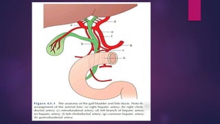
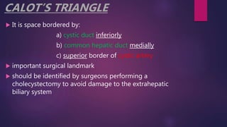
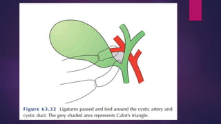
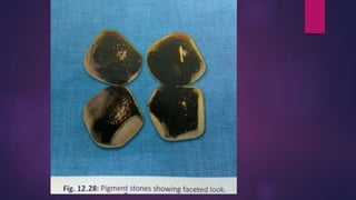
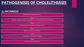
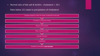
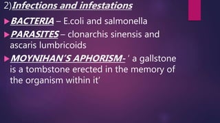
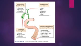
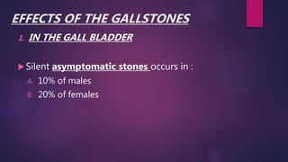
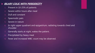
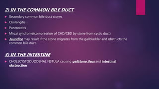
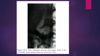
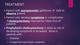
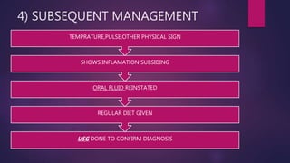
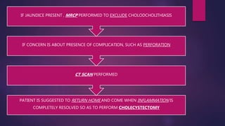
CHOLELITHIASIS
SATEESH
MBBS FINAL PROFF
ANATOMY OF GALL BLADDER
 Pear shaped structure
 Lies on the underside of the liver in the main liver scissura
at the junction of right and left lobe of the liver
 7.5-12 cm long
 NORMAL CAPACITY  25-30 ml
 Anatomically divided into three parts:
a) FUNDUS
b) BODY
c) NECK
Muscle fibres in the wall of the gall bladder
are arranged in crisscross manner, being
particularly well developed in neck.
Cystic duct
1. 3cm in length
2. Lumen is 1-3cm in length
3. Mucosa of cystic duct arranged in variable
folds known as “VALVES OF HEISTER”
4) Wall of cystic duct surrounded by sphincteric structure
called “VALVES OF HEISTER”
 Common hepatic duct
1. usually 2.5cm long
2. Formed by union of right and left hepatic ducts
 Common bile duct
1. 7.5 cm long
2. Formed by junction of cystic and common hepatic ducts
CALOT’S TRIANGLE
 It is space bordered by:
a) cystic duct inferiorly
b) common hepatic duct medially
c) superior border of cystic artery
 important surgical landmark
 should be identified by surgeons performing a
cholecystectomy to avoid damage to the extrahepatic
biliary system
FUNCTIONS OF GALL BLADDER
Reservoir for bile
Concentration of bile by active
absorption of water, sodium chloride and
bicarbonate by the mucous membrane of
the gall bladder.
Secretion of mucous approx 20ml/day
GALL STONES(CHOLELITHIASIS)
 Most common biliary pathology.
 Affects 10-15% of the population in western
societies.
 Asymptomatic in more then 80% of cases
 1-2% of asymptomatic patients will develop
symptoms requiring surgery per year.
 It makes cholecystectomy one of the most
common operation performed by general
surgeons
TYPES OF GALLSTONES
 There are three types of gallstones:
1. CHOLESTROL STONES (6%) : Radiating crystal
like appearance
2. MIXED STONES(90%) : Contains cholesterol ,
calcium salts of phosphate & carbonate
,proteins , bile acid, bile pigments,
phospholipids
3. PIGMENT STONES : Small, black or greenish
black, may be soft & sludge like or hard & coral
like.
INCIDENCE
5F’S
1. FAT
2. FERTILE
3. FLATULENT
4. FEMALE
5. FEMALE OF FIFTY
Can occur in BOTH SEXES
Quite often in EARLY AGES(even in
childhood)
More common in OLD AGES
MULTIFACTORIAL
Common in western countries and
northern INDIA
PATHOGENISIS OF CHOLELITHIASIS
1) METABOLIC
Stone formation takes place.
Precipitation of insoluble cholesterol
Reduces micelle concentration
Altered levels of cholesterol, bile salts, lecithin
Solubility is determined by concentration of cholesterol, bile salts and lecithin
Cholesterol is synthesized in liver.
• Normal ratio of bile salt & lecithin : cholesterol = 25:1
Ratio below 13:1 leads to precipitation of cholesterol
Cholesterol monohydrate stone formation
Crystallization
Insoluble cholesterol
Makes inadequate micelle
Bile gets supersaturated
If cholesterol component increases
Insoluble cholesterol is within the soluble micelle(lecithin & bile salts)
2)Infections and infestations
BACTERIA – E.coli and salmonella
PARASITES – clonarchis sinensis and
ascaris lumbricoids
MOYNIHAN’S APHORISM- ‘ a gallstone
is a tombstone erected in the memory of
the organism within it’
3) BILE STAISIS
Occurs due to :
a) ESTROGEN therapy
b) PREGNANCY
c) VAGOTOMY
d) Patient who are on LONG TERM I.V
FLUID
4) INCREASED BILIRUBIN
PRODUCTION
 Due to any of the causes of haemolysis as in :
A. HERIDITARY SPHEROCYTOSIS
B. SICKEL CELL ANEMIA
C. THALESSEMIA
D. MALARIA
E. CIRRHOSIS
 Rarely centre of the stone contains radiolucent gas, it
is either
i. TRIRADIATE ( MERCEDES BENZ SIGN)
ii. BIRADIATE (SEAGULL SIGN)
 1) 10% GALLSTONES are RADIO-OPAQUE
2) 90% GALLSTONES are RADIOLUCENT
 In some of the cases gallbladder may be filled with
‘TOOTHPASTE’ like material i.e. mixture of calcium
carbonate and phosphate, which on X-RAY(plain)
looks like an OPACIFIED gallbladder( also called
LIMEY GALLBLADDER)
CASUAL FACTORS IN GALLSTONE
FORMATION
PIGMENT STONE contain less than
30% cholestrol.
It is of two types:
i. BLACK stones
ii. BROWN stones
BLACK STONES
 Composed of insoluble bilirubin pigment polymer , mixed with
calcium phosphate and calcium bicarbonate.
 20%-30% stones are black
 Incidence rise with age
 BLACK stones are associated with :
a. Haemolysis
b. Usually hereditary
c. Spherocytosis
d. Sickle cell disease
 Patient with CIRRHOSIS have a higher instance of PIGMENTED
STONES
BROWN STONES
 Contains:
i. Calcium bilirubinate
ii. Calcium palmitate
iii. Calcium stearate
iv. Cholesterol
 Rare in gall bladder
 Form in bile duct
 Also associated with the presence of foreign bodies with
the bile ducts such as parasites like ascaris lumbricoids.
EFFECTS OF THE GALLSTONES
1. IN THE GALL BLADDER
 Silent asymptomatic stones occurs in :
A. 10% of males
B. 20% of females
 BILARY COLIC WITH PERIODICITY
1. Present in 10-25% of patients
2. Severe within hours after meal
3. Dull and constant
4. Spasmodic pain
5. Severe in nature
6. In right upper quadrant and epigastrium, radiating towards chest and
shoulder
7. Generally starts at night, wakes the patient.
8. Precipitated by heavy meal
9. Fever and increased WBC count may be observed.
2) IN THE COMMON BILE DUCT
 Secondary common bile duct stones
 Cholangitis
 Pancreatitis
 Mirzzi syndrome(compression of CHD/CBD by stone from cystic duct)
 Jaundice may result if the stone migrates from the gallbladder and obstructs the
common bile duct.
3) IN THE INTESTINE
 CHOLECYSTODUODENAL FISTULA causing gallstone ileus and intestinal
obstruction
EFFECTS AND COMPLICATIONS OF
GALLSTONES
 BILARY COLIC
 ACUTE CHOLECYSTITIS
 CHRONIC CHOLECYSTITIS
 GALLSTONE PANCREATITIS
 OBSTRUCTIVE JAUNDICE
 ACUTE CHOLANGITIS
 INTESTINAL OBSTRUCTION ( gallstone ileus)
 MUCOCELE / EMPYEMA OF GALL BLADDER
BILARY COLIC
 Episodic pain in epigastrium
 Pain radiates to lower pole of scapula
 Patients suffers from :
1. Sweating
2. Nausea
3. Vomiting
 Intermittent jaundice with pale stool and dark
urine.
Differential diagnosis
1. Renal colic
2. Intestinal obstruction
3. Angina
Pain episode may resolve
1. when stone is passed into common bile duct
2. When stone falls back into the gall bladder
ACUTE CHOLECYSTITIS
 Severe
 Constant
 Localized right hypochondrium pain
 CLINICAL FEATURES :
a. Fever
b. Toxaemia
c. Rigors
d. Leucocytosis
e. Tenderness in right hypochondrium
f. Murphy’s sign
g. Palpable gallbladder
CHRONIC CHOLECYSTITIS
 Repeated inflammation resulting in fibrosis and
thickening of gallbladder
 Longstanding dyspepsia with episodic
cholecystitis
 DIFFERENTIAL DIAGNOSIS
1. Peptic ulcer
2. Hiatus hernia
3. Angina
 COMPLICATIONS
a) Empyema
b) Perforation
c) Obstructive jaundice
d) Acalculous cholecystitis
 MURPHY’S SIGN : In acute phase, the patient may
have right upper quadrant tenderness that is
exacerbated during inspiration by the examiner’s
right subcoastal palpation.
 Minor episodes of same discomfort may intermittently
during the day.
 Dyspeptic symptoms may coexist and may be worse after
such an attack
 As pain resolves, patient improves and is able to eat and
drink again
 Other symptoms include :
1. Dyspepsia
2. Food intolerance
3. Alteration in biliary frequency
GALLSTONE PANCREATITIS
Due to transient blocking of
ampulla of vater by stone.
Especially when stones are small
and numerous.
OBSTRUCTIVE JAUNDICE
 Acute in onset
 Patient having History of pain
 Non palpable gallbladder
 Courvoisier’s law( Courvoisier’s sign): it states that
in presence of palpably enlarged gallbladder which is
non tender & accompanied with mild painless
jaundice, the cause is unlikely to be gallstones.
ACUTE CHOLANGITIS
Infection of the bile duct
usually caused by bacteria ascending from
its junction with the duodenum.
 tends to occur if the bile duct is already
partially obstructed by gallstones.
CAUSES
1. FEVER
2. JAUNDICE
PREDISPOSING FACTORS
1. Stone in common bile duct
2. Biliary stricture
3. Post-ERCP
4. Post- biliary reconstructive procedure
Antibiotics and resuscitation followed by
decompression of biliary tree.
GALLSTONE ILEUS
 Obstruction of small bowel by a large gallstone
 SYMPTOMS
a) Vomiting
b) Abdominal pain
c) Distention
 SIGNS
a) Abdominal distention
b) Obstructive bowel sounds
TREATMENT
Laparotomy
Removal of stone from small bowel
DIAGNOSIS OF CHOLELITHIASIS
 Diagnosis is based on:
1. History
2. Physical examination
3. Confirmatory radiological studies .i.e.
1. transabdominal ultrasonography
2. radionuclide scan
 A positive murphy’s sign suggests
1. Acute inflammation
2. Leucocytosis
3. Elevated liver function tests
A mass may be palpable as the
omentum walls of an inflamed gall
bladder
If resolution does not occur, an
empyema of gall bladder may result.
Walls of gall bladder may become
necrotic and perforate , with
development of localised peritonitis.
INVESTIGATION
 ULTASOUND ABDOMEN
 PLAIN X-RAY
 LIVER FUNCTION TEST
 WBC COUNT
 CT SCAN ABDOMEN
 LAPROSCOPIC CHOLECYSTECTOMY
 OPEN CHOLECYSTECTOMY
TREATMENT
 Patient with asymptomatic gallstones  Safe to
observe patient
 Patient who develop symptoms or complication
cholecystectomy reserved for these kind of
patients
 Prophylactic cholecystectomy is done as risk of
developing symptoms is increased , done in
patients with:
a) Diabetes
b) Congenital haemolytic anaemia
Cholecystectomy is treatment of
choice in patients with :
i. Biliary colic
ii. Cholecystitis
In more than 90% of cases :
Symptoms of acute cholecystitis
subside with conservative measures
NON – OPERATIVE TREATMENT
Based on four principles :
1)NIL PER MOUTH (NPO) & I.V
FLUID administration until pain
resolves
2)Administration of ANALGESICS
3) ADMINISTRATION OF ANTIBIOTICS
 CYSTIC DUCT is blocked in most cases
 concentration of antibiotics in serum is more
important than its concentration in bile.
 Broad spectrum antibiotics is more effective
against gram –ve bacteria
eg: a) gentamicin
b) cefazolin
c) cefuroxime
4) SUBSEQUENT MANAGEMENT
USG DONE TO CONFIRM DIAGNOSIS
REGULAR DIET GIVEN
ORAL FLUID REINSTATED
SHOWS INFLAMATION SUBSIDING
TEMPRATURE,PULSE,OTHER PHYSICAL SIGN
PATIENT IS SUGGESTED TO RETURN HOME AND COME WHEN INFLAMMATION IS
COMPLETELY RESOLVED SO AS TO PERFORM CHOLECYSTECTOMY
CT SCAN PERFORMED
IF CONCERN IS ABOUT PRESENCE OF COMPLICATION, SUCH AS PERFORATION
IF JAUNDICE PRESENT , MRCP PERFORMED TO EXCLUDE CHOLODCHOLITHIASIS
CHOLECYSTECTOMY
 PREPRATION FOR OPERATION
1. Full blood count
2. Renal profile & liver function test
3. Prothrombin time
4. Chest X-RAY & ECG(if over 45yr or medically
indicated)
5. Antibiotic prophylaxis
6. Deep vein thrombosis prophylaxis
7. Informed consent
LAPROSCOPIC CHOLECYSTECTOMY
 Preparation and indications are same, either performed by laparoscopy or
open technique
 It is procedure of choice for majority of patients.
 Key is :
1. Open surgery
2. Identification and safe dissection of CALOT’S TRIANGLE
OPEN CHOLECYSTECTOMY
For patient in whom a laparoscopic
approach is not indicated.
For patient whom conversion from
laparoscopic approach is required an
open cholecystectomy is performed.
It is done through right subcoastal
KOCHER’S INCISION.
COMPLICATIONS OF
CHOLECYSTECTOMY
EARLY BILIARY COMPLICATIONS
1. Bile leak
2. Hematoma
3. Abscess
4. Dropped stones
5. Inadequate cholecystectomy
LATE BILIARY COMPLICATIONS
1. PORT SITE HERNIA
2. POSTOPERATIVE PAIN
3. CHRONIC INFLAMMATION
MECHANICAL FACTORS
1. Common bile duct stricture
2. Retained stones
3. Fistula formation
4. Papillary disorders
THANKS

Cholelithiasis

  • 1.
  • 2.
    ANATOMY OF GALLBLADDER  Pear shaped structure  Lies on the underside of the liver in the main liver scissura at the junction of right and left lobe of the liver  7.5-12 cm long  NORMAL CAPACITY  25-30 ml  Anatomically divided into three parts: a) FUNDUS b) BODY c) NECK
  • 3.
    Muscle fibres inthe wall of the gall bladder are arranged in crisscross manner, being particularly well developed in neck. Cystic duct 1. 3cm in length 2. Lumen is 1-3cm in length 3. Mucosa of cystic duct arranged in variable folds known as “VALVES OF HEISTER”
  • 4.
    4) Wall ofcystic duct surrounded by sphincteric structure called “VALVES OF HEISTER”  Common hepatic duct 1. usually 2.5cm long 2. Formed by union of right and left hepatic ducts  Common bile duct 1. 7.5 cm long 2. Formed by junction of cystic and common hepatic ducts
  • 6.
    CALOT’S TRIANGLE  Itis space bordered by: a) cystic duct inferiorly b) common hepatic duct medially c) superior border of cystic artery  important surgical landmark  should be identified by surgeons performing a cholecystectomy to avoid damage to the extrahepatic biliary system
  • 8.
    FUNCTIONS OF GALLBLADDER Reservoir for bile Concentration of bile by active absorption of water, sodium chloride and bicarbonate by the mucous membrane of the gall bladder. Secretion of mucous approx 20ml/day
  • 9.
    GALL STONES(CHOLELITHIASIS)  Mostcommon biliary pathology.  Affects 10-15% of the population in western societies.  Asymptomatic in more then 80% of cases  1-2% of asymptomatic patients will develop symptoms requiring surgery per year.  It makes cholecystectomy one of the most common operation performed by general surgeons
  • 11.
    TYPES OF GALLSTONES There are three types of gallstones: 1. CHOLESTROL STONES (6%) : Radiating crystal like appearance 2. MIXED STONES(90%) : Contains cholesterol , calcium salts of phosphate & carbonate ,proteins , bile acid, bile pigments, phospholipids 3. PIGMENT STONES : Small, black or greenish black, may be soft & sludge like or hard & coral like.
  • 13.
    INCIDENCE 5F’S 1. FAT 2. FERTILE 3.FLATULENT 4. FEMALE 5. FEMALE OF FIFTY
  • 14.
    Can occur inBOTH SEXES Quite often in EARLY AGES(even in childhood) More common in OLD AGES MULTIFACTORIAL Common in western countries and northern INDIA
  • 15.
    PATHOGENISIS OF CHOLELITHIASIS 1)METABOLIC Stone formation takes place. Precipitation of insoluble cholesterol Reduces micelle concentration Altered levels of cholesterol, bile salts, lecithin Solubility is determined by concentration of cholesterol, bile salts and lecithin Cholesterol is synthesized in liver.
  • 16.
    • Normal ratioof bile salt & lecithin : cholesterol = 25:1 Ratio below 13:1 leads to precipitation of cholesterol Cholesterol monohydrate stone formation Crystallization Insoluble cholesterol Makes inadequate micelle Bile gets supersaturated If cholesterol component increases Insoluble cholesterol is within the soluble micelle(lecithin & bile salts)
  • 17.
    2)Infections and infestations BACTERIA– E.coli and salmonella PARASITES – clonarchis sinensis and ascaris lumbricoids MOYNIHAN’S APHORISM- ‘ a gallstone is a tombstone erected in the memory of the organism within it’
  • 18.
    3) BILE STAISIS Occursdue to : a) ESTROGEN therapy b) PREGNANCY c) VAGOTOMY d) Patient who are on LONG TERM I.V FLUID
  • 19.
    4) INCREASED BILIRUBIN PRODUCTION Due to any of the causes of haemolysis as in : A. HERIDITARY SPHEROCYTOSIS B. SICKEL CELL ANEMIA C. THALESSEMIA D. MALARIA E. CIRRHOSIS
  • 20.
     Rarely centreof the stone contains radiolucent gas, it is either i. TRIRADIATE ( MERCEDES BENZ SIGN) ii. BIRADIATE (SEAGULL SIGN)  1) 10% GALLSTONES are RADIO-OPAQUE 2) 90% GALLSTONES are RADIOLUCENT  In some of the cases gallbladder may be filled with ‘TOOTHPASTE’ like material i.e. mixture of calcium carbonate and phosphate, which on X-RAY(plain) looks like an OPACIFIED gallbladder( also called LIMEY GALLBLADDER)
  • 21.
    CASUAL FACTORS INGALLSTONE FORMATION PIGMENT STONE contain less than 30% cholestrol. It is of two types: i. BLACK stones ii. BROWN stones
  • 22.
    BLACK STONES  Composedof insoluble bilirubin pigment polymer , mixed with calcium phosphate and calcium bicarbonate.  20%-30% stones are black  Incidence rise with age  BLACK stones are associated with : a. Haemolysis b. Usually hereditary c. Spherocytosis d. Sickle cell disease  Patient with CIRRHOSIS have a higher instance of PIGMENTED STONES
  • 23.
    BROWN STONES  Contains: i.Calcium bilirubinate ii. Calcium palmitate iii. Calcium stearate iv. Cholesterol  Rare in gall bladder  Form in bile duct  Also associated with the presence of foreign bodies with the bile ducts such as parasites like ascaris lumbricoids.
  • 25.
    EFFECTS OF THEGALLSTONES 1. IN THE GALL BLADDER  Silent asymptomatic stones occurs in : A. 10% of males B. 20% of females
  • 26.
     BILARY COLICWITH PERIODICITY 1. Present in 10-25% of patients 2. Severe within hours after meal 3. Dull and constant 4. Spasmodic pain 5. Severe in nature 6. In right upper quadrant and epigastrium, radiating towards chest and shoulder 7. Generally starts at night, wakes the patient. 8. Precipitated by heavy meal 9. Fever and increased WBC count may be observed.
  • 27.
    2) IN THECOMMON BILE DUCT  Secondary common bile duct stones  Cholangitis  Pancreatitis  Mirzzi syndrome(compression of CHD/CBD by stone from cystic duct)  Jaundice may result if the stone migrates from the gallbladder and obstructs the common bile duct. 3) IN THE INTESTINE  CHOLECYSTODUODENAL FISTULA causing gallstone ileus and intestinal obstruction
  • 28.
    EFFECTS AND COMPLICATIONSOF GALLSTONES  BILARY COLIC  ACUTE CHOLECYSTITIS  CHRONIC CHOLECYSTITIS  GALLSTONE PANCREATITIS  OBSTRUCTIVE JAUNDICE  ACUTE CHOLANGITIS  INTESTINAL OBSTRUCTION ( gallstone ileus)  MUCOCELE / EMPYEMA OF GALL BLADDER
  • 29.
    BILARY COLIC  Episodicpain in epigastrium  Pain radiates to lower pole of scapula  Patients suffers from : 1. Sweating 2. Nausea 3. Vomiting  Intermittent jaundice with pale stool and dark urine.
  • 30.
    Differential diagnosis 1. Renalcolic 2. Intestinal obstruction 3. Angina Pain episode may resolve 1. when stone is passed into common bile duct 2. When stone falls back into the gall bladder
  • 31.
    ACUTE CHOLECYSTITIS  Severe Constant  Localized right hypochondrium pain  CLINICAL FEATURES : a. Fever b. Toxaemia c. Rigors d. Leucocytosis e. Tenderness in right hypochondrium f. Murphy’s sign g. Palpable gallbladder
  • 32.
    CHRONIC CHOLECYSTITIS  Repeatedinflammation resulting in fibrosis and thickening of gallbladder  Longstanding dyspepsia with episodic cholecystitis  DIFFERENTIAL DIAGNOSIS 1. Peptic ulcer 2. Hiatus hernia 3. Angina
  • 33.
     COMPLICATIONS a) Empyema b)Perforation c) Obstructive jaundice d) Acalculous cholecystitis  MURPHY’S SIGN : In acute phase, the patient may have right upper quadrant tenderness that is exacerbated during inspiration by the examiner’s right subcoastal palpation.
  • 34.
     Minor episodesof same discomfort may intermittently during the day.  Dyspeptic symptoms may coexist and may be worse after such an attack  As pain resolves, patient improves and is able to eat and drink again  Other symptoms include : 1. Dyspepsia 2. Food intolerance 3. Alteration in biliary frequency
  • 36.
    GALLSTONE PANCREATITIS Due totransient blocking of ampulla of vater by stone. Especially when stones are small and numerous.
  • 37.
    OBSTRUCTIVE JAUNDICE  Acutein onset  Patient having History of pain  Non palpable gallbladder  Courvoisier’s law( Courvoisier’s sign): it states that in presence of palpably enlarged gallbladder which is non tender & accompanied with mild painless jaundice, the cause is unlikely to be gallstones.
  • 38.
    ACUTE CHOLANGITIS Infection ofthe bile duct usually caused by bacteria ascending from its junction with the duodenum.  tends to occur if the bile duct is already partially obstructed by gallstones. CAUSES 1. FEVER 2. JAUNDICE
  • 39.
    PREDISPOSING FACTORS 1. Stonein common bile duct 2. Biliary stricture 3. Post-ERCP 4. Post- biliary reconstructive procedure Antibiotics and resuscitation followed by decompression of biliary tree.
  • 40.
    GALLSTONE ILEUS  Obstructionof small bowel by a large gallstone  SYMPTOMS a) Vomiting b) Abdominal pain c) Distention  SIGNS a) Abdominal distention b) Obstructive bowel sounds
  • 41.
  • 42.
    DIAGNOSIS OF CHOLELITHIASIS Diagnosis is based on: 1. History 2. Physical examination 3. Confirmatory radiological studies .i.e. 1. transabdominal ultrasonography 2. radionuclide scan  A positive murphy’s sign suggests 1. Acute inflammation 2. Leucocytosis 3. Elevated liver function tests
  • 43.
    A mass maybe palpable as the omentum walls of an inflamed gall bladder If resolution does not occur, an empyema of gall bladder may result. Walls of gall bladder may become necrotic and perforate , with development of localised peritonitis.
  • 44.
    INVESTIGATION  ULTASOUND ABDOMEN PLAIN X-RAY  LIVER FUNCTION TEST  WBC COUNT  CT SCAN ABDOMEN  LAPROSCOPIC CHOLECYSTECTOMY  OPEN CHOLECYSTECTOMY
  • 47.
    TREATMENT  Patient withasymptomatic gallstones  Safe to observe patient  Patient who develop symptoms or complication cholecystectomy reserved for these kind of patients  Prophylactic cholecystectomy is done as risk of developing symptoms is increased , done in patients with: a) Diabetes b) Congenital haemolytic anaemia
  • 48.
    Cholecystectomy is treatmentof choice in patients with : i. Biliary colic ii. Cholecystitis In more than 90% of cases : Symptoms of acute cholecystitis subside with conservative measures
  • 49.
    NON – OPERATIVETREATMENT Based on four principles : 1)NIL PER MOUTH (NPO) & I.V FLUID administration until pain resolves 2)Administration of ANALGESICS
  • 50.
    3) ADMINISTRATION OFANTIBIOTICS  CYSTIC DUCT is blocked in most cases  concentration of antibiotics in serum is more important than its concentration in bile.  Broad spectrum antibiotics is more effective against gram –ve bacteria eg: a) gentamicin b) cefazolin c) cefuroxime
  • 51.
    4) SUBSEQUENT MANAGEMENT USGDONE TO CONFIRM DIAGNOSIS REGULAR DIET GIVEN ORAL FLUID REINSTATED SHOWS INFLAMATION SUBSIDING TEMPRATURE,PULSE,OTHER PHYSICAL SIGN
  • 52.
    PATIENT IS SUGGESTEDTO RETURN HOME AND COME WHEN INFLAMMATION IS COMPLETELY RESOLVED SO AS TO PERFORM CHOLECYSTECTOMY CT SCAN PERFORMED IF CONCERN IS ABOUT PRESENCE OF COMPLICATION, SUCH AS PERFORATION IF JAUNDICE PRESENT , MRCP PERFORMED TO EXCLUDE CHOLODCHOLITHIASIS
  • 53.
    CHOLECYSTECTOMY  PREPRATION FOROPERATION 1. Full blood count 2. Renal profile & liver function test 3. Prothrombin time 4. Chest X-RAY & ECG(if over 45yr or medically indicated) 5. Antibiotic prophylaxis 6. Deep vein thrombosis prophylaxis 7. Informed consent
  • 54.
    LAPROSCOPIC CHOLECYSTECTOMY  Preparationand indications are same, either performed by laparoscopy or open technique  It is procedure of choice for majority of patients.  Key is : 1. Open surgery 2. Identification and safe dissection of CALOT’S TRIANGLE
  • 55.
    OPEN CHOLECYSTECTOMY For patientin whom a laparoscopic approach is not indicated. For patient whom conversion from laparoscopic approach is required an open cholecystectomy is performed. It is done through right subcoastal KOCHER’S INCISION.
  • 57.
    COMPLICATIONS OF CHOLECYSTECTOMY EARLY BILIARYCOMPLICATIONS 1. Bile leak 2. Hematoma 3. Abscess 4. Dropped stones 5. Inadequate cholecystectomy
  • 58.
    LATE BILIARY COMPLICATIONS 1.PORT SITE HERNIA 2. POSTOPERATIVE PAIN 3. CHRONIC INFLAMMATION
  • 59.
    MECHANICAL FACTORS 1. Commonbile duct stricture 2. Retained stones 3. Fistula formation 4. Papillary disorders
  • 60.