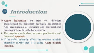
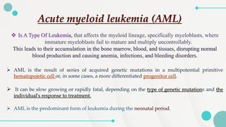
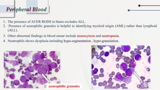
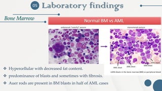
By :
Fatima A. Al-mansouri
Acute Myeloid
Leukemias
CONTENT LIST :
Introduction Etiology
pathophysiology
A. Peripheral Blood
B. Bone marrow
01 02
03
Laboratory findings:
05
Other Laboratory findings
06
Classifications: WHO, FAB
07
Clinical Features
04
Introduction
01
➢ Acute leukemia's are stem cell disorders
characterized by malignant neoplastic proliferation
And accumulation of immature and non-functional
hematopoietic cells in the bone marrow.
➢ The neoplastic cells show increased proliferation and
decreased apoptosis.
➢ If the defect primarily affects the common myeloid
progenitor (CMP) then it is called Acute myeloid
leukemia.
➢ AML is the result of series of acquired genetic mutations in a multipotential primitive
hematopoietic cell or, in some cases, a more differentiated progenitor cell.
➢ It can be slow growing or rapidly fatal, depending on the type of genetic mutations and the
individual's response to treatment.
➢ AML is the predominant form of leukemia during the neonatal period.
Acute myeloid leukemia (AML)
❖ Is A Type Of Leukemia, that affects the myeloid lineage, specifically myeloblasts, where
immature myeloblasts fail to mature and multiply uncontrollably.
This leads to their accumulation in the bone marrow, blood, and tissues, disrupting normal
blood production and causing anemia, infections, and bleeding disorders.
(Multipotent
Progenitor Cells)
Lymphoid-Myeloid
Progenitor Cells
(Common Myeloid
Progenitor)
Etiology
02
1.Growth Factors: Proteins that stimulate cell growth and division.
2.Growth Factor Receptors: Proteins on the cell surface that respond to growth signals.
3.Signaling Pathways: Systems of molecules that communicate signals from the cell surface to its
interior, affecting cell behavior.
4.Transcription Factors: Proteins that help control the expression of specific genes involved in
cell survival and growth.
➢ These mutations disrupt the normal regulation of these processes, leading to the uncontrolled
proliferation of immature cells, which is a hallmark of AML.
Excessive Proliferation Results From Mutations that
Affecting :
Etiology
02
➢ Smoking can also increase the risk
➢ DRUGS:
Anticancer drugs are the leading cause of therapy - associated AML.
➢ EXPOSURES TO RADIATIONS.
➢ CHEMICAL :
Exposure to benzene, petroleum products and pesticides can
increase the risk.
Others
pathophysiology
03
➢ More than half of AML cases involve genetic abnormalities in the chromosomes, mainly in
the form of balanced reciprocal translocations, where segments of chromosomes exchange
places.
➢ These translocations create fusion genes, which produce abnormal fusion proteins. These
proteins interfere with the normal function of affected genes, leading to uncontrolled cell
growth (proliferation), failure of cells to mature (differentiation), and increased cell survival.
➢ Additionally, other genetic changes, such as epigenetic modifications, interact with these
chromosomal mutations, further contributing to the complete transformation of normal cells
into leukemic cells.
For example: (DNA Methylation)
A chemical group called a methyl group (-CH3) is added to the DNA.
When this happens to genes responsible for preventing cancer (Tumor Suppressor Genes), these genes
become silenced, allowing cells to multiply uncontrollably.
CLINICAL FEATURES
04
➢ SYMPTOMS :
Present with nonspecific symptoms initially.
❖ Fatigue Is the first symptom.
❖ Fever with or without infection will be present in approximately 10% patients
❖ Bleeding, easy bruising
❖ occasionally, bone pain, lymphadenopathy, nonspecific cough, headache, or
diaphoresis
❖ Rarely patients may present with symptoms from a myeloid sarcoma,
a tumor.
CLINICAL FEATURES
04
➢ PHYSICAL FINDINGS :
• Splenomegaly, Hepatomegaly, Lymphadenopathy, Sternal
tenderness.
• Evidence of infection and haemorrhage.
• Severe gastrointestinal bleeding, intrapulmonary haemorrhage, or
intracranial haemorrhage occurs mostly In APL.
• Infiltration of the gingivae, skin, soft tissues, or meninges with
leukemic blasts is characteristic of the monocytic subtypes.
Laboratory Findings
Peripheral Blood
05
➢ LEUCOCYTES:
1. Elevated leukocyte counts.
2. 50% of cases will have decreased or within reference
interval.
3. Counts ranges from <1000 to >100000/ml.
4. Presence of blasts > 20% suggests AL diagnosis.
➢ The current WHO definition of
AL requires 20% or more blasts in
the peripheral blood or bone
marrow.
Laboratory findings
Peripheral Blood
05
- Typically, decreased and macrocytic Or normocytic normochromic.
- Hemoglobin value less than 10 g/dL is common.
‐ RDW is elevated.
‐ Erythrocyte inclusions such as Howell-Jolly bodies, Pappenheimer bodies and basophilic
stippling is seen.
‐ Nucleated RBCs can be present in proportion to anaemia or marrow damage.
➢ ERYTHROCYTES:
- Typically decreased.
- Hypo-granular forms of platelets : Platelets appear lacking granules, indicating abnormal maturation.
- Qualitative platelet defect : Even if platelet count is normal, their function is impaired.
➢ PLATELETS:
Peripheral Blood
1. The presence of AUER RODS in blasts excludes ALL.
2. Presence of azurophilic granules is helpful in identifying myeloid origin (AML) rather than lymphoid
(ALL).
3. Other abnormal findings in blood smear include monocytosis and neutropenia.
4. Neutrophils shows dysplasia including hypo-segmentation , hypo-granulation.
azurophilic granules
05
Bone Marrow
Laboratory findings
❖ Hypercellular with decreased fat content.
❖ predominance of blasts and sometimes with fibrosis.
❖ Auer rods are present in BM blasts in half of AML cases
06
Other Laboratory findings
After the initial diagnosis, cases must be classified using a combination of different tests,
including:
1. Immunologic Tests (Flow Cytometry & Immunophenotyping):
➢ These tests help identify surface markers (CD markers) present on leukemia cells.
➢ Different types of leukemia express distinct CD markers, which help distinguish between Acute Myeloid Leukemia
(AML) and Acute Lymphoblastic Leukemia (ALL).
For example:
• AML blasts often express CD13 , CD33.
• ALL blasts typically express CD10 , CD19
➢ These tests detect chromosomal abnormalities, including translocations, deletions, or duplications .
Some translocations are strongly associated with specific AML subtypes:
▪ t (15;17) → Found in Acute Promyelocytic Leukemia (APL) .
▪ Inv(16) or t(16;16) → Associated with AML with abnormal eosinophils.
2. Cytogenetic Tests (Chromosomal Analysis):
06
Other Laboratory findings
These tests identify specific gene mutations that affect leukemia cell function,
proliferation, and response to treatment.
3. Molecular Genetic Tests (Mutation Analysis):
❑Common mutations in AML include
• FLT3-ITD mutation →Linked to poor prognosis due to increased proliferation and
relapse risk.
• TP53 mutation →Indicates a very poor prognosis due to Resistance to chemotherapy.
06
Other Laboratory findings
4. Cytochemical Tests (Special Stains):
These tests use chemical stains to identify specific cell types based on their enzymatic
activity.
❑One of the most important stains is Myeloperoxidase (MPO):
• MPO-positive →Confirms myeloid lineage, strongly suggesting AML.
• MPO-negative →Suggests ALL or undifferentiated leukemia
❑Other stains include: Sudan Black B (SBB) → Stains lipids in myeloid blasts.
• Positive SBB: Stained cells appear black or dark brown due to the interaction of the stain with
granule components.
• Negative SBB : do not stain .
✓ Differentiates AML from ALL by detecting myeloid granules.
✓ Used alongside Myeloperoxidase (MPO) staining for higher diagnostic accuracy.
CLASSIFICATIONS
Identification of Cell Lineage
07
➢ Because blast cells are immature cells, they are often difficult to identify by
morphology alone using light microscopy.
➢ Cytochemistry, molecular testing, and immunophenotyping give additional
information that can help define cell lineage.
❖ The 2016 World Health Organization (WHO) classification is an update to
the 2008 classification of hematologic malignancies, expanding the criteria
used for diagnosis.
WHO classification of AML and related neoplasms
FAB classification of AML
Hematologic Malignancies : Acute myeloid leukemia (AML).pdf

Hematologic Malignancies : Acute myeloid leukemia (AML).pdf

  • 1.
    By : Fatima A.Al-mansouri Acute Myeloid Leukemias
  • 2.
    CONTENT LIST : IntroductionEtiology pathophysiology A. Peripheral Blood B. Bone marrow 01 02 03 Laboratory findings: 05 Other Laboratory findings 06 Classifications: WHO, FAB 07 Clinical Features 04
  • 3.
    Introduction 01 ➢ Acute leukemia'sare stem cell disorders characterized by malignant neoplastic proliferation And accumulation of immature and non-functional hematopoietic cells in the bone marrow. ➢ The neoplastic cells show increased proliferation and decreased apoptosis. ➢ If the defect primarily affects the common myeloid progenitor (CMP) then it is called Acute myeloid leukemia.
  • 4.
    ➢ AML isthe result of series of acquired genetic mutations in a multipotential primitive hematopoietic cell or, in some cases, a more differentiated progenitor cell. ➢ It can be slow growing or rapidly fatal, depending on the type of genetic mutations and the individual's response to treatment. ➢ AML is the predominant form of leukemia during the neonatal period. Acute myeloid leukemia (AML) ❖ Is A Type Of Leukemia, that affects the myeloid lineage, specifically myeloblasts, where immature myeloblasts fail to mature and multiply uncontrollably. This leads to their accumulation in the bone marrow, blood, and tissues, disrupting normal blood production and causing anemia, infections, and bleeding disorders.
  • 5.
  • 6.
    Etiology 02 1.Growth Factors: Proteinsthat stimulate cell growth and division. 2.Growth Factor Receptors: Proteins on the cell surface that respond to growth signals. 3.Signaling Pathways: Systems of molecules that communicate signals from the cell surface to its interior, affecting cell behavior. 4.Transcription Factors: Proteins that help control the expression of specific genes involved in cell survival and growth. ➢ These mutations disrupt the normal regulation of these processes, leading to the uncontrolled proliferation of immature cells, which is a hallmark of AML. Excessive Proliferation Results From Mutations that Affecting :
  • 7.
    Etiology 02 ➢ Smoking canalso increase the risk ➢ DRUGS: Anticancer drugs are the leading cause of therapy - associated AML. ➢ EXPOSURES TO RADIATIONS. ➢ CHEMICAL : Exposure to benzene, petroleum products and pesticides can increase the risk. Others
  • 8.
    pathophysiology 03 ➢ More thanhalf of AML cases involve genetic abnormalities in the chromosomes, mainly in the form of balanced reciprocal translocations, where segments of chromosomes exchange places. ➢ These translocations create fusion genes, which produce abnormal fusion proteins. These proteins interfere with the normal function of affected genes, leading to uncontrolled cell growth (proliferation), failure of cells to mature (differentiation), and increased cell survival. ➢ Additionally, other genetic changes, such as epigenetic modifications, interact with these chromosomal mutations, further contributing to the complete transformation of normal cells into leukemic cells. For example: (DNA Methylation) A chemical group called a methyl group (-CH3) is added to the DNA. When this happens to genes responsible for preventing cancer (Tumor Suppressor Genes), these genes become silenced, allowing cells to multiply uncontrollably.
  • 9.
    CLINICAL FEATURES 04 ➢ SYMPTOMS: Present with nonspecific symptoms initially. ❖ Fatigue Is the first symptom. ❖ Fever with or without infection will be present in approximately 10% patients ❖ Bleeding, easy bruising ❖ occasionally, bone pain, lymphadenopathy, nonspecific cough, headache, or diaphoresis ❖ Rarely patients may present with symptoms from a myeloid sarcoma, a tumor.
  • 10.
    CLINICAL FEATURES 04 ➢ PHYSICALFINDINGS : • Splenomegaly, Hepatomegaly, Lymphadenopathy, Sternal tenderness. • Evidence of infection and haemorrhage. • Severe gastrointestinal bleeding, intrapulmonary haemorrhage, or intracranial haemorrhage occurs mostly In APL. • Infiltration of the gingivae, skin, soft tissues, or meninges with leukemic blasts is characteristic of the monocytic subtypes.
  • 11.
    Laboratory Findings Peripheral Blood 05 ➢LEUCOCYTES: 1. Elevated leukocyte counts. 2. 50% of cases will have decreased or within reference interval. 3. Counts ranges from <1000 to >100000/ml. 4. Presence of blasts > 20% suggests AL diagnosis. ➢ The current WHO definition of AL requires 20% or more blasts in the peripheral blood or bone marrow.
  • 12.
    Laboratory findings Peripheral Blood 05 -Typically, decreased and macrocytic Or normocytic normochromic. - Hemoglobin value less than 10 g/dL is common. ‐ RDW is elevated. ‐ Erythrocyte inclusions such as Howell-Jolly bodies, Pappenheimer bodies and basophilic stippling is seen. ‐ Nucleated RBCs can be present in proportion to anaemia or marrow damage. ➢ ERYTHROCYTES: - Typically decreased. - Hypo-granular forms of platelets : Platelets appear lacking granules, indicating abnormal maturation. - Qualitative platelet defect : Even if platelet count is normal, their function is impaired. ➢ PLATELETS:
  • 13.
    Peripheral Blood 1. Thepresence of AUER RODS in blasts excludes ALL. 2. Presence of azurophilic granules is helpful in identifying myeloid origin (AML) rather than lymphoid (ALL). 3. Other abnormal findings in blood smear include monocytosis and neutropenia. 4. Neutrophils shows dysplasia including hypo-segmentation , hypo-granulation. azurophilic granules
  • 14.
    05 Bone Marrow Laboratory findings ❖Hypercellular with decreased fat content. ❖ predominance of blasts and sometimes with fibrosis. ❖ Auer rods are present in BM blasts in half of AML cases
  • 15.
    06 Other Laboratory findings Afterthe initial diagnosis, cases must be classified using a combination of different tests, including: 1. Immunologic Tests (Flow Cytometry & Immunophenotyping): ➢ These tests help identify surface markers (CD markers) present on leukemia cells. ➢ Different types of leukemia express distinct CD markers, which help distinguish between Acute Myeloid Leukemia (AML) and Acute Lymphoblastic Leukemia (ALL). For example: • AML blasts often express CD13 , CD33. • ALL blasts typically express CD10 , CD19 ➢ These tests detect chromosomal abnormalities, including translocations, deletions, or duplications . Some translocations are strongly associated with specific AML subtypes: ▪ t (15;17) → Found in Acute Promyelocytic Leukemia (APL) . ▪ Inv(16) or t(16;16) → Associated with AML with abnormal eosinophils. 2. Cytogenetic Tests (Chromosomal Analysis):
  • 16.
    06 Other Laboratory findings Thesetests identify specific gene mutations that affect leukemia cell function, proliferation, and response to treatment. 3. Molecular Genetic Tests (Mutation Analysis): ❑Common mutations in AML include • FLT3-ITD mutation →Linked to poor prognosis due to increased proliferation and relapse risk. • TP53 mutation →Indicates a very poor prognosis due to Resistance to chemotherapy.
  • 17.
    06 Other Laboratory findings 4.Cytochemical Tests (Special Stains): These tests use chemical stains to identify specific cell types based on their enzymatic activity. ❑One of the most important stains is Myeloperoxidase (MPO): • MPO-positive →Confirms myeloid lineage, strongly suggesting AML. • MPO-negative →Suggests ALL or undifferentiated leukemia ❑Other stains include: Sudan Black B (SBB) → Stains lipids in myeloid blasts. • Positive SBB: Stained cells appear black or dark brown due to the interaction of the stain with granule components. • Negative SBB : do not stain . ✓ Differentiates AML from ALL by detecting myeloid granules. ✓ Used alongside Myeloperoxidase (MPO) staining for higher diagnostic accuracy.
  • 18.
    CLASSIFICATIONS Identification of CellLineage 07 ➢ Because blast cells are immature cells, they are often difficult to identify by morphology alone using light microscopy. ➢ Cytochemistry, molecular testing, and immunophenotyping give additional information that can help define cell lineage. ❖ The 2016 World Health Organization (WHO) classification is an update to the 2008 classification of hematologic malignancies, expanding the criteria used for diagnosis.
  • 19.
    WHO classification ofAML and related neoplasms
  • 20.
    WHO classification ofAML and related neoplasms
  • 21.