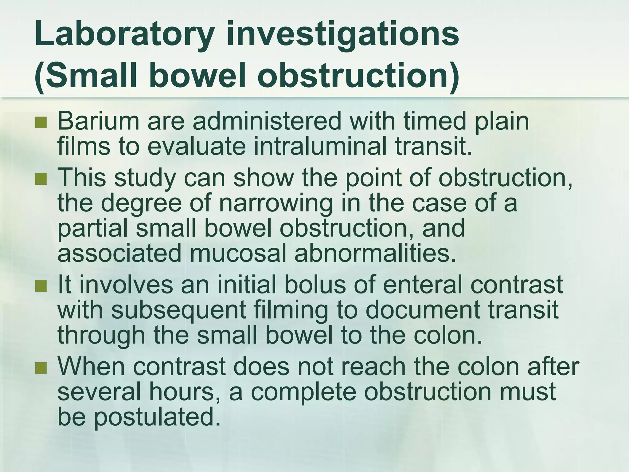
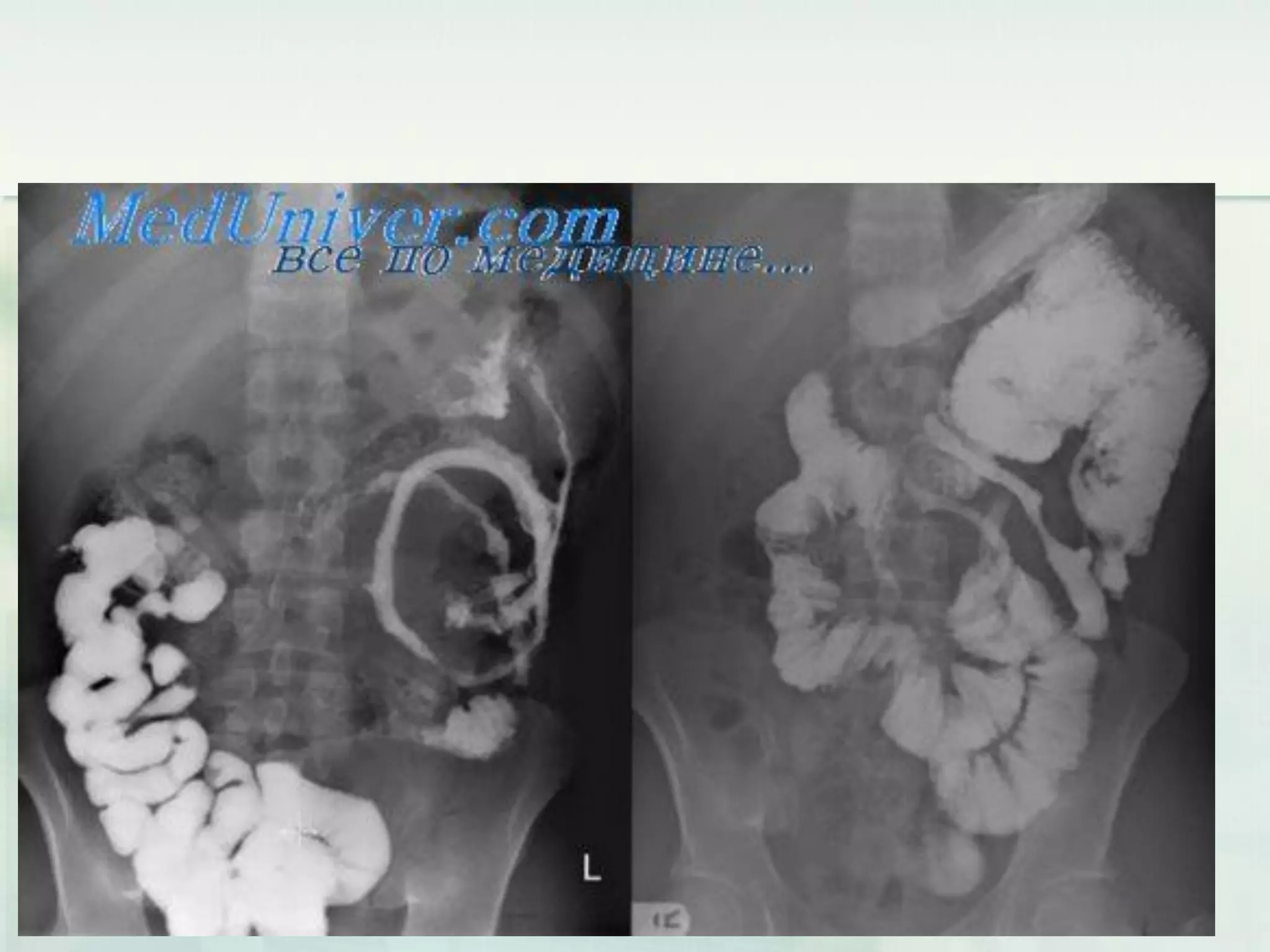
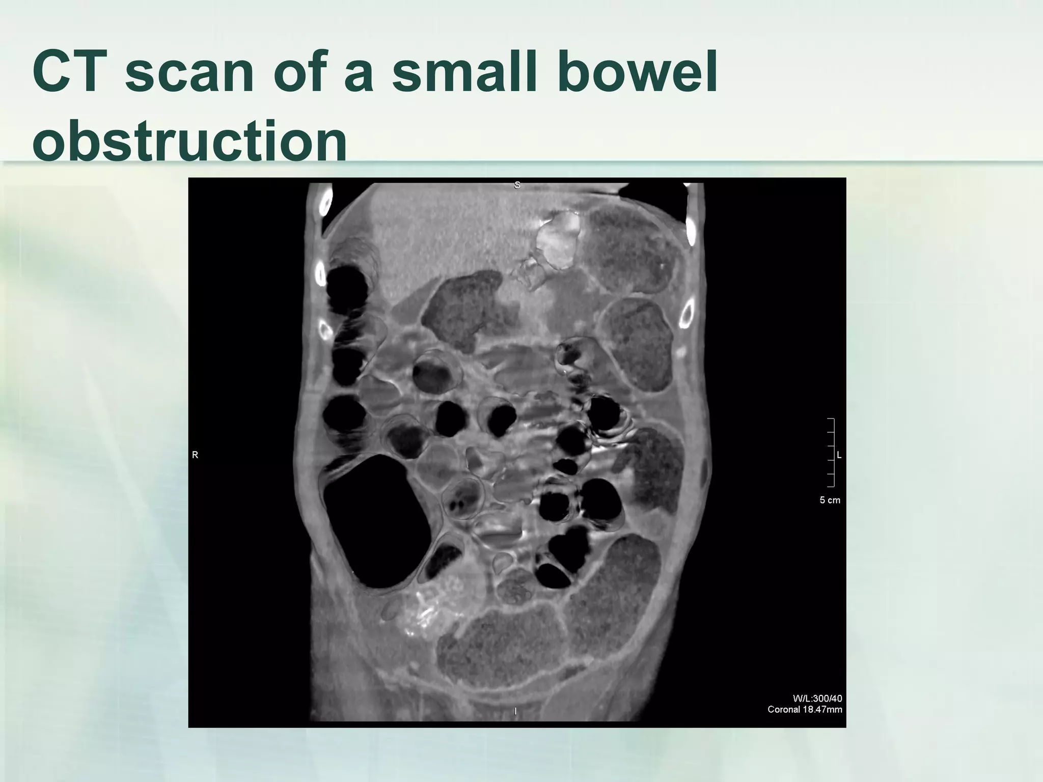
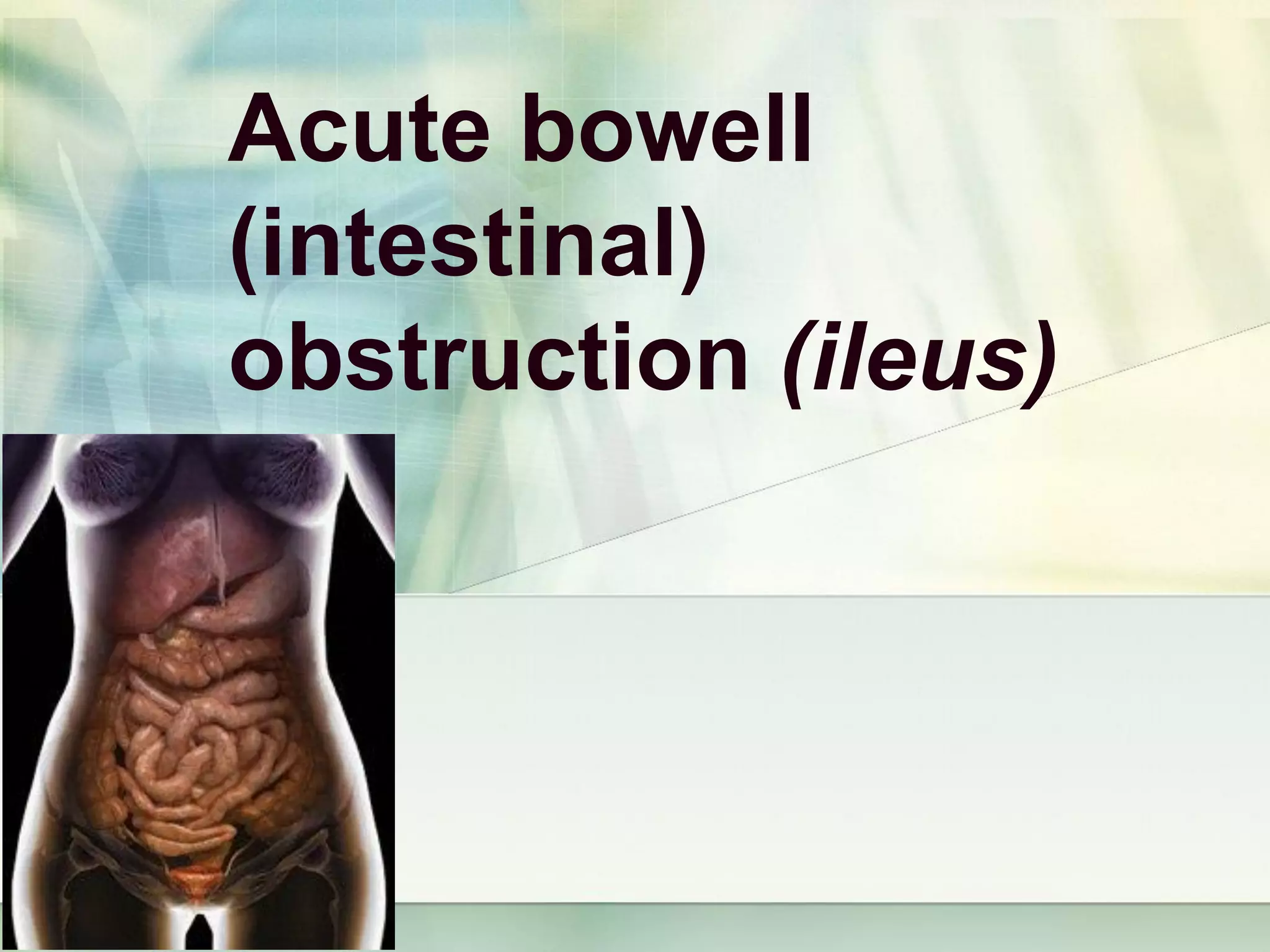
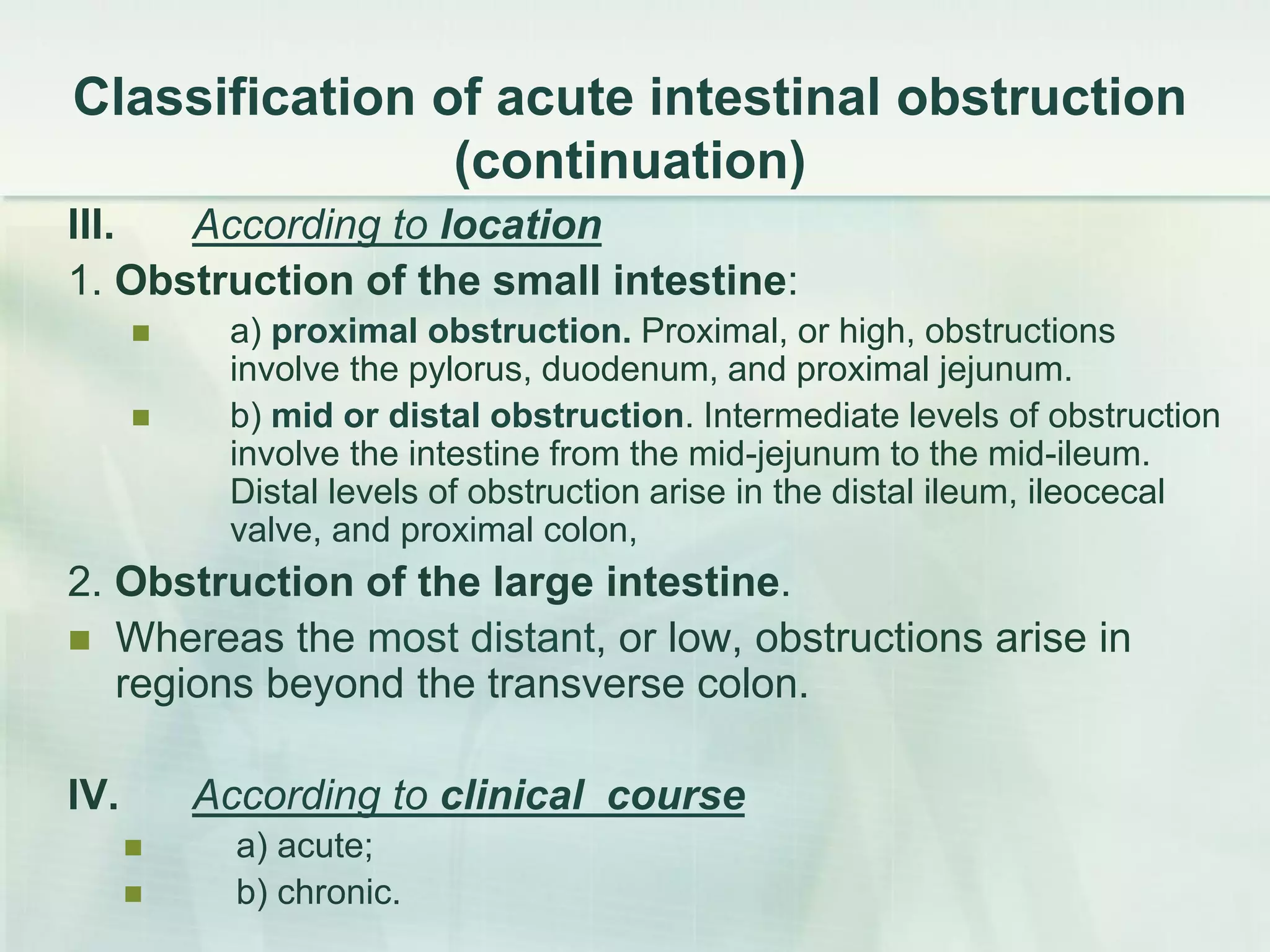
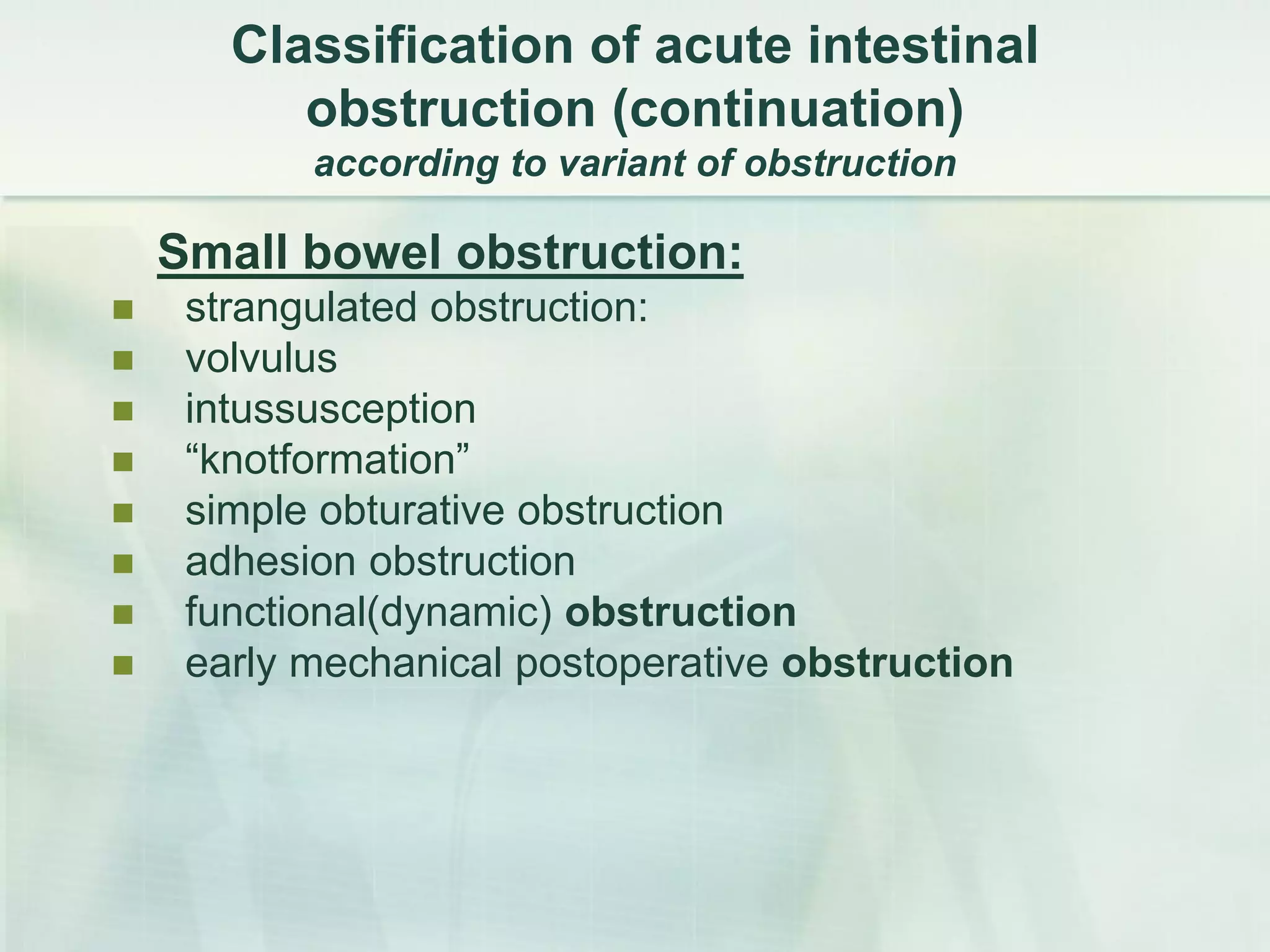
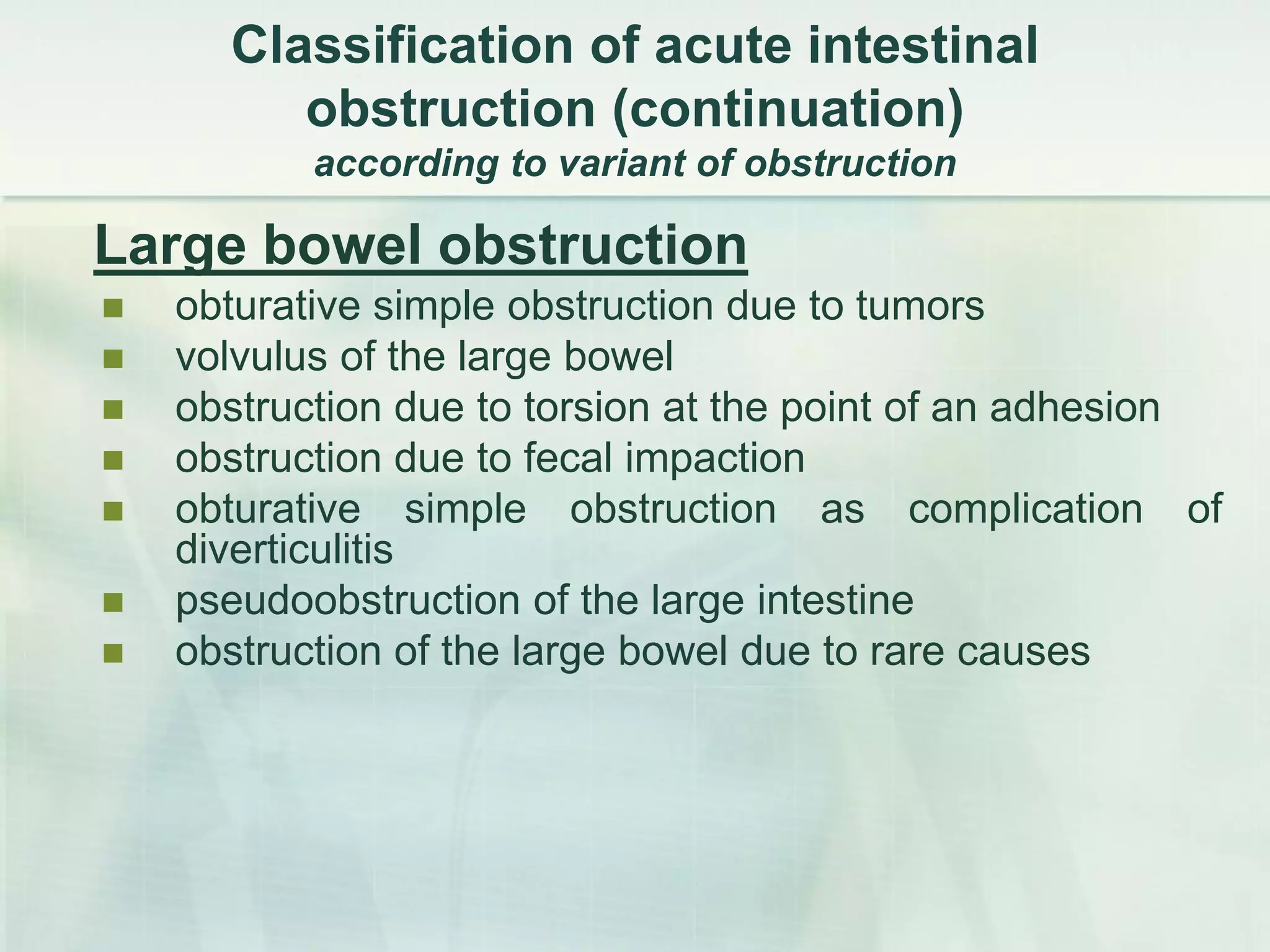
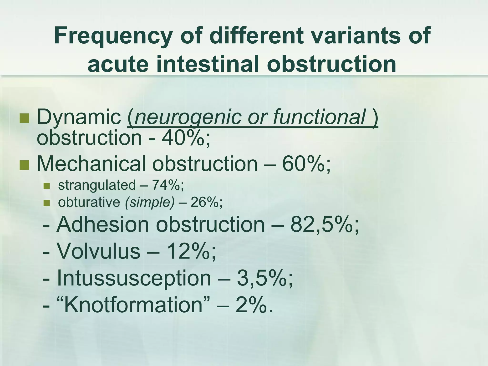
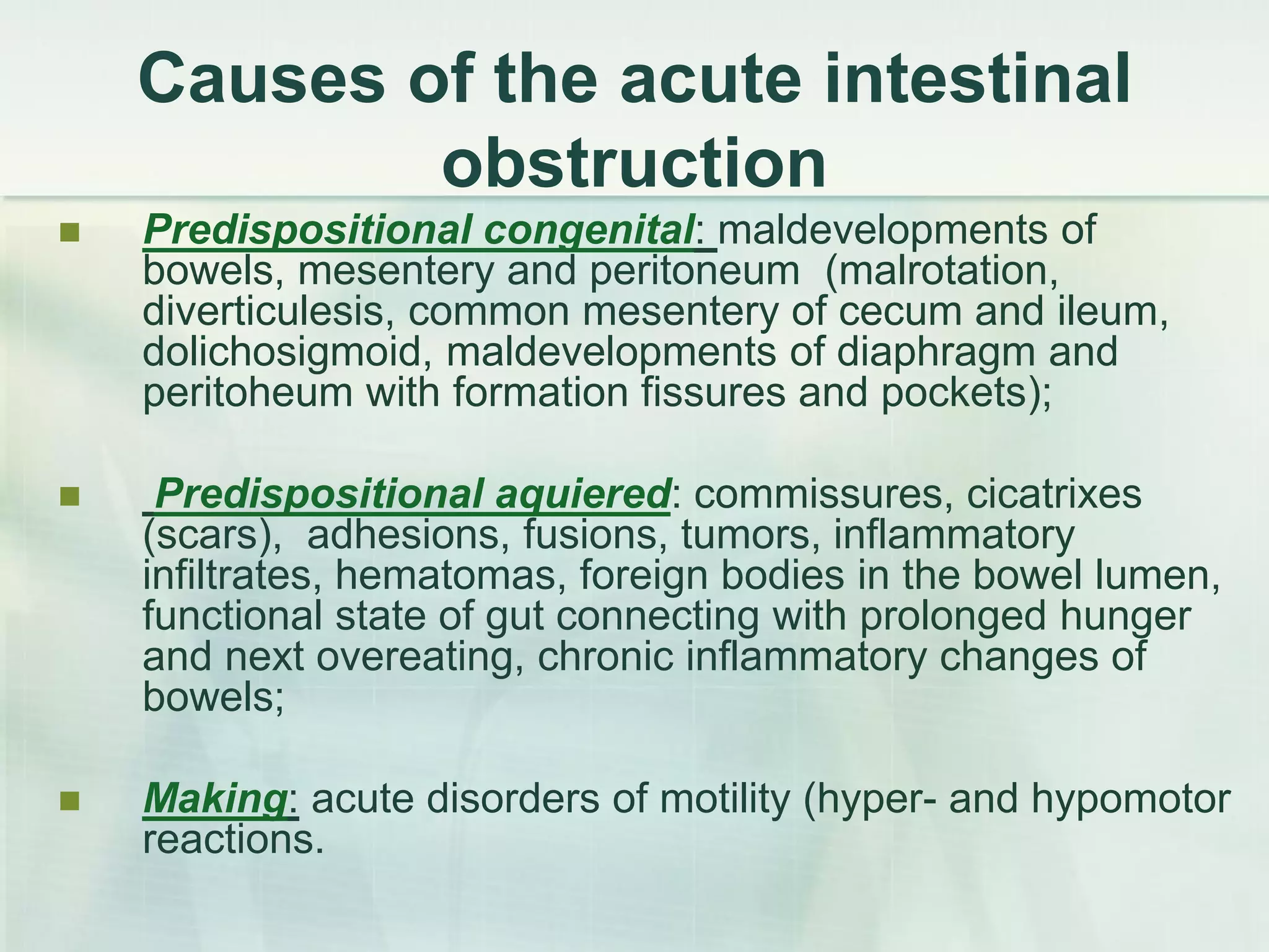
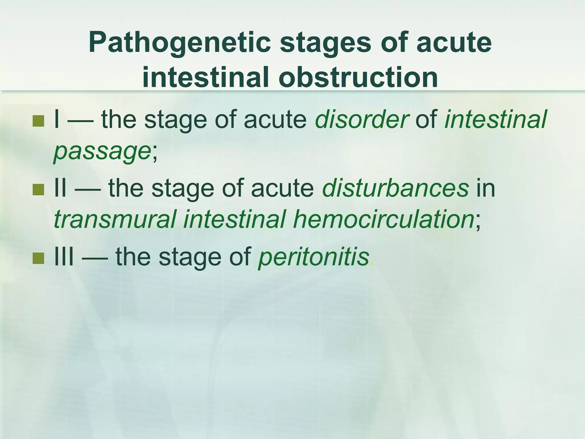
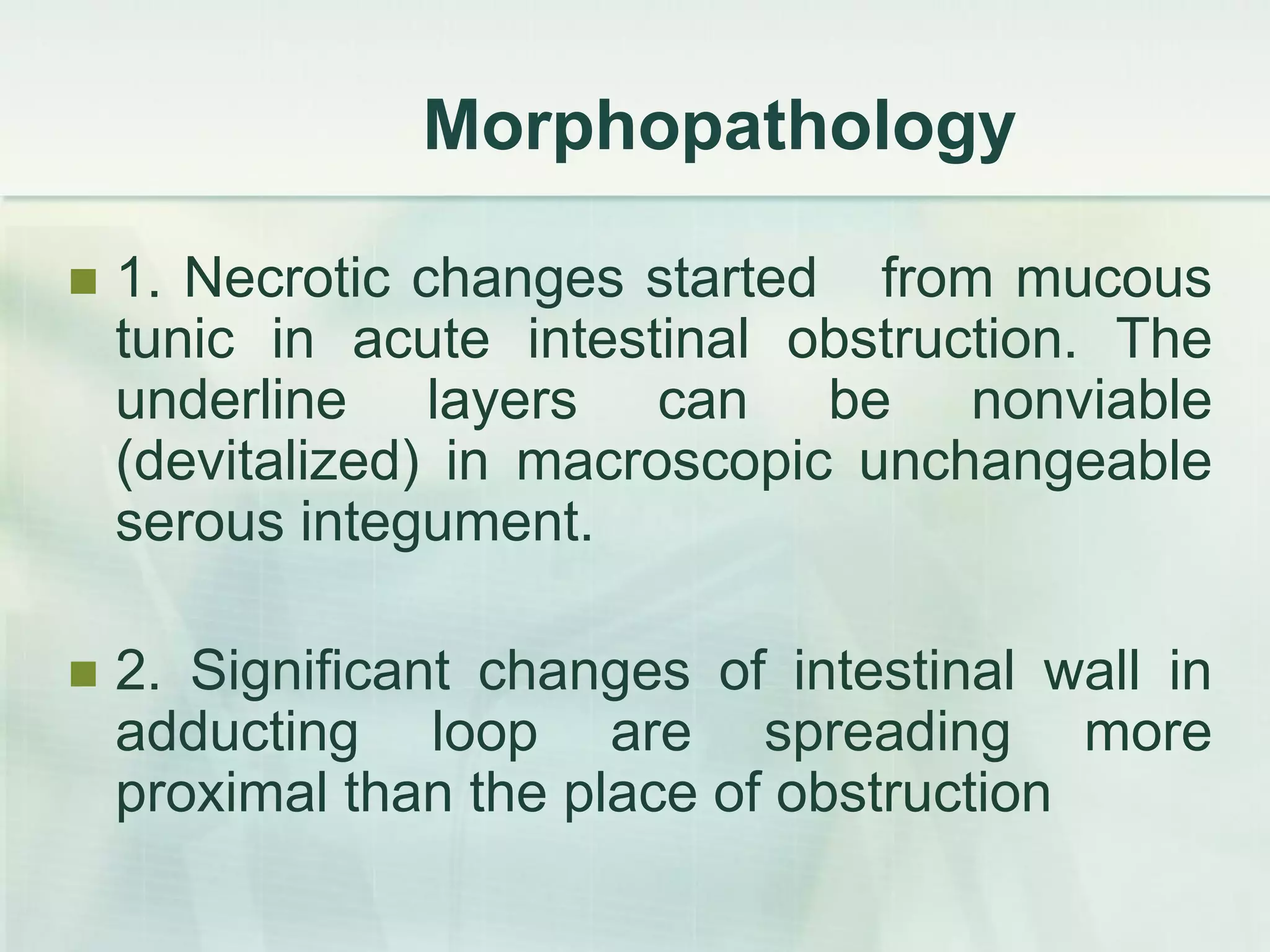
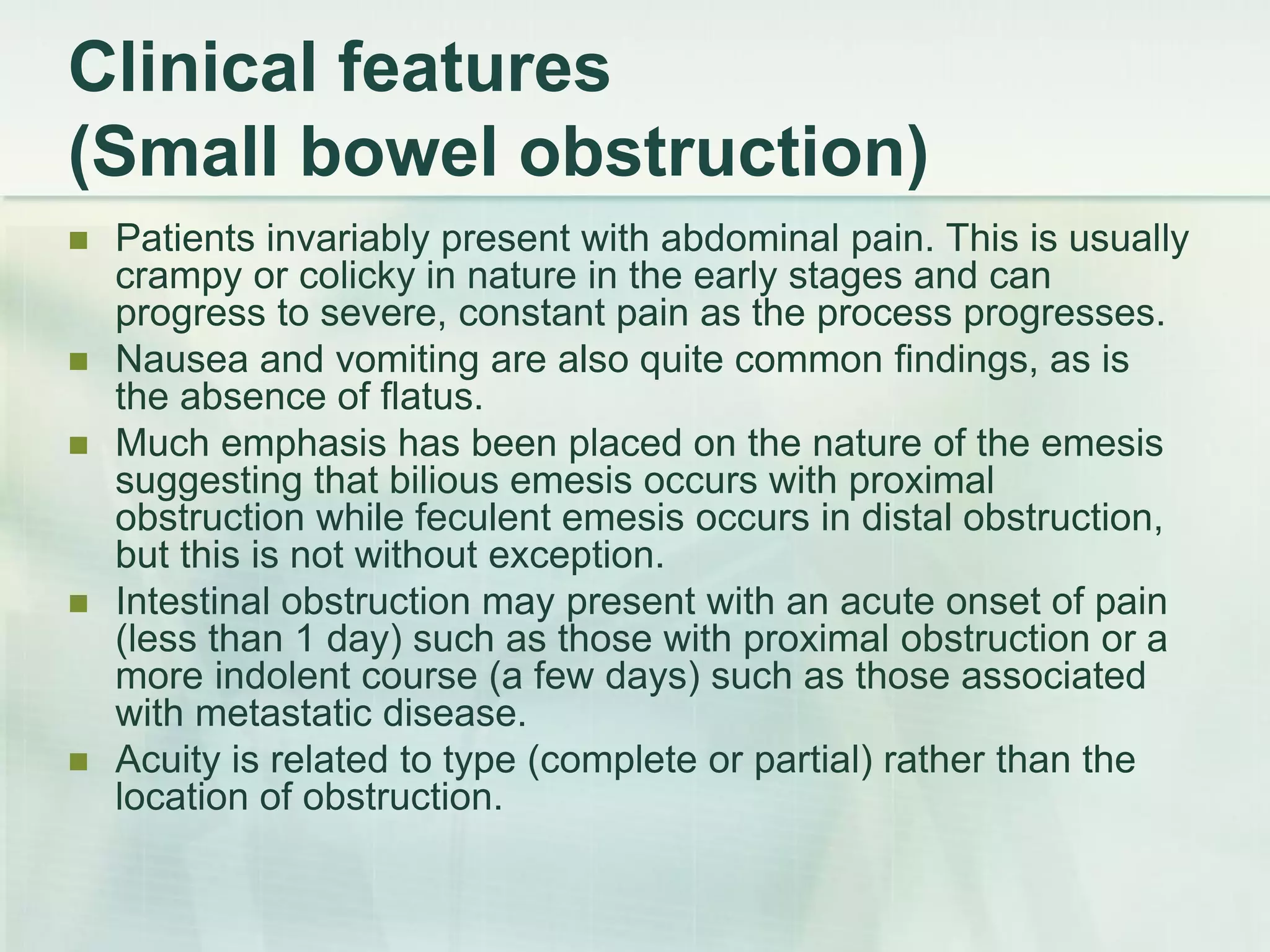
This document discusses acute intestinal obstruction, including classifications, causes, clinical features, investigations and treatments. It classifies obstruction based on origin, method of occurrence, blood flow and clinical course. Mechanical obstruction is distinguished from functional obstruction. Signs of small bowel obstruction include abdominal pain, nausea, vomiting and distention. Large bowel obstruction causes dull abdominal cramps and distention. Treatment involves decompression, fluid resuscitation, antibiotics and surgery to remove obstructions and non-viable bowel segments.
This document discusses intestinal obstruction, its causes, symptoms, diagnosis and treatment. It notes that an obstruction can occur anywhere along the small or large intestine and can be partial or complete. Common causes include birth defects, scar tissue from prior surgery, hernias and tumors. Symptoms include abdominal cramping, bloating and vomiting. Treatment typically involves passing a tube through the nose to remove material and fluids above the blockage, administering IV fluids and electrolytes, and sometimes surgery.
This document discusses intestinal obstruction, including its definition, causes, clinical features, investigations and management. Intestinal obstruction occurs when bowel contents cannot pass through normally due to a mechanical or functional blockage. Clinical features depend on the location and cause of obstruction and may include pain, vomiting, distension and constipation.
Intestinal Obstruction
Prepared by : A.A.A
Under supervision : Dr Mohemed Hazim
Objective
To understand:
The Pathophysiology of dynamic and Adynamic intestinal obstruction
The Cardinal features on history and examination
• The Causes of small and large bowel obstruction
• The Indications for surgery and other treatment options in bowel obstruction
CLASSIFICATION classified into two types:
Dynamic
Adynamic
PATHOPHYSIOLOGY
Gas
Fluid
STRANGULATION
Causes of strangulation
■ Direct pressure on the bowel wall Hernial orifices Adhesions/bands
■ Interrupted mesenteric blood flow Volvulus Intussusception
■ Increased intraluminal pressure Closed-loop obstruction
SPECIAL TYPES OF MECHANICAL INTESTINAL OBSTRUCTION
Internal hernia
Obstruction from enteric strictures
Bolus obstruction : Gallstones , food , Trychobezoars and phytobezoars , Stercolith and worms.
Obstruction by adhesions and bands
Acute intussusception
This occurs when one portion of the gut invaginates into an immediately adjacent segment; almost invariably, it is the proximal into the distal.
Volvulus
C/F OF INTESTINAL OBSTRUCTION
C/F of strangulation
C/F of Intussusception
‘Redcurrant Jelly’ Stool
Imaging
TREATMENT
ADYNAMIC OBSTRUCTION
Varieties of Paralytic Ileus :
• Postoperative
• Infection
• Reflex ileus
• Metabolic
Pseudo-Obstruction
Thank You
Intestinal obstruction caused by volvulus by dr basilBasil Tumaini
This document discusses intestinal obstruction caused by volvulus, beginning with an introduction that defines intestinal obstruction and its causes. It then covers the historical background, pathophysiology, clinical presentation, and management of volvulus. Volvulus is caused by twisting of the intestine on itself, most commonly occurring in the sigmoid colon. It can lead to bowel obstruction and ischemia. Treatment involves surgical intervention to untwist the intestine and potentially resect nonviable sections.
Intestinal obstruction occurs when the downward movement of intestinal contents is arrested. It can be classified based on its pathological cause, level of obstruction, onset and course. Common causes include adhesions, hernias, tumors and strictures. Symptoms depend on the level and type of obstruction and may include pain, distension, vomiting and constipation. Examination findings can provide clues to whether the obstruction is simple, strangulated or closed loop. Further testing is needed to determine the specific cause and appropriate management.
A 50-year-old man presented with abdominal pain, distension, vomiting, and constipation. Imaging showed findings suggestive of bowel obstruction. The document discusses the evaluation, causes, and management of bowel obstructions, focusing on distinguishing between small vs. large bowel obstruction and determining the etiology and complications like strangulation. Initial management involves resuscitation, decompression, correction of electrolyte abnormalities, and antibiotics while determining need for surgery.
This document discusses small bowel obstruction, including its pathophysiology, clinical presentation, diagnosis, management, and prevention. The pathophysiology section explains how obstruction leads to accumulation of gas and fluid in the bowel above the site of obstruction. The clinical presentation section outlines common symptoms like colicky abdominal pain and nausea/vomiting, as well as signs seen on examination. Diagnosis involves distinguishing mechanical obstruction from ileus, determining the etiology, and discriminating between partial and complete or simple versus strangulating obstruction, often using radiological exams. Management depends on whether the obstruction is simple or strangulated. With conservative treatment, the majority of patients with adhesive small bowel obstruction are readmitted in less than 20% of cases over 5
The document discusses different types of intestinal obstruction including dynamic, adynamic, and paralytic ileus. Dynamic obstruction can be intraluminal, intramural, or extramural. Common causes are adhesions, hernias, strictures, and tumors. Clinical features of dynamic obstruction are colicky pain, distension, vomiting, and constipation. Treatment involves nasogastric decompression, IV fluids, antibiotics, and surgery to relieve the obstruction. Paralytic ileus is a failure of peristalsis due to neuromuscular dysfunction. It can be treated by addressing the underlying cause, nasogastric decompression, and IV fluids. Acute intussusception occurs when one segment of
This document provides an overview of small intestinal obstruction. It begins by defining small intestinal obstruction as a blockage of the small intestine. It then discusses the epidemiology, pathophysiology, signs and symptoms, causes, complications, diagnosis and treatment of small intestinal obstruction. The main points are that adhesion and hernia are the most common causes, and treatment involves correcting fluid and electrolyte imbalances, nasogastric decompression, antibiotics and potentially surgery for cases of strangulation or complete blockage. The document provides details on evaluating and managing both mechanical and paralytic forms of small intestinal obstruction.
Acute Intestinal obstruction by Dr. Daniel B. YidanaDaniel Yidana
This document discusses acute intestinal obstruction (AIO), defined as a sudden stoppage of intestinal contents. AIO is a common surgical emergency. It classifies AIO by site (high vs low) and nature (simple, strangulated, closed loop). The pathophysiology involves proximal dilation and distension due to gas, fluid, and impaired blood flow in some cases. Common causes are adhesions, hernias, tumors. Clinical features are abdominal pain, distension, vomiting, and constipation. Management involves resuscitation, NG tubes, antibiotics, and surgery if conservative measures fail to resolve the obstruction within 72 hours.
Large bowel obstruction is an emergent condition that requires prompt surgical intervention. It can result from infectious, inflammatory, neoplastic, or mechanical causes such as volvulus or incarcerated hernia. Symptoms include abdominal pain, distention, nausea, vomiting, and constipation. Diagnosis involves physical exam, imaging studies like CT scan, and lab tests. Treatment involves resuscitation, nasogastric decompression, and surgical resection of the obstructing lesion with proximal diversion such as colostomy. Complications can include perforation, sepsis, and death if not treated early. Prognosis depends on the underlying cause, with cancer outcomes varying based on the specific carcinoma.
This document discusses obstructive jaundice and intestinal obstruction. It provides details on a case of a 78-year-old man admitted with abdominal pain and jaundice. Investigation showed gallstones and elevated bilirubin. Management of obstructive jaundice may include cholecystectomy, ERCP, or stenting. Complications include sepsis and liver/renal failure. Intestinal obstruction can be dynamic or adynamic, and may be caused by adhesions, hernias, tumors or impacted feces. Treatment involves decompression, IV fluids, and surgery to remove obstructions or bypass affected areas.
This presentation explains in detail the definition, pathophysiology, signs & symptoms, management, and prognosis of intestinal obstruction, ileus, and volvulus.
This document provides an overview of small bowel obstruction, including classification, common causes, clinical features, investigation, and treatment. It discusses how to determine if a patient has bowel obstruction or ileus, and how to investigate and manage the patient. The main causes of small bowel obstruction are discussed, including adhesions, hernias, volvulus, and intussusception. Indications for surgery include generalized peritonitis, failure to improve with conservative treatment, and unclear diagnosis. Initial management focuses on resuscitation, decompression, and monitoring for signs of strangulation or perforation that would require surgery.
Bowel obstruction occurs when the intestines become blocked, preventing normal movement of digestive contents. There are several types, including small and large bowel obstruction. Causes include adhesions from prior surgery, hernias, tumors, inflammatory bowel disease, and foreign bodies. Symptoms depend on the location but commonly include abdominal pain, distension, vomiting, and constipation. Diagnosis involves medical history, physical exam, imaging tests like CT scans, and sometimes endoscopy or surgery to determine the specific cause. Treatment aims to resolve the blockage through conservative management with NG tubes and IV fluids or potentially surgery. Complications can include dehydration, electrolyte imbalances, infection, and bowel perforation if not properly treated
Intestinal obstruction is a blockage of the bowel that prevents contents from passing through. There are two main types: mechanical obstruction from pressure on the bowel wall, and functional obstruction where the bowel muscles cannot propel contents. Causes of small bowel obstruction include adhesions, intussusception, volvulus, and tumors. Causes of large bowel obstruction include carcinoma, diverticulitis, and inflammatory bowel disorders. Treatment involves decompressing the bowel, fluid replacement, and usually surgery to relieve the obstruction.
Intestinal obstruction is a partial or complete blockage of the bowel that prevents contents from passing through. It most commonly occurs in children ages 1-5 years old infected with Ascaris lumbricoides. Common causes include abdominal or pelvic surgery which can lead to adhesions, Crohn's disease thickening the intestine walls, and abdominal cancer. Symptoms include abdominal swelling, fever, bloody stools, vomiting, inability to pass gas or stool. Treatment depends on the severity but may include surgery to remove the obstructed part of the intestine or create an anastomosis.
Paralytic ileus is the inability of the intestines to conduct peristalsis, which can lead to obstruction. It most often occurs after surgery but can be caused by inflammation, electrolyte abnormalities, or spinal fractures. Symptoms include colicky abdominal pain, nausea, vomiting, abdominal distension, and constipation. Diagnostic tests like CT scans and x-rays are used, and treatment focuses on resting the bowel with NPO, IV fluids for hydration, and sometimes NG tube placement. Patient education emphasizes reporting changes in symptoms and the importance of treatment compliance.
Intestinal obstruction occurs when the intestine is blocked, preventing normal movement of its contents. It can be caused by mechanical blockages such as tumors or hernias, or functional issues like neuromuscular problems. Symptoms include abdominal pain, vomiting, distension and constipation. Treatment involves fluid resuscitation, antibiotics, pain management, and sometimes surgery to remove the obstruction. Proper diagnosis and timely treatment are important to prevent complications like perforation and sepsis.
Autors: Richard M. Gore, MD*, Robert I. Silvers, MD, Kiran H. Thakrar, MD,
Daniel R. Wenzke, MD, Uday K. Mehta, MD, Geraldine M. Newmark, MD,
Jonathan W. Berlin, MD
This document discusses acquired intestinal ileus, which can be paralytic or mechanical in nature. Paralytic ileus is caused by medications, surgery, infection, or other insults and results in paralysis of intestinal movement. Mechanical obstruction can be caused by hernias, adhesions, tumors or other structural issues that physically block intestinal contents. Symptoms include abdominal pain, distension and inability to pass gas or stool. Diagnosis involves physical exam, imaging and labs. Treatment focuses on restoring bowel motility with decompression, fluids and electrolyte replacement. The document also discusses specific causes like intussusception, adhesions and their signs, symptoms, diagnosis and management.
This document defines and describes mechanical and functional ileus. Mechanical ileus is caused by an obstruction blocking intestinal contents, which can be due to adhesions, hernias, tumors or inflammation. Functional ileus involves reduced bowel wall contractions and can occur after surgery or due to drugs, metabolic issues or poor perfusion. The document discusses evaluation, conservative treatment including bowel rest and contrast studies, and indications for surgery such as failure of conservative measures or signs of strangulation. It also covers specific types of functional ileus like postoperative or opioid-induced ileus, and intestinal pseudo-obstruction.
Small intestine perforation- Easy ppt for student nurses
definition
causes
clinical manifestations
diagnostic tests
management of small intestine perforation
This document discusses intestinal obstruction, including its causes, clinical presentation, diagnosis and treatment. It describes different types of intestinal obstruction including mechanical obstruction, paralytic ileus and strangulation obstruction. The clinical exam, abdominal x-rays, CT scans and laboratory tests used in diagnosis are outlined. Treatment involves fluid resuscitation, decompression of the intestine, monitoring of electrolytes and timely surgery depending on the severity and duration of the obstruction.
This document discusses bowel obstruction, including classification, common causes, clinical features, investigations, and treatment. Bowel obstruction can be dynamic or mechanical, and is classified as partial or complete. Common causes include adhesions, hernias, volvulus, and tumors. Clinical features include colicky pain, vomiting, abdominal distension, and constipation. Investigations may include blood tests, abdominal x-rays, CT scans, and contrast studies. Treatment involves resuscitation, decompression, antibiotics, and surgery to remove the obstruction or affected bowel segment. Complications can include bleeding, infection, leakage, and recurrent obstruction.
An intestinal obstruction occurs when your small or large intestine is blocked. The blockage can be partial or total, and it prevents passage of fluids and digested food. If intestinal obstruction happens, food, fluids, gastric acids, and gas build up behind the site of the blockage.
Case presentation volvulus in geriatric patientReynel Dan
1. The document presents a case of intestinal obstruction in a geriatric patient, discussing the etiology, pathophysiology, signs and symptoms, and nursing care for intestinal obstruction.
2. Intestinal obstruction can be caused by adhesions, hernias, tumors, or volvulus and results in a blockage of intestinal contents that increases pressure and risk of ischemia in the bowel.
3. Nursing care focuses on pain management, fluid resuscitation, monitoring for complications like peritonitis, and supportive care until the obstruction can be resolved medically or surgically.
This document summarizes bowel motility disorders including ileus, bowel obstruction, and strangulation. It defines ileus as a disruption of normal gastrointestinal motility without mechanical causes, while mechanical bowel obstruction results from structural abnormalities. Patients with bowel obstruction present difficult diagnostic and treatment challenges. Obstructions are classified based on their morphology, function, level, and other factors. Causes, clinical manifestations, diagnostic imaging findings, and complications of various types of bowel obstructions are described in detail.
This document discusses intestinal obstruction, including its definition, classification, etiology, pathophysiology, symptoms, signs, diagnosis, and management. Intestinal obstruction can be dynamic or mechanical in nature. Mechanical obstructions are further classified as luminal, intramural, or extrinsic lesions. Common causes include adhesions, hernias, tumors, and gallstones. Symptoms include abdominal cramps, vomiting, and constipation or diarrhea. Diagnosis involves examination of the abdomen and imaging tests. Initial management is conservative with NG tube, IV fluids, and antibiotics. Surgery is indicated for peritonism, deterioration, or failure of conservative treatment and may involve lysis of adhesions, resection, or
This document discusses intestinal obstruction, including:
1) Intestinal obstruction occurs when air and secretions cannot pass through the intestines due to mechanical compression or gastrointestinal paralysis.
2) Clinical evaluation of a patient with suspected intestinal obstruction involves assessing their history of present illness, previous surgeries or illnesses, and performing a physical exam.
3) Key physical exam findings that suggest intestinal obstruction include abdominal pain, distention, nausea, vomiting, and failure to pass gas. The pattern and severity of pain can provide clues to the level and type of obstruction.
This document provides an overview of intestinal obstruction. It begins with an introduction defining intestinal obstruction and its causes. It then covers the classification of intestinal obstruction including location, degree, and specific causes. Risk factors and pathophysiology are discussed. Clinical presentation includes symptoms like pain, vomiting, and distension. Investigations involve imaging studies like abdominal x-rays and CT scans. Management is outlined, differentiating conservative treatment from surgical intervention depending on factors like failure to resolve or signs of strangulation. Surgical procedures aim to relieve the obstruction and resect non-viable bowel.
This document provides an overview of small intestinal obstruction. It begins by defining small intestinal obstruction as a blockage of the small intestine. It then discusses the epidemiology, pathophysiology, signs and symptoms, causes, complications, diagnosis and treatment of small intestinal obstruction. The main points are that adhesion and hernia are the most common causes, and treatment involves correcting fluid and electrolyte imbalances, nasogastric decompression, antibiotics and potentially surgery for cases of strangulation or complete blockage. Both non-operative and surgical treatments are discussed.
Evaluation and management of intestinal obstructionImad Zoukar
This document discusses the evaluation and management of intestinal obstruction. Key points include:
- Intestinal obstruction is most commonly caused by adhesions, malignancy, or herniation and presents with abdominal pain, nausea/vomiting, and inability to pass gas/stool.
- Diagnostic testing includes abdominal x-rays, which show dilated bowel loops in 60% of cases, and CT scan, which is more sensitive and can identify the level and cause of obstruction.
- Management involves fluid resuscitation, bowel rest with nasogastric decompression, and surgery if there is evidence of vascular compromise, perforation, or failure to resolve with conservative measures.
This document discusses bowel obstruction, including its causes, symptoms, diagnosis, and treatment. A bowel obstruction occurs when the bowel becomes partially or fully blocked, preventing normal movement of digested products. Small bowel obstructions are more common and a common reason for small intestine surgery. Causes include adhesions from prior abdominal surgery, hernias, and cancer. Diagnosis is typically via CT scan. Treatment depends on severity but may include decompression via nasogastric tube or surgery to remove blockages. Prompt treatment is important to prevent complications like perforation and sepsis.
1. Bowel obstruction occurs when the bowel becomes blocked, preventing food and liquids from passing through the intestines. This can affect either the small or large intestine.
2. There are different types of bowel obstruction including small or large intestine obstruction, partial or complete obstruction, and mechanical or functional obstruction.
3. Symptoms of bowel obstruction include abdominal pain, bloating, vomiting, constipation, and loss of appetite. Diagnosis involves imaging tests and physical examination to locate the blockage.
4. Treatment depends on the severity and includes managing symptoms, surgery to remove or bypass the blockage, and nursing care during recovery. Complications can include infection, sepsis, and short bowel syndrome.
Intestinal obstruction by Dr.Usman HaqqaniUsman Haqqani
This document discusses intestinal obstruction, including its classification, etiology, causes, symptoms, diagnosis and management. It classifies obstruction by site (small vs large bowel), presentation (acute, chronic, acute on chronic), and blood flow (simple vs strangulated). Common causes are adhesions, hernias, tumors, strictures and volvulus. Diagnosis involves history, exam, labs, imaging like abdominal X-rays and CT scans. Treatment depends on the severity and includes resuscitation, NG decompression, IV fluids, antibiotics if needed, and surgery for severe cases to remove the obstruction. Surgical procedures vary based on the site and nature of the obstruction.
Intestinal obstruction occurs when the intestine is blocked partially or completely, preventing contents from passing through. It can be classified as dynamic, adynamic, small bowel, or large bowel obstruction.
Clinical presentation depends on the location and severity of the obstruction. Symptoms often include colicky abdominal pain, vomiting, distention, and constipation.
Common causes are adhesions, hernias, volvulus, intussusceptions, gallstones, and tumors. Strangulated obstruction with compromised blood flow is a surgical emergency.
Diagnosis involves blood tests, abdominal exams, imaging studies like abdominal x-rays and CT scans to detect air-
Intestinal obstruction is a partial or complete blockage of the bowel that prevents bowel contents from passing through. There are several types including simple mechanical obstruction, strangulating obstruction where blood flow is compromised, and closed loop obstruction where two points in the bowel are obstructed forming a closed loop. Common causes are hernias, adhesions from prior surgery or inflammation, tumors, gallstones, and intussusception. Clinical features depend on the location and severity of the blockage and may include colicky abdominal pain, nausea, vomiting, distention, and constipation. Treatment involves fluid resuscitation, decompressing the bowel, and potentially surgery to remove any obstructions.
1. Intestinal obstruction can be classified as dynamic or adynamic and can have various causes such as hernias, adhesions, tumors, or strangulation.
2. Clinical features include abdominal pain, distension, vomiting, and constipation. Imaging shows bowel dilation and fluid levels.
3. Treatment involves resuscitation, nasogastric decompression, and surgery if conservative measures fail or if there are signs of strangulation or ischemia. Surgical options depend on the cause and may include adhesiolysis, resection, or bypass procedures.
This document discusses intestinal obstruction, including its causes, classifications, symptoms, diagnosis and treatment. Some key points:
- Intestinal obstruction can be caused by adhesions, hernias, tumors, strictures and more. It is classified by the obstructed site and presence of blood flow issues.
- Symptoms include pain, vomiting, constipation and distension. Signs depend on obstruction location and duration. Strangulated obstructions require urgent surgery to prevent tissue death.
- Diagnosis involves medical history, physical exam, imaging like x-rays and CT scans. Treatment involves resuscitation, nasogastric drainage, and surgery to relieve the obstruction and address the underlying cause. S
This document discusses intestinal obstruction, which occurs when the normal flow of intestinal contents is impaired by a blockage. There are several types, including mechanical obstruction by adhesions, tumors, or foreign bodies; paralytic ileus involving impaired intestinal motility; and strangulation obstruction involving compromised blood supply. Symptoms include abdominal pain, distention, vomiting, and constipation or diarrhea. Diagnosis involves imaging and labs. Treatment involves correcting fluid/electrolytes, decompressing the bowel, and sometimes surgery to remove the obstruction. Nursing care focuses on pain relief, maintaining fluid/electrolyte balance, and monitoring for complications like peritonitis.
Large bowel obstruction is a medical emergency that requires early identification and treatment. It occurs when the flow of intestinal contents is mechanically blocked or the colon dilates abnormally without an anatomical cause. Common causes include cancer, volvulus, diverticulitis, and inflammatory bowel diseases. Symptoms include vomiting, bloating, constipation or lack of gas, and abdominal pain. Diagnosis involves abdominal x-rays, CT scans, barium studies, and blood tests. Initial treatment is insertion of a nasogastric tube. Surgery may be needed to inspect the cause and remove any damaged tissue through laparotomy. Complications can include dehydration, shock, strangulation, gangrene, and perforation if
Similar to Acute bowel obstuction (lecture mogilevec e.v) (20)
Acute appendicitis is an inflammation of the appendix that can lead to necrosis or perforation. It is one of the most common surgical conditions of the abdomen. The presentation can vary greatly from mild abdominal pain to symptoms mimicking many other conditions. A definitive diagnosis can be difficult without imaging or surgery. Treatment is an appendectomy, though the surgical approach may vary. Complications can include appendiceal masses, abscesses, and peritonitis if not treated surgically. Both pre-operative and post-operative complications are possible if perforation or infection occurs.
This document discusses varicose veins of the lower limbs, including:
- Varicose veins affect 1 in 5 women and 1 in 15 men worldwide.
- They are often caused by genetic factors and weakness of the venous walls and valves.
- Diagnosis involves functional tests to evaluate venous valves and detect incompetent perforator veins.
- Treatment ranges from compression therapy and sclerotherapy to surgical procedures like vein stripping.
- Surgery is recommended for decompensated or complicated cases to prevent issues like bleeding, skin changes, and deep vein thrombosis.
Suppurative diseases of lungs and pleura (lecture vasilevsky v.p.)Сяржук Батаеў
1. The document discusses various suppurative lung diseases including pulmonary abscesses, gangrene, bronchiectasis, and empyema.
2. Empyema is defined as pus within the pleural cavity and can be acute or chronic. Treatment options depend on whether it is an acute or chronic case and include thoracentesis, tube thoracostomy, thoracoscopic debridement, and various thoracotomy procedures.
3. The document provides detailed classifications, presentations, and treatment approaches for different suppurative lung conditions. Surgery is indicated in cases of bleeding, foreign objects, or treatment failure, while chronic cases may require more invasive approaches like thoracoscopic procedures or rib resections
Peritonitis is inflammation of the peritoneum lining the abdominal cavity. It can be caused by infection, injury, or chemical irritation. Acute peritonitis requires prompt treatment to eliminate the infectious source and reduce bacterial load. Treatment involves intravenous antibiotics, surgery to resolve the underlying cause, and intensive care as needed. Prognosis depends on the severity and cause of peritonitis. More severe or generalized cases with organ dysfunction carry a high risk of complications and mortality.
Obliterative diseases of the lower limbs (lecture vasilevsky v.pСяржук Батаеў
This document discusses obliterative atherosclerosis of the lower limbs. It begins with definitions of atherosclerosis and discusses prevalence rates that increase with age. Two main theories of pathogenesis are described. Risk factors include disorders of the vessel wall, receptor changes, immunity, homocysteinemia, and genetics. Clinical manifestations include pain, numbness, weakness, and color/texture changes in the limbs. Stages of the disease are classified using the Fontaine system. Diagnostic tools mentioned are reovasography, ultrasound, duplex scanning, angiography, MRI/CT, and transcutaneous oxygen tension measurements. Palpation of lower limb arteries is also described.
Diseases of aorta and its branches (lecture vasilevsky v.p.) Сяржук Батаеў
This document discusses the etiology, diagnosis, classification, and management of non-specific aorto-arteritis (NAA) and impairment of the aorta and its branches. It provides details on the following:
- Causes of NAA including atherosclerosis, non-specific aorto-arteritis, pathological twisting, and fibro-muscular dysplasia.
- Classification of NAA based on disease stage, morphological changes, localization, and organ ischemia.
- Diagnosis involves physical exams, lab tests, and imaging. General principles for management include conservative treatments like anti-inflammatory therapies and surgery for significant damage or complications.
The document discusses complications of peptic ulcers, including perforation, hemorrhage, gastric outlet obstruction, penetration, and malignant change. Perforation is a common complication that occurs when a weak spot in the stomach or duodenal wall ruptures. Symptoms include severe abdominal pain. Treatment involves surgery to repair the perforation along with antibiotics and resuscitation. Bleeding is another major complication that can range from mild to life-threatening. Symptoms include vomiting blood or black stools. Treatment involves endoscopic methods to stop bleeding along with fluid replacement and medications. Surgery may be needed if bleeding cannot be controlled otherwise or for other complications like perforation.
Cholelitiasis and its complications. (lecture vasilevsky v.p.)Сяржук Батаеў
1) Gallstones affect over 20 million Americans and cause 300,000 operations and 6,000 deaths annually from complications. The risk increases with age, with 20% of women and 5% of men over 50 affected.
2) Symptoms of gallstones/cholecystitis include pain in the right upper abdomen and back, nausea, vomiting, fever, and tenderness when pressing on the right costal margin or xiphoid process.
3) Treatment options include stone dissolution, lithotripsy, laparoscopic cholecystectomy (removal of the gallbladder through small incisions), and open or laparoscopic surgery on the biliary ducts to remove stones or correct strictures.
Acute pancreatitis is an inflammatory disease of the pancreas caused by activation of pancreatic enzymes. The document discusses the definition, etiology, clinical presentation, treatment and surgical management of acute pancreatitis. Conservative treatment focuses on pain management, reducing pancreatic stimulation and enzyme inactivation. Surgical intervention is indicated for infected pancreonecrosis, organ failure despite conservative care, or pancreatic parenchymal necrosis over 50%. The goal of surgery is adequate drainage of the abdominal and retroperitoneal spaces.
NVBDCP.pptx Nation vector borne disease control programSapna Thakur
NVBDCP was launched in 2003-2004 . Vector-Borne Disease: Disease that results from an infection transmitted to humans and other animals by blood-feeding arthropods, such as mosquitoes, ticks, and fleas. Examples of vector-borne diseases include Dengue fever, West Nile Virus, Lyme disease, and malaria.
- Video recording of this lecture in English language: https://youtu.be/kqbnxVAZs-0
- Video recording of this lecture in Arabic language: https://youtu.be/SINlygW1Mpc
- Link to download the book free: https://nephrotube.blogspot.com/p/nephrotube-nephrology-books.html
- Link to NephroTube website: www.NephroTube.com
- Link to NephroTube social media accounts: https://nephrotube.blogspot.com/p/join-nephrotube-on-social-media.html
Local Advanced Lung Cancer: Artificial Intelligence, Synergetics, Complex Sys...Oleg Kshivets
Overall life span (LS) was 1671.7±1721.6 days and cumulative 5YS reached 62.4%, 10 years – 50.4%, 20 years – 44.6%. 94 LCP lived more than 5 years without cancer (LS=2958.6±1723.6 days), 22 – more than 10 years (LS=5571±1841.8 days). 67 LCP died because of LC (LS=471.9±344 days). AT significantly improved 5YS (68% vs. 53.7%) (P=0.028 by log-rank test). Cox modeling displayed that 5YS of LCP significantly depended on: N0-N12, T3-4, blood cell circuit, cell ratio factors (ratio between cancer cells-CC and blood cells subpopulations), LC cell dynamics, recalcification time, heparin tolerance, prothrombin index, protein, AT, procedure type (P=0.000-0.031). Neural networks, genetic algorithm selection and bootstrap simulation revealed relationships between 5YS and N0-12 (rank=1), thrombocytes/CC (rank=2), segmented neutrophils/CC (3), eosinophils/CC (4), erythrocytes/CC (5), healthy cells/CC (6), lymphocytes/CC (7), stick neutrophils/CC (8), leucocytes/CC (9), monocytes/CC (10). Correct prediction of 5YS was 100% by neural networks computing (error=0.000; area under ROC curve=1.0).
Muktapishti is a traditional Ayurvedic preparation made from Shoditha Mukta (Purified Pearl), is believed to help regulate thyroid function and reduce symptoms of hyperthyroidism due to its cooling and balancing properties. Clinical evidence on its efficacy remains limited, necessitating further research to validate its therapeutic benefits.
Basavarajeeyam is a Sreshta Sangraha grantha (Compiled book ), written by Neelkanta kotturu Basavaraja Virachita. It contains 25 Prakaranas, First 24 Chapters related to Rogas& 25th to Rasadravyas.
Osteoporosis - Definition , Evaluation and Management .pdfJim Jacob Roy
Osteoporosis is an increasing cause of morbidity among the elderly.
In this document , a brief outline of osteoporosis is given , including the risk factors of osteoporosis fractures , the indications for testing bone mineral density and the management of osteoporosis
Here is the updated list of Top Best Ayurvedic medicine for Gas and Indigestion and those are Gas-O-Go Syp for Dyspepsia | Lavizyme Syrup for Acidity | Yumzyme Hepatoprotective Capsules etc
Cell Therapy Expansion and Challenges in Autoimmune DiseaseHealth Advances
There is increasing confidence that cell therapies will soon play a role in the treatment of autoimmune disorders, but the extent of this impact remains to be seen. Early readouts on autologous CAR-Ts in lupus are encouraging, but manufacturing and cost limitations are likely to restrict access to highly refractory patients. Allogeneic CAR-Ts have the potential to broaden access to earlier lines of treatment due to their inherent cost benefits, however they will need to demonstrate comparable or improved efficacy to established modalities.
In addition to infrastructure and capacity constraints, CAR-Ts face a very different risk-benefit dynamic in autoimmune compared to oncology, highlighting the need for tolerable therapies with low adverse event risk. CAR-NK and Treg-based therapies are also being developed in certain autoimmune disorders and may demonstrate favorable safety profiles. Several novel non-cell therapies such as bispecific antibodies, nanobodies, and RNAi drugs, may also offer future alternative competitive solutions with variable value propositions.
Widespread adoption of cell therapies will not only require strong efficacy and safety data, but also adapted pricing and access strategies. At oncology-based price points, CAR-Ts are unlikely to achieve broad market access in autoimmune disorders, with eligible patient populations that are potentially orders of magnitude greater than the number of currently addressable cancer patients. Developers have made strides towards reducing cell therapy COGS while improving manufacturing efficiency, but payors will inevitably restrict access until more sustainable pricing is achieved.
Despite these headwinds, industry leaders and investors remain confident that cell therapies are poised to address significant unmet need in patients suffering from autoimmune disorders. However, the extent of this impact on the treatment landscape remains to be seen, as the industry rapidly approaches an inflection point.
TEST BANK For Community Health Nursing A Canadian Perspective, 5th Edition by...Donc Test
TEST BANK For Community Health Nursing A Canadian Perspective, 5th Edition by Stamler, Verified Chapters 1 - 33, Complete Newest Version Community Health Nursing A Canadian Perspective, 5th Edition by Stamler, Verified Chapters 1 - 33, Complete Newest Version Community Health Nursing A Canadian Perspective, 5th Edition by Stamler Community Health Nursing A Canadian Perspective, 5th Edition TEST BANK by Stamler Test Bank For Community Health Nursing A Canadian Perspective, 5th Edition Pdf Chapters Download Test Bank For Community Health Nursing A Canadian Perspective, 5th Edition Pdf Download Stuvia Test Bank For Community Health Nursing A Canadian Perspective, 5th Edition Study Guide Test Bank For Community Health Nursing A Canadian Perspective, 5th Edition Ebook Download Stuvia Test Bank For Community Health Nursing A Canadian Perspective, 5th Edition Questions and Answers Quizlet Test Bank For Community Health Nursing A Canadian Perspective, 5th Edition Studocu Test Bank For Community Health Nursing A Canadian Perspective, 5th Edition Quizlet Test Bank For Community Health Nursing A Canadian Perspective, 5th Edition Stuvia Community Health Nursing A Canadian Perspective, 5th Edition Pdf Chapters Download Community Health Nursing A Canadian Perspective, 5th Edition Pdf Download Course Hero Community Health Nursing A Canadian Perspective, 5th Edition Answers Quizlet Community Health Nursing A Canadian Perspective, 5th Edition Ebook Download Course hero Community Health Nursing A Canadian Perspective, 5th Edition Questions and Answers Community Health Nursing A Canadian Perspective, 5th Edition Studocu Community Health Nursing A Canadian Perspective, 5th Edition Quizlet Community Health Nursing A Canadian Perspective, 5th Edition Stuvia Community Health Nursing A Canadian Perspective, 5th Edition Test Bank Pdf Chapters Download Community Health Nursing A Canadian Perspective, 5th Edition Test Bank Pdf Download Stuvia Community Health Nursing A Canadian Perspective, 5th Edition Test Bank Study Guide Questions and Answers Community Health Nursing A Canadian Perspective, 5th Edition Test Bank Ebook Download Stuvia Community Health Nursing A Canadian Perspective, 5th Edition Test Bank Questions Quizlet Community Health Nursing A Canadian Perspective, 5th Edition Test Bank Studocu Community Health Nursing A Canadian Perspective, 5th Edition Test Bank Quizlet Community Health Nursing A Canadian Perspective, 5th Edition Test Bank Stuvia
2. Intestinal obstruction
Intestinal obstruction is the disturbance
of passage (blockage) which precludes
intestinal contents from moving in the
usual oral to anal progression.
Obstruction is the most common
surgical disorder of the small intestine.
3. There are some pathological processes of
the same type and irrespective of the
cause of disorder in human organism due
to bowel obstruction.
Terminal early conservative or surgical
removal of functional and objective
morphological problems are most
important in reducing morbidity and
mortality of mechanical intestinal
obstruction and ileus.
4. Сlassification of acute intestinal
obstruction
There are four signs in the bases of all
today classifications of acute intestinal
obstruction: origin; method of occurrence;
blood flow state; clinical course.
I. According to origin а) congenital;
b) acquired.
5. Сlassification of acute intestinal
obstruction
II. According to morphofunctional nature
(etiology)
1. Mechanical obstruction of the intestine
The term mechanical obstruction means that
luminal contents cannot pass through the gut tube
because the lumen is blocked.
6. Сlassification of acute intestinal
obstruction
1. Mechanical obstruction of the intestine
a) In simple obstruction, the intestinal lumen is partially or
completely occluded without compromise of intestinal blood
flow.
Simple obstructions can be complete, meaning that the
lumen is totally occluded, or incomplete, meaning
that the lumen is narrowed but permits distal passage of
some fluid and air.
Simple obstruction is most often due to adhesion, groin
hernia, or neoplasm, intraluminal foreign bodies or
gallstones . The hernia can act as a tourniquet, causing a
closed-loop obstruction and strangulation.
7. Сlassification of acute intestinal
obstruction
1. Mechanical obstruction of the intestine
b) strangulated obstruction (volvulus,
“knotformation”, incarceration); In strangulated
obstruction, blood flow to the obstructed segment
is compromised, and tissue necrosis and
gangrene are imminent. Strangulation usually
implies that the obstruction is complete, but some
forms of partial obstruction can also be
complicated by strangulation.
c) mixed forms (adhesions, intussusception)
8. Сlassification of acute intestinal
obstruction (continuation)
2. Description of the previous form contrasts with
neurogenic or functional obstruction, in which luminal
contents fail to pass because of disturbances in gut
motility that prevent coordinated peristalsis from one
region of the gut to the next. This latter form of
obstruction is commonly referred to as ileus in the
small intestine and pseudoobstruction in the large
intestine in the US-medical literature.
a) adynamic (paralytic);
b) spastic.
9. Сlassification of acute intestinal obstruction
(continuation)
III. According to location
1. Obstruction of the small intestine:
а) proximal obstruction. Proximal, or high, obstructions
involve the pylorus, duodenum, and proximal jejunum.
b) mid or distal obstruction. Intermediate levels of obstruction
involve the intestine from the mid-jejunum to the mid-ileum.
Distal levels of obstruction arise in the distal ileum, ileocecal
valve, and proximal colon,
2. Obstruction of the large intestine.
Whereas the most distant, or low, obstructions arise in
regions beyond the transverse colon.
IV. According to clinical course
а) acute;
b) chronic.
10. Сlassification of acute intestinal
obstruction (continuation)
according to variant of obstruction
Small bowel obstruction:
strangulated obstruction:
volvulus
intussusception
“knotformation”
simple obturative obstruction
adhesion obstruction
functional(dynamic) obstruction
early mechanical postoperative obstruction
11. Сlassification of acute intestinal
obstruction (continuation)
according to variant of obstruction
Large bowel obstruction
obturative simple obstruction due to tumors
volvulus of the large bowel
obstruction due to torsion at the point of an adhesion
obstruction due to fecal impaction
obturative simple obstruction as complication of
diverticulitis
pseudoobstruction of the large intestine
obstruction of the large bowel due to rare causes
12. Frequency of different variants of
acute intestinal obstruction
Dynamic (neurogenic or functional )
obstruction - 40%;
Mechanical obstruction – 60%;
strangulated – 74%;
obturative (simple) – 26%;
- Adhesion obstruction – 82,5%;
- Volvulus – 12%;
- Intussusception – 3,5%;
- “Knotformation” – 2%.
13. Causes of the acute intestinal
obstruction
Predispositional congenital: maldevelopments of
bowels, mesentery and peritoneum (malrotation,
diverticulesis, common mesentery of cecum and ileum,
dolichosigmoid, maldevelopments of diaphragm and
peritoheum with formation fissures and pockets);
Predispositional aquiered: commissures, cicatrixes
(scars), adhesions, fusions, tumors, inflammatory
infiltrates, hematomas, foreign bodies in the bowel lumen,
functional state of gut connecting with prolonged hunger
and next overeating, chronic inflammatory changes of
bowels;
Making: acute disorders of motility (hyper- and hypomotor
reactions.
14. Pathogenetic stages of acute
intestinal obstruction
I — the stage of acute disorder of intestinal
passage;
II — the stage of acute disturbances in
transmural intestinal hemocirculation;
III — the stage of peritonitis.
15. Morphopathology
1. Necrotic changes started from mucous
tunic in acute intestinal obstruction. The
underline layers can be nonviable
(devitalized) in macroscopic unchangeable
serous integument.
2. Significant changes of intestinal wall in
adducting loop are spreading more
proximal than the place of obstruction
16. Clinical features
(Small bowel obstruction)
Patients invariably present with abdominal pain. This is usually
crampy or colicky in nature in the early stages and can
progress to severe, constant pain as the process progresses.
Nausea and vomiting are also quite common findings, as is
the absence of flatus.
Much emphasis has been placed on the nature of the emesis
suggesting that bilious emesis occurs with proximal
obstruction while feculent emesis occurs in distal obstruction,
but this is not without exception.
Intestinal obstruction may present with an acute onset of pain
(less than 1 day) such as those with proximal obstruction or a
more indolent course (a few days) such as those associated
with metastatic disease.
Acuity is related to type (complete or partial) rather than the
location of obstruction.
17. Clinical features
(Small bowel obstruction)
Physical examination must include assessment of
vital signs.
Abdominal examination may reveal distension,
evidence of prior surgery, or incarcerated hernias.
Abdominal auscultation may reveal high-pitched
bowel sounds with obstruction, though the
presence or absence of bowel sounds is not an
absolute indicator of abdominal pathology.
Abdominal pain may be elicited with or without
signs of peritoneal irritation.
A rectal examination should be performed not only
to check for occult blood but also to rule out an
obstructing rectal mass.
18. Clinical features
(Large bowel obstruction)
The manifestations of an obstructed colon
can occur insidiously or less often rapidly,
superimposed on chronic complaints.
Frequently the abdomen distends gradually
and there is progressive constipation and
finally obstipation.
The severe, crampy abdominal pain
characteristic of small-bowel obstruction is
not a common feature; most patients have
dull, lower abdominal cramps, which may
radiate to the hypogastrium if the ascending
or transverse colon is involved.
19. Clinical features
(Large bowel obstruction)
The distended abdomen is tympanitic, and high-
pitched tinkles or more prolonged, low-pitched
peristaltic sounds may be present.
There is local tenderness in the right lower
quadrant over the cecum if perforation is
impending.
The rectum tends to feel empty and capacious;
rarely, a rectal tumor is palpable on digital rectal
examination.
Traces of blood suggest a possible tumor or bowel
ischemia.
The patient is carefully examined for the presence
of hernias.
20. Clinical features
(Large bowel obstruction)
The clinical presentation can be more
fulminant when there is complete
obstruction and/or perforation of the colon.
Abdominal distension is frequently
pronounced; abdominal tenderness,
guarding and rebound are found.
Hypovolemia can lead to hypotension and
oliguria.
21. Laboratory investigations
(Small bowel obstruction)
All laboratory and radiographic tests must be
interpreted in the context of the history and physical
findings.
Laboratory tests are helpful in determining the severity
of illness, though they are not specific for small bowel
obstruction (Patients can manifest leukocytosis with
bandemia in cases of intestinal ischemia;
Hemoconcentration can occur with severe volume
depletion; electrolyte abnormalities commonly occur
from protracted vomiting, an elevated level of blood
urea nitrogen signifies intravascular volume depletion
ect.)
These tests may suggest that obstruction should be
included in the differential diagnosis, though they are
not absolute indicators.
22. Laboratory investigations
(Small bowel obstruction)
An abdominal plain film is usually performed in the
upright position.
Signs of small bowel obstruction include bowel
dilatation proximal to the site of obstruction, air–fluid
levels, paucity of large bowel gas, bowel wall
thickening, a fixed loop, and ground glass appearance
signifying intraluminal fluid.
In early small intestinal obstruction, however, there
may still be gas in the large bowel due to incomplete
evacuation of contents distal to the point of obstruction.
Air–fluid levels may suggest small bowel obstruction in
a patient with a consistent history, though this finding
can be present in any illness which decreases bowel
motility resulting in ileus. Plain films can also appear
normal in the setting of small bowel obstruction.
23.
24.
25. Laboratory investigations
(Small bowel obstruction)
Barium are administered with timed plain
films to evaluate intraluminal transit.
This study can show the point of obstruction,
the degree of narrowing in the case of a
partial small bowel obstruction, and
associated mucosal abnormalities.
It involves an initial bolus of enteral contrast
with subsequent filming to document transit
through the small bowel to the colon.
When contrast does not reach the colon after
several hours, a complete obstruction must
be postulated.
26.
27. Laboratory investigations
(Small bowel obstruction)
Computed tomography (CT) is playing a
growing role in the diagnosis of intestinal
obstruction.
Signs of obstruction by CT scan include
proximal dilatation with transition point and
closed-loop obstruction with a 'beak' sign.
Small bowel strangulation can be shown as
circumferential thickening of the bowel wall,
increased small bowel attenuation,
pneumatosis, and 'target sign' secondary to
thickening.
29. Laboratory investigations
(Small bowel obstruction)
Contrast administration is helpful, though
fluid-filled loops of small bowel often act as
their own contrast medium.
Rectal contrast is useful in ruling out large
bowel obstruction as the etiology of small
bowel obstructive symptoms.
Current recommendations include utilizing CT
scan in cases where plain films are non-
diagnostic, there is a disparity between
clinical and radiographic findings, there is
postoperative small bowel obstruction, and
cases where neoplasms are suspected.
30. Laboratory investigations
(Small bowel obstruction)
CT scans are useful in the diagnosis of
complete as opposed to partial small
bowel obstruction in the postoperative
period, with metastatic disease, in
inflammatory bowel disease, where a
bowel malignancy is suspected, and in
patients with a history of a chronic partial
bowel obstruction.
31. Laboratory investigations
(Large bowel obstruction)
Abdominal films provide useful information:
they may suggest or confirm the diagnosis
and site of obstruction, and the degree of
cecal distension can be assessed from
them.
32.
33.
34. Laboratory investigations
(Large bowel obstruction)
The site of obstruction and its severity can
be detected with a retrograde contrast
study; the diagnosis of pseudo-obstruction
is also ruled out.
Computed tomographic (CT) scans are of
value: they may illustrate the transitional
area of colonic obstruction, as well as
extracolonic abnormalities and more subtle
degrees of pneumoperitoneum.
35.
36. Treatment
(conservative)
Decompression with a nasogastric or long
intestinal tube and evacuation of intestinal
content;
Fluid and electrolyte resuscitation,
parenteral nutrition (feeding) with
spasmolytics injection;
Bilateral paranephral novocaine (procaine)
blokade;
Siphon enema.
37. Treatment
(surgical)
The modern approach to intestinal obstruction and
ileus has paralleled the development of techniques
for safe abdominal surgery.
The indications for operation of acute intestinal
obstruction are established differentiatedly,
according to the variant of obstruction and terms of
it’s development.
Preoperative antibiotics to cover bowel and skin
flora should be administered.
1. Anesthesia – intubation narcosis with AVL
2. Approach – midline laparotomy.
38. 3. The goal of the operation and
sequence of actions:
Elimination of intestinal obstruction;
Bowel involved in obstruction is carefully examined for
viability. If frankly necrotic, the bowel should be resected.
Primary anastamosis can then be performed, either with
stapled or hand-sewn techniques. The determinination of
bowel viability can also be aided by intraoperative
inspection after either a waiting period or fluoroscein
injection with Woods lamp detection.
An intestinal bypass may be necessary in cases of diffuse
metastatic disease. Irrigation is then used to clean the
abdominal cavity.
Abolition (if it is possible) the main pathology, which is the
cause of acute intestinal obstruction, and reversal the
reason of obstruction recurrence;
Sanation and drainage of abdominal cavity in cases of
peritonitis presense.
39. Three major types of operations
are done (large bowel obstruction):
Three major types of operations are done:
decompressive procedures such as loop ileostomy;
colon resections; and
bypass procedures.
At times, different types of procedures are
combined.
Resections are safest done under the following
circumstances: when
the ileocecal valve is incompetent;
the obstructed colonic segment can be removed in its
entirety; and
the bowel can be decompressed to a normal size
without vascular compromise.
40. Prognosis of treatment acute
intestinal obstruction
Nonstrangulating obstruction has a death rate of
about 2%; most of these deaths occur in the
elderly.
Strangulation obstruction has a mortality rate of
approximately 8% if operation is performed
within 36 hours of the onset of symptoms and
25% if operation is delayed beyond 36 hours.