Downloaded 598 times


![Therapy
• Supportive care: begin promptly
– Bowel Rest and NG decompression (esp in NEC)
– IV fluids
– Blood products (FFPs, RCC)
• Specific care
– Barrier agents (sucralfate)
– H2 receptor antagonists (cimetidine, ranitidine, etc.)
– Proton pump inhibitors (omeprazole, lansoprazole)
– Vasoconstrictors (somatostatin analogue[Octreotide], vasopressin,
Beta Blockers)
– Inj Vitamin K (HDN)
– Stool Softeners (Anal Fissure)
– Prokinetics (to reduce vomiting)
– Antibiotics (for enteritis, Cl. Difficile ass. Colitis)
– Withdrawl of offending milk protein (in cases of milk protein allergy)
– H.pylori Eradication ( triple therapy)](https://image.slidesharecdn.com/gibleedpedsawais-111027142053-phpapp01/85/Gi-bleed-peds-awais-39-320.jpg)


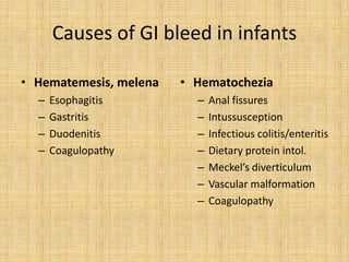
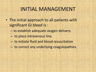
This document discusses gastrointestinal (GI) bleeding in infants and children. It covers: 1. Definitions of different types of GI bleeding like melena, hematochezia, and hematemesis. 2. Pathophysiology of GI bleeding including consequences of blood loss, higher risk in children, and compensatory mechanisms. 3. Causes of GI bleeding in different age groups ranging from neonates to adolescents. Common causes include esophagitis, gastritis, ulcers, varices, and infections. 4. Evaluation involves a thorough history, physical exam, lab tests like CBC and coagulation studies, and endoscopic procedures to identify the source of bleeding. Management is then


