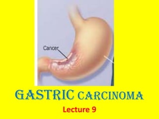
L9 gastric carcinoma f
•Download as PPTX, PDF•
14 likes•2,926 views

Gastric adenocarcinoma is the most common type of stomach cancer, comprising over 90% of cases. Risk factors include H. pylori infection, smoking, low fruit/vegetable diet, and family history. It is classified based on growth pattern (exophytic, flat, excavated), depth of invasion (early vs. advanced), and histology (intestinal vs. diffuse). Early detection through screening endoscopy and surgical resection offer the best chance of cure, with 5-year survival rates over 90% for early cancer but below 20% for advanced cases. Prevention focuses on modifiable lifestyle risk factors and mass endoscopic screening programs.

Recommended
More Related Content
What's hot
What's hot (20)
Viewers also liked
Viewers also liked (20)
Similar to L9 gastric carcinoma f
Similar to L9 gastric carcinoma f (20)
More from Mohammad Manzoor
More from Mohammad Manzoor (20)
Recently uploaded
Recently uploaded (20)
L9 gastric carcinoma f
- 2. GASTRIC ADENOCARCINOMA Adenocarcinoma is the most common 90% -95% • Lymphomas 4% • Carcinoid 3% • Stromal tumors 2% malignancy of the stomach, comprising over of all gastric cancers.
- 3. Epidemiology & Classification • GC is the second leading cause of cancer-related deaths in the world after lung cancer. • In Japan, Chile, Costa Rica, and Eastern Europe the incidence is up to 20-fold Africa, and Southeast Asia. higher than in North America, northern Europe,
- 4. In the United States, gastric cancer rates over 85% dropped by during the twentieth century. Similar declines have been reported in many other Western countries, suggesting that environmental and dietary factors are responsible. One possible explanation is decreased consumption of dietary carcinogens. Intake of green, leafy vegetables and citrus fruits. the
- 5. more common in lower socioeconomic groups and in individuals with multifocal mucosal atrophy and intestinal metaplasia. Gastric cancer is
- 6. • Although overall incidence of gastric adenocarcinoma is falling, •cancer of the gastric cardia is on the rise.
- 7. Classification: GC show two morphologic types, called intestinal & diffuse. I.The intestinal type • arise from gastric mucous cells that have undergone intestinal metaplasia in the setting of chronic gastritis. • better differentiated • the more common type in high risk populations.
- 8. • occurs primarily after age years 2: 1 male predominance. The incidence of intestinal –type carcinoma has progressively diminished in the US. • with a • 50
- 9. II.The diffuse variant arise de novo from native gastric mucous cells, is not associated with chronic gastritis, poorly differentiated. occurs at an earlier age with female predominance. The incidence of diffuse GA has not changed significantly in the past 60 years and now constitutes approximately half of gastric carcinomas in the US.
- 10. • The intestinal and diffuse forms of gastric carcinomas can be considered as distinct entities, although their clinical outcome is similar.
- 11. The incidence of diffuse gastric cancer across countries , there are is relatively uniform no identified precursor lesions, and similar frequencies in males and females. the disease occurs at
- 12. Risk Factors • Gender -- men have more than double the risk of getting stomach cancer than women. • Race -- being African-American or Asian may increase your risk. • Genetics -- genetic abnormalities and some inherited cancer syndromes may increase your risk • Geography -- stomach cancer is more common in Japan, the former Soviet Union, and parts of Central America and South America. • Blood type -- individuals with blood group A may be at increased risk.
- 13. • Advanced age -- stomach cancer occurs more often around ages 70 and 74 in men and women, respectively. • Family history of gastric cancer can double or triple the risk of stomach cancer. • Lifestyle factors such as smoking, drinking alcohol, and eating a diet low in fruits and vegetables or high in salted, smoked, or nitrate-preserved foods may increase your risk • Helicobacter pylori • Certain health conditions including chronic gastritis, pernicious anemia, gastric polyps, intestinal metaplasia, and prior stomach surgery. • Work-related exposure due to coal mining, nickel refining, and rubber and timber processing and asbestos exposure.
- 14. • Diffuse Carcinoma (Risk factors) • Risk factors undefined, except for a rare inherited mutation of E-cadherin • Infection with H. pylori and chronic gastritis often absent
- 15. Pathogenesis • The mechanisms of neoplastic transformation are not entirely clear. • Chronic inflammation induced by H. pylori • may release reactive oxygen species, • which eventually cause DNA damage, • leading to an imbalance between cell proliferation and apoptosis, particularly in areas of tissue repair .
- 16. Morphology • The location of gastric carcinoma within the stomach is as follows: • Pylorus and antrum, 50% to 60%; • Cardia 25%; and • the remainder (15-25%) in the body and fundus. • The lesser curvature is involved in about 40% and the greater curvature in 12%. • Thus, a favored location is the lesser curvature of the antropyloric region.
- 17. Morphology cont. • Though less frequent, an ulcerative lesion on the greater curvature malignant than benign. is more likely to be
- 18. Morphology cont. • GC is classified on the basis of • depth of invasion, • Macroscopic growth pattern, and • Histologic subtype.
- 19. Morphology cont. Classification on the basis of depth of Invasion • Early gastric carcinoma mucosa and submucosa, is defined as a lesion confined to the regardless of the presence or absence of perigastric lymph node metastases.
- 20. Morphology Classification on the basis of depth of Invasion cont. • Advanced gastric carcinoma is a neoplasm that has extended below the submucosa into the muscular wall and has perhaps spread more widely.
- 21. Morphology cont. Gastric mucosal dysplasia the presumed precursor lesion of early gastric cancer, which then in turn progresses to advanced lesions. is
- 22. •Part II
- 23. Morphology cont. Classification on the basis of macroscopic growth patterns • The three macroscopic growth patterns of gastric carcinoma which may be evident at both the early and advanced stages, are 1.Exophytic with protrusion of a tumor mass into the lumen; 2.Flat or depressed, 3.Excavated, crater in which there is no obvious tumor mass within the mucosa; and whereby a shallow or deeply erosive is present in the wall of the stomach.
- 24. Morphology cont. • Exophytic tumors may contain portions of an adenoma. • Flat or depressed l effacement malignancy presents only as regiona of the normal surface mucosal pattern. • Excavated cancers may mimic, in size and appearance, chronic peptic ulcers, although more advanced cases show heaped-up margins.
- 25. Morphology cont. Classification on the basis of histology intestinal & diffuse.
- 26. Morphology cont. The intestinal variant is composed of malignant cells forming neoplastic intestinal glands those of resembling colonic adenocarcinoma.
- 27. • Gastric tumors with an intestinal morphology tend to form bulky tumors
- 28. Morphology cont. • The diffuse variant is composed of gastric –type mucous cells that generally do not form glands but rather permeate the mucosa and wall as scattered individual • signet-ring cells or • small clusters in an infiltrative growth pattern.
- 29. Morphology cont. A mass may be difficult to appreciate in diffuse gastric cancer, but these infiltrative tumors often evoke a Desmoplastic reaction that stiffens the gastric wall and may provide a valuable diagnostic clue.
- 30. Morphology cont. The rigid and thickened stomach is termed a leather bottle stomach, or linitis plastica due to desmoplastic reaction ( in diffuse variant). Brinton's disease
- 31. • Whatever the histologic variant, all gastric carcinomas eventually penetrate the wall to serosa, spread to regional and more distant lymph nodes, metastasize and widely. involve the
- 32. For obscure reasons, In advanced cases gastric carcinoma the earliest lymph node metastasis may sometimes involve a Supraclavicular lymph node (Virchow node).
- 33. Rudolf Karl Virchow Virchow - German pathologist (1821-1902)
- 34. Gastric tumors can also metastasize to the to form a periumbilical region subcutaneous nodule, Sister termed a Mary Joseph nodule after the nurse who first noted this lesion as a marker of , metastatic carcinoma.
- 35. • Another somewhat unusual mode of intraperitoneal spread in females is to both the ovaries, giving rise to the so called Krukenburg tumor.
- 36. Local invasion into the duodenum, pancreas, Retroperitoneum
- 37. Diffuse type Gastric adenocarcinoma. A, Intestinal-type adenocarcinoma consisting of an elevated mass with heaped-up borders and central ulceration. B, Linitis plastica. The gastric wall is markedly thickened, and rugal folds are partially lost.
- 38. Clinical features Early stage: • • • • • Indigestion and stomach discomfort A bloated feeling after eating Mild nausea Loss of appetite Heartburn
- 39. Clinical features Advanced stage: • Discomfort in the upper or middle part of the abdomen. • Blood in the stool (which appears as black, tarry stools). • Vomiting or vomiting blood. • Weight loss. • Pain or bloating in the stomach after eating. • Weakness or fatigue associated with mild anemia (a deficiency in red blood cells).
- 40. Diagnosis • • • • • • • • • • • • • Signs& symptoms Medical History & Physical exam Upper endoscopy Biopsy Testing Biopsy Imaging tests Endoscopic Ultrasound CT scan MRI PET Chest X-ray Laparoscopy Lab tests
- 41. Treatment Chemotherapy or radiation therapy palliative care. surgical resection treatment and However, when possible, for gastric adenocarcinoma. remains the preferred
- 42. Prognosis • After surgical resection, the 5-year survival rate of can early gastric cancer exceed 90 %, even if lymph node metastases are present.
- 43. Prognosis cont . • In contrast, the 5-year survival rate for advanced gastric cancer remains below 20%.
- 44. Prevention General Measures Screening (Mass endoscopic screening programs) The only hope for cure is early detection and surgical removal, because the most important prognostic indicator is stage of the tumor at the time of resection.