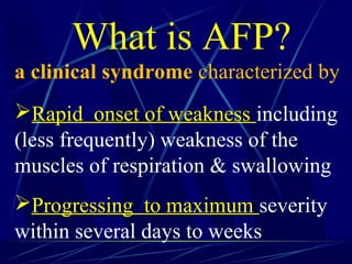
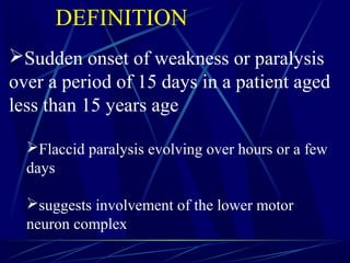
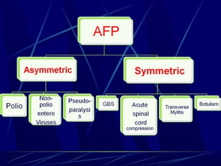
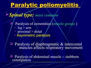
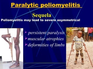
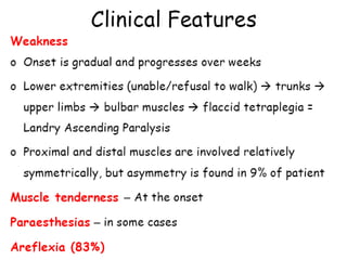
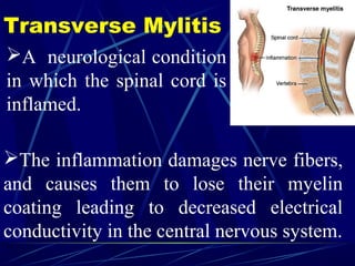
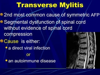
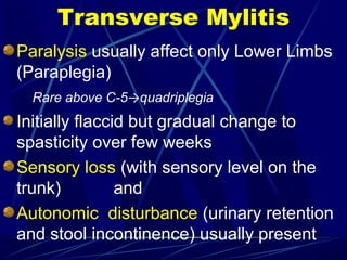
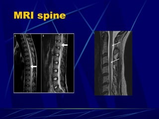
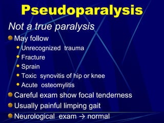
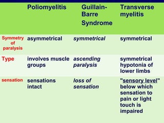
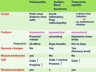
ACUTE FLACID PARALYSIS
Dr. Muhammad Sajjad Sabir
MBBS, DCH , MCPS, FCPS
Assist Prof of Paediatrics
ACUTE- Rapid in onset
FLACCID
PARALYSIS
ACUTE
FLACCID-
PARALYSIS
Drooping
without spasticity
ACUTE
FLACCID
PARALYSIS-Loss of ability to
move a body part
What is AFP?
a clinical syndrome characterized by
Rapid onset of weakness including
(less frequently) weakness of the
muscles of respiration & swallowing
Progressing to maximum severity
within several days to weeks
Sudden onset of weakness or paralysis
over a period of 15 days in a patient aged
less than 15 years age
Flaccid paralysis evolving over hours or a few
days
suggests involvement of the lower motor
neuron complex
DEFINITION
Anterior horn cells AHC (spinal cord)
 acute poliomyelitis
acute transverse myelitis
Peripheral Nerves
roots: GBS (post-infectious)
toxins: Diphteria, porphyria
N-M junction:
botulinum toxin , tick toxin
Metabolic: Periodic paralysis
Muscular: myositis (rare)
A lesion compressing the spinal cord must be ruled out
Syndromes Presenting as AFP (DD)
Most common
 Poliomyelitis
 Guillain-Barre Syndrome
Transverse myelitis
Acute Flaccid Paralysis(AFP)
Differential diagnosis
AFP in <15yrs age child is
notify-able disease to WHO
Polio surveillance officers—all
districts
Poliomyeliti
s
Poliomyelitis
Polio= gray matter
Myelitis= inflammation of the spinal cord
Most affects children < the age of 5
Global eradication in near future
Etiology
Enterovirus (RNA)
Three serotypes: 1, 2, 3
Clinical Classification of Poliomyelitis
Asymptomatic infection (Inapparent) (90- 95%)
Abortive poliomyelitis (5%)
 Non- paralytic poliomyelitis (aseptic meningitis) (1- 4%)
 Paralytic poliomyelitis (0.1-1%)
 Spinal form
 Bulbar form
 Bulbospinal form
 Encephalitic form
Poliovirus infections may follow one of several courses
Paralytic poliomyelitis
 Prodromal period
 Preparalytic period
 Paralytic period
 Convalescent period
 Sequela period
Commonly known as Polio
Only 1/1,000 to 1/100 (0.1% to 1%) infected
individuals develop paralytic disease
Preparalytic period
 High fever
 Cutaneous hyperesthesia or paresthesia in the
extremities
 Muscular pain is common
 Muscles are tender even to gentle palpation
 Severe headache & vomiting
 Lethargy
 Signs of meningeal irritation
(neck stiffness, Kerning & Brudzinski signs positive)
Paralytic poliomyelitis
Weakness of one or more muscle groups (spinal or
cranial)
Loose & Floppy Limbs, hypotonia-- Flaccid paralysis
Asymmetric paralysis
Paralysis is complete – fever normalizes
Weakness persists
Respiratory & vasomotor changes
Bladder & bowel dysfunction
Tendon reflexes are absent with paralysis.
Sensory defects do not occur in poliomyelitis
Paralytic poliomyelitis
Paralytic period
Spinal type: most common
 Paralysis of extremities (muscle groups )
 leg > arm
 proximal > distal
 Asymmetric paralysis
 Paralysis of diaphragmatic & intercostal
muscles affects respiratory movement
 Paralysis of abdominal muscle -- stubborn
constipation
Paralytic poliomyelitis
Sequela
Poliomyelitis may lead to severe asymmetrical
 persistent paralysis
muscular atrophies
deformities of limbs
Paralytic poliomyelitis
Diagnosis
WHO recommends diagnosis of poliomyelitis be
confirmed by isolation and identification of poliovirus in
the stool, with specific identification of wild-type and
vaccine type strains
Polioviruses may be isolated from 80 to 90% of acutely
ill patients
whereas less than 20% may yield virus within 3-4 wk
after onset of paralysis
Identification of poliovirus in stool
Stool examination
Two samples 24 hr apart
Within 14 days of onset of paralysis
8-10 grams or thumb size
Collected in a clean wide mouth bottle –
(plastic or glass) with screw cap
Sample stored below 8°C
No dessication or leakage till received
at WHO Accredited Lab
Collection of sample
Laboratory Findings
Cerebrospinal fluid
aseptic menigitis
 pleocytosis: 50~500×109
/L
 protein: normal or slightly increase
 glucose and chloride; normal
 albuminocytologic dissociation
Serological Tests
Neutralizing antibodies (IgG)
Antibodies to C antigen (IgM)
 Anti-D antibodies
Management
Isolation
Rest in bed
Neutral positioning of the limbs
Good nursing
No I.M Injection or surgery
Relief of pain and spasm of muscles
Physiotherapy
Treatment symptomatic and supportive
Prevention
control of source of infection:
 isolation: 40 days after the onset of illness
protection of susceptible population-
vaccination :
 attenuated live vaccine by
Oral -- OPV
 killed virus vaccine
Injectable -- IPV
Guillain-Barre Syndrome
(GBS)
Guillain-Barre Syndrome
(GBS)
Post-infectious
acute, rapidly progressing
ascending
potentially fatal form of polyneuritis
involving mainly motor but sometimes also
sensory and autonomic nerves
also known as: Acute inflammatory demyelinating
polyneuropathy (AIDP)
Transverse Mylitis
A neurological condition
in which the spinal cord is
inflamed.
The inflammation damages nerve fibers,
and causes them to lose their myelin
coating leading to decreased electrical
conductivity in the central nervous system.
2nd most common cause of symmetric AFP
Segmental dysfunction of spinal cord
without evidence of spinal cord
compression
Cause is either:
a direct viral infection
or
an autoimmune disease
Transverse Mylitis
Transverse Mylitis
Paralysis usually affect only Lower Limbs
(Paraplegia)
Rare above C-5→quadriplegia
Initially flaccid but gradual change to
spasticity over few weeks
Sensory loss (with sensory level on the
trunk) and
Autonomic disturbance (urinary retention
and stool incontinence) usually present
MRI spine
Treatment
Intravenous Steroids
High-dose I.V methyl-prednisolone for 3-5 days
Plasma Electrophoresis
Prolonged course
Complete recovery in 60% of cases
Botulism
Botulism
It’s a toxic neuromuscular blockade
caused by Clostridium botulinum
(anaerobic gram positive)
According to the type of infection, there
are 3 types
Infant B. due to exposure to soil , honey
Food-borne B. due to ingestion improperly
home preserved foods containing the toxins
Wound B. due to wound contamination with
the organism
All three types of botulism result in
symmetric
descending
Flaccid paralysis of motor and
autonomic nerves
always beginning with cranial nerves
These symptoms preceded by
constipation (infant botulism)
If untreated - paralysis of respiratory →
death
Botulism
Pseudoparalysis
Pseudoparalysis
Not a true paralysis
May follow
Unrecognized trauma
Fracture
Sprain
Toxic synovitis of hip or knee
Acute osteomylitis
Careful exam show focal tenderness
Usually painful limping gait
Neurological exam → normal
Acute spinal cord
compression
Trauma to the back
Spinal epidural abscess
Vascular anomalies of the cord
Spinal cord tumors
Clinically difficult to differentiate from
Transverse Mylitis
CT scan/ MRI spine:
 Sensitive
 Can show nature of obstruction
Acute spinal cord compression
Prognosis & complications of AFP
According to cause
Poliomyelitis
Respiratory
death
Limb atrophy
GBS
Respiratory
autonomic crises
residual weakness
death
Acute transverse myelitis
Residual deficits --bowel and bladder
dysfunction & weakness - lower Limbs
Poliomyelitis Guillain-
Barre
Syndrome
Transverse
myelitis
Symmetry
of
paralysis
asymmetrical symmetrical symmetrical
Type involves muscle
groups
ascending
paralysis
symmetrical
hypotonia of
lower limbs
sensation sensations
intact
loss of
sensation
"sensory level"
below which
sensation to
pain or light
touch is
impaired
Poliomyelitis Guillain-
Barre
Syndrome
Transverse
myelitis
Cause Polio virus
Anterior horn
cells
Acute
inflamatory
Poly-
radiculopathy
a direct viral
infection
or
an autoimmune
disease
Paralysis Asymmetric symmetrical symmetrical
Proximal
>distal
ascending Hypotonic lower
limbs
Progression 24-48hrs Days-4weeks Hrs to days
Sensory changes No Yes Yes
(Sensory level)
Muscle tenderness yes No No
CSF Cells ↑
Proteins ↑
Cells = normal
Proteins ↑
Cells ↑
Residual paralysis yes + yes
ACUTE FLACCID PARALYSIS MBBS Lecture

ACUTE FLACCID PARALYSIS MBBS Lecture

  • 2.
    ACUTE FLACID PARALYSIS Dr.Muhammad Sajjad Sabir MBBS, DCH , MCPS, FCPS Assist Prof of Paediatrics
  • 3.
    ACUTE- Rapid inonset FLACCID PARALYSIS
  • 4.
  • 5.
  • 6.
    What is AFP? aclinical syndrome characterized by Rapid onset of weakness including (less frequently) weakness of the muscles of respiration & swallowing Progressing to maximum severity within several days to weeks
  • 7.
    Sudden onset ofweakness or paralysis over a period of 15 days in a patient aged less than 15 years age Flaccid paralysis evolving over hours or a few days suggests involvement of the lower motor neuron complex DEFINITION
  • 8.
    Anterior horn cellsAHC (spinal cord)  acute poliomyelitis acute transverse myelitis Peripheral Nerves roots: GBS (post-infectious) toxins: Diphteria, porphyria N-M junction: botulinum toxin , tick toxin Metabolic: Periodic paralysis Muscular: myositis (rare) A lesion compressing the spinal cord must be ruled out Syndromes Presenting as AFP (DD)
  • 10.
    Most common  Poliomyelitis Guillain-Barre Syndrome Transverse myelitis Acute Flaccid Paralysis(AFP) Differential diagnosis
  • 11.
    AFP in <15yrsage child is notify-able disease to WHO Polio surveillance officers—all districts
  • 12.
  • 13.
    Poliomyelitis Polio= gray matter Myelitis=inflammation of the spinal cord Most affects children < the age of 5 Global eradication in near future Etiology Enterovirus (RNA) Three serotypes: 1, 2, 3
  • 14.
    Clinical Classification ofPoliomyelitis Asymptomatic infection (Inapparent) (90- 95%) Abortive poliomyelitis (5%)  Non- paralytic poliomyelitis (aseptic meningitis) (1- 4%)  Paralytic poliomyelitis (0.1-1%)  Spinal form  Bulbar form  Bulbospinal form  Encephalitic form Poliovirus infections may follow one of several courses
  • 15.
    Paralytic poliomyelitis  Prodromalperiod  Preparalytic period  Paralytic period  Convalescent period  Sequela period Commonly known as Polio Only 1/1,000 to 1/100 (0.1% to 1%) infected individuals develop paralytic disease
  • 16.
    Preparalytic period  Highfever  Cutaneous hyperesthesia or paresthesia in the extremities  Muscular pain is common  Muscles are tender even to gentle palpation  Severe headache & vomiting  Lethargy  Signs of meningeal irritation (neck stiffness, Kerning & Brudzinski signs positive) Paralytic poliomyelitis
  • 17.
    Weakness of oneor more muscle groups (spinal or cranial) Loose & Floppy Limbs, hypotonia-- Flaccid paralysis Asymmetric paralysis Paralysis is complete – fever normalizes Weakness persists Respiratory & vasomotor changes Bladder & bowel dysfunction Tendon reflexes are absent with paralysis. Sensory defects do not occur in poliomyelitis Paralytic poliomyelitis Paralytic period
  • 18.
    Spinal type: mostcommon  Paralysis of extremities (muscle groups )  leg > arm  proximal > distal  Asymmetric paralysis  Paralysis of diaphragmatic & intercostal muscles affects respiratory movement  Paralysis of abdominal muscle -- stubborn constipation Paralytic poliomyelitis
  • 19.
    Sequela Poliomyelitis may leadto severe asymmetrical  persistent paralysis muscular atrophies deformities of limbs Paralytic poliomyelitis
  • 20.
    Diagnosis WHO recommends diagnosisof poliomyelitis be confirmed by isolation and identification of poliovirus in the stool, with specific identification of wild-type and vaccine type strains Polioviruses may be isolated from 80 to 90% of acutely ill patients whereas less than 20% may yield virus within 3-4 wk after onset of paralysis Identification of poliovirus in stool
  • 21.
    Stool examination Two samples24 hr apart Within 14 days of onset of paralysis 8-10 grams or thumb size Collected in a clean wide mouth bottle – (plastic or glass) with screw cap Sample stored below 8°C No dessication or leakage till received at WHO Accredited Lab Collection of sample
  • 22.
    Laboratory Findings Cerebrospinal fluid asepticmenigitis  pleocytosis: 50~500×109 /L  protein: normal or slightly increase  glucose and chloride; normal  albuminocytologic dissociation Serological Tests Neutralizing antibodies (IgG) Antibodies to C antigen (IgM)  Anti-D antibodies
  • 23.
    Management Isolation Rest in bed Neutralpositioning of the limbs Good nursing No I.M Injection or surgery Relief of pain and spasm of muscles Physiotherapy Treatment symptomatic and supportive
  • 24.
    Prevention control of sourceof infection:  isolation: 40 days after the onset of illness protection of susceptible population- vaccination :  attenuated live vaccine by Oral -- OPV  killed virus vaccine Injectable -- IPV
  • 25.
  • 26.
    Guillain-Barre Syndrome (GBS) Post-infectious acute, rapidlyprogressing ascending potentially fatal form of polyneuritis involving mainly motor but sometimes also sensory and autonomic nerves also known as: Acute inflammatory demyelinating polyneuropathy (AIDP)
  • 31.
    Transverse Mylitis A neurologicalcondition in which the spinal cord is inflamed. The inflammation damages nerve fibers, and causes them to lose their myelin coating leading to decreased electrical conductivity in the central nervous system.
  • 32.
    2nd most commoncause of symmetric AFP Segmental dysfunction of spinal cord without evidence of spinal cord compression Cause is either: a direct viral infection or an autoimmune disease Transverse Mylitis
  • 33.
    Transverse Mylitis Paralysis usuallyaffect only Lower Limbs (Paraplegia) Rare above C-5→quadriplegia Initially flaccid but gradual change to spasticity over few weeks Sensory loss (with sensory level on the trunk) and Autonomic disturbance (urinary retention and stool incontinence) usually present
  • 34.
  • 35.
    Treatment Intravenous Steroids High-dose I.Vmethyl-prednisolone for 3-5 days Plasma Electrophoresis Prolonged course Complete recovery in 60% of cases
  • 36.
  • 37.
    Botulism It’s a toxicneuromuscular blockade caused by Clostridium botulinum (anaerobic gram positive) According to the type of infection, there are 3 types Infant B. due to exposure to soil , honey Food-borne B. due to ingestion improperly home preserved foods containing the toxins Wound B. due to wound contamination with the organism
  • 38.
    All three typesof botulism result in symmetric descending Flaccid paralysis of motor and autonomic nerves always beginning with cranial nerves These symptoms preceded by constipation (infant botulism) If untreated - paralysis of respiratory → death Botulism
  • 39.
  • 40.
    Pseudoparalysis Not a trueparalysis May follow Unrecognized trauma Fracture Sprain Toxic synovitis of hip or knee Acute osteomylitis Careful exam show focal tenderness Usually painful limping gait Neurological exam → normal
  • 41.
  • 42.
    Trauma to theback Spinal epidural abscess Vascular anomalies of the cord Spinal cord tumors Clinically difficult to differentiate from Transverse Mylitis CT scan/ MRI spine:  Sensitive  Can show nature of obstruction Acute spinal cord compression
  • 43.
    Prognosis & complicationsof AFP According to cause Poliomyelitis Respiratory death Limb atrophy GBS Respiratory autonomic crises residual weakness death Acute transverse myelitis Residual deficits --bowel and bladder dysfunction & weakness - lower Limbs
  • 44.
    Poliomyelitis Guillain- Barre Syndrome Transverse myelitis Symmetry of paralysis asymmetrical symmetricalsymmetrical Type involves muscle groups ascending paralysis symmetrical hypotonia of lower limbs sensation sensations intact loss of sensation "sensory level" below which sensation to pain or light touch is impaired
  • 45.
    Poliomyelitis Guillain- Barre Syndrome Transverse myelitis Cause Poliovirus Anterior horn cells Acute inflamatory Poly- radiculopathy a direct viral infection or an autoimmune disease Paralysis Asymmetric symmetrical symmetrical Proximal >distal ascending Hypotonic lower limbs Progression 24-48hrs Days-4weeks Hrs to days Sensory changes No Yes Yes (Sensory level) Muscle tenderness yes No No CSF Cells ↑ Proteins ↑ Cells = normal Proteins ↑ Cells ↑ Residual paralysis yes + yes