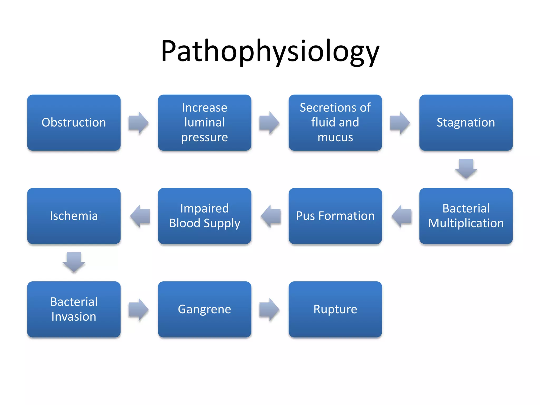
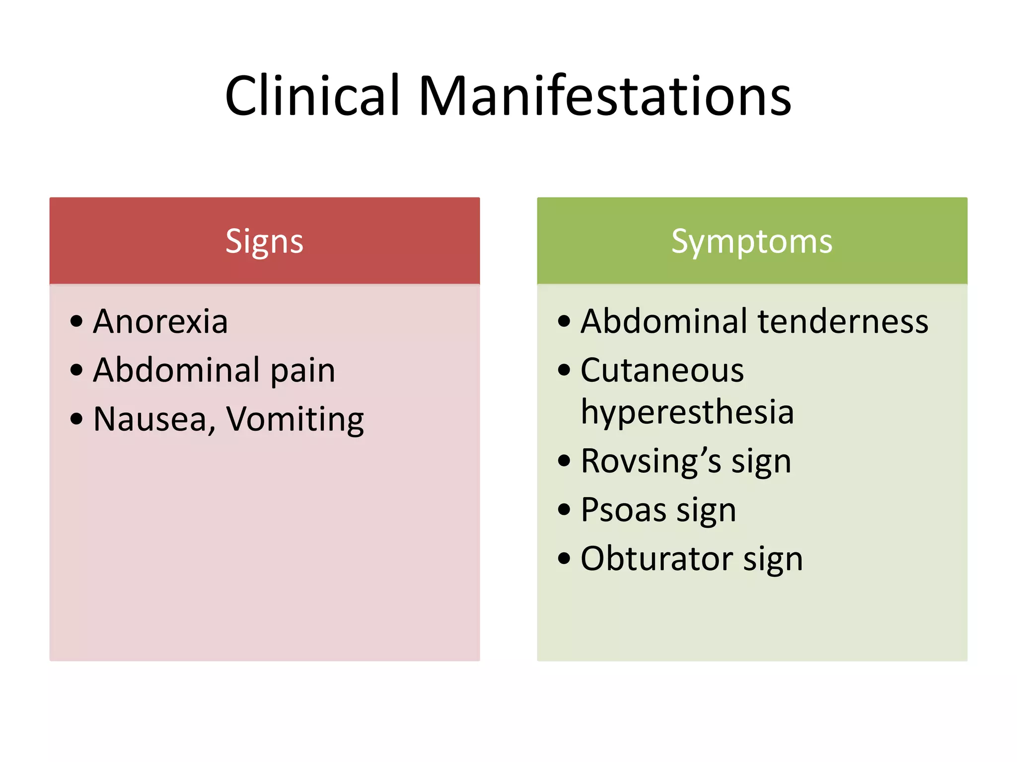
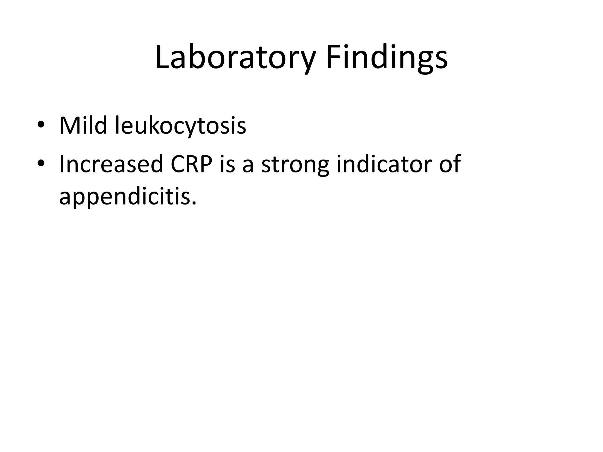
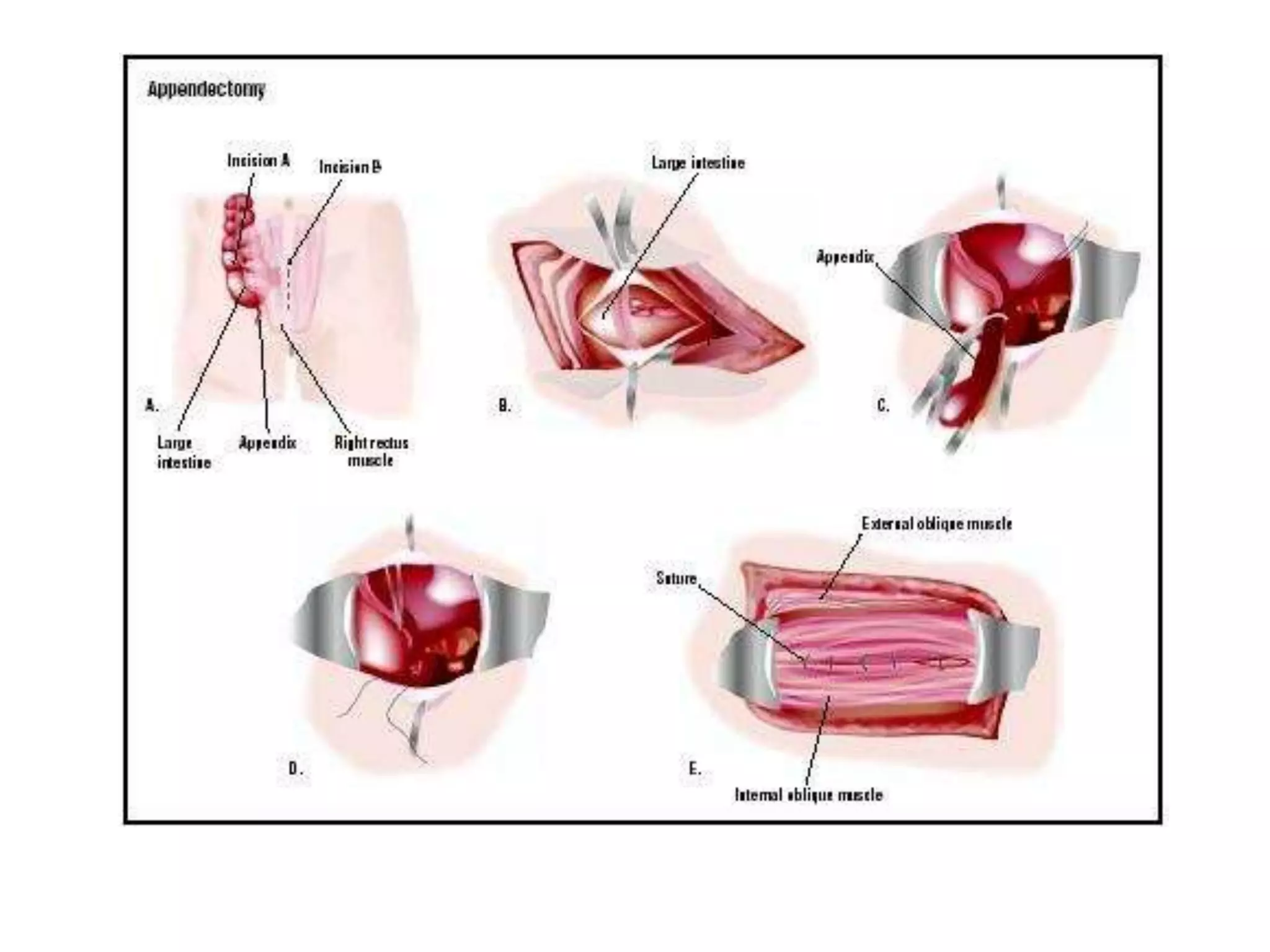
POKHREL, BHARAT M.D.
ACUTE APPENDICITIS
The Appendix
• A thin, wormlike, tubular organ
• located at the inferior part of the cecum
• Length: <1 cm to >30 cm; most appendices are
6 to 9 cm long
ANATOMY
• Arterial supply: Appendicular branch of
Iliocolic artery
• Innervation derived from sympathetic
elements contributed by superior mesenteric
plexus (T10-L1)
• Afferents from parasympathetic elements via
Vagus nerves
Locations of Appendix
Physiology
• An immunologic organ
• Secretion of immunoglobulins (IgA)
• Inverse association between appendectomy
and development of ulcerative colitis.
Incidence
• One of the most common surgical
emergencies
• Yearly incidence rate 100 per 100,000.
• Lifetime risk for males 8.7%, females 6.7%
• Highest incidence in second decade of life
Pathophysiology
Obstruction
Increase
luminal
pressure
Secretions of
fluid and
mucus
Stagnation
Bacterial
Multiplication
Pus Formation
Impaired
Blood Supply
Ischemia
Bacterial
Invasion
Gangrene Rupture
Clinical Manifestations
Signs
• Anorexia
• Abdominal pain
• Nausea, Vomiting
Symptoms
• Abdominal tenderness
• Cutaneous
hyperesthesia
• Rovsing’s sign
• Psoas sign
• Obturator sign
Laboratory Findings
• Mild leukocytosis
• Increased CRP is a strong indicator of
appendicitis.
Alvarado Scale
Imaging studies
• Ultrasonography and CT scan are the most
commonly used tests.
Treatment
Open Appendectomy
• Muscle splitting
incisions at the RLQ
– McBurney’s (Oblique)
– Rocky-Davis (Transverse)
• Incision is centered over
either the point of
maximal tenderness or
palpable mass
Open Appendectomy
• Typically under GA, patient in supine position
• Abdomen prepped and draped
• Incision at McBurney’s point
• If appendix is not easily identified, cecum
should be located.
• Tracing the taenia libera, the most visible of 3
taeniae coli, distally, the base of the appendix
can be identified.
Laparoscopic Appendectomy
• Under GA, patient in supine position
• Uses 3 ports.
• 10-12mm port at umbilicus, 5mm port at
suprapubic and in LLQ
• Appendix should be identified by tracking
taeniae libera/coli
• Appendix is removed through infraumbilical
trocar in a retrieval bag.
ANTIBIOTICS
• Simple Acute Appendicitis
• 24 hours antibiotic coverage
• Perforated or Gangrenous
• Continued until patient is afebrile
• Normal WBC
References
• Schwartz’s Principles of Surgery, Tenth edition
THANK YOU

Acute appendicitis

Editor's Notes

  • #8 Secretions of fluids and mucus – distention which cause dull, diffuse pain Bacteria – Marked distention – reflex nausea/vomiting Impaired blood supply: venous outflow obstruction and decrease in arterial inflow - ischemia Occluded capillary and veins – RLQ Pain
  • #10 Inflammatory process in the appendeix presents as PAIN, initially diffuse visceral progressing to localized. GI symptoms that develop before the onset of pain suggest different etiology such as AGE Many have OBSTIPATION, feels that defecation will relief the pain Diarrhea may occur in association with perforation.
  • #12 Patients with scores of 9 or 10 are almost certain to have appendicitis; there is little advantage in further work-up, and they should go to the operating room. Patients with scores of 7 or 8 have a high likelihood of appendicitis, whereas scores of 5 or 6 are compatible with, but not diagnostic of, appendicitis. CT scanning is certainly appropriate for patients with Alvarado scores of 5 and 6, and a case can be built for imaging for those with scores of 7 and 8. On the other hand, it is difficult to justify the expense, radiation exposure, and possible complications of CT scanning in patients whose scores of 0 to 4 make it extremely unlikely (but not impossible) that they have appendicitis.
  • #15 Equivocal Appendicitis – (+) RLQ Pain with atyipical history and PE. Uncomplicated Appendicitis – acutely inflamed, phlegmonous, suppurative, mildly inflamed appendix with or without peritonitis Complicated Appendicitis – gangrenous appendicitis, perforated appe, localized purulent collection at operation, generalized peritonitis and periappendiceal abscess Sonography: inexpensive/no contrast medium; (+) if non compressible appendix/ >6mm AP diameter; highly suggestive = thickening of appendiceal wall/periappendiceal fluid CT Scans: Focus – only RLQ; Non focused – whole abdomen Enhanced – rectally administered contrast Non enhanced – dye allergy CONS: expensive, radiation exposure Alvarado score of 5-6 Alvarado – scoring system 9-10 = almost certain; 7-8 high likelihood; 5-6 compatible with’ 0-4 extremely unlikely SYMPTOMS: migration of pain (2), Anorexia (1), Vom/Nas (1); Signs: RLQ Tenderness (2), Rebound (1), Fever (1) Labs: Leukocytosis (2), Left Shift (1)
  • #16 McBurney’s – 2- 5 cm above ASIS and continues to a point one-third of the way to the umbilicus (McBurney’s Point); incision is parallel to the external oblique muscle which allows the muscle to be split in the direction of fibers, decreasing healing times and scar formation Rocky-Davis – straight transverse at the skin; GRIDIRON
  • #17 Layers of Muscles and Tissue are cut and large intestine or colon is visualized (LAYERS: Skin, Fats Campers/Scarpas, Muscles (EO, IO, Rectus Abdominis, Transverse Abdominal Muscle, Pyramidalis Muscle) Fascia, Peritoneum Appendix is located: convergence of the taenia can be followed to the base of the appendix; a sweeping lateral to medial motion can aid in delivering the appediceal tip; mobilized by dividing the mesoappendix; ligate the appendiceal artery Tied and removed: stump can be managed by simple ligation and inversion with either a purse string-or z stitch Muscle and tissue are stitched; peritoneal cavity is irrigated and the wound is closed by layers