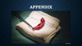
The document provides information on the appendix, including its history, anatomy, embryology, physiology, acute appendicitis, neoplasms, and variants. Some key points include: the appendix was first depicted by Leonardo da Vinci in 1492; acute appendicitis is caused by obstruction leading to distention and infection, with symptoms like migrating right lower quadrant pain; imaging like CT can help diagnose appendicitis; complications include perforation; and neoplasms like carcinoid tumors or adenocarcinomas can rarely affect the appendix.
The incidence of biliary injury after laparoscopic cholecystectomy (LC) has shown a declining trend though it may still be twice that as with open cholecystectomy. Major biliary or vasculobiliary injury is associated with significant morbidity. As prevention is the best strategy, the concept of a culture of safe cholecystectomy has been recently introduced to educate surgeons and apprise them of basic tenets of safe performance of LC. Various aspects of safe cholecystectomy include: (1) thorough knowledge of relevant anatomy, various anatomical landmarks, and anatomical variations; (2) an understanding of the mechanisms involved in biliary/vascular injury, the most important being the misidentification injury; (3) identification of various preoperative and intraoperative predictors of difficult cholecystectomy; (4) proper gallbladder retraction; (5) safe use of various energy devices; (6) understanding the critical view of safety, including its doublet view and documentation; (7) awareness of various error traps (e.g., fundus first technique); (8) use of various bailout strategies (e.g., subtotal cholecystectomy) in difficult gallbladder cases; (9) use of intraoperative imaging techniques (e.g., intraoperative cholangiogram) to ascertain correct anatomy; and (10) understanding the concept of time-out. Surgeons should be facile with these aspects of this culture of safety in cholecystectomy in an attempt to reduce the incidence of biliary/vascular injury during LC.
The incidence of biliary injury after laparoscopic cholecystectomy (LC) has shown a declining trend though it may still be twice that as with open cholecystectomy. Major biliary or vasculobiliary injury is associated with significant morbidity. As prevention is the best strategy, the concept of a culture of safe cholecystectomy has been recently introduced to educate surgeons and apprise them of basic tenets of safe performance of LC. Various aspects of safe cholecystectomy include: (1) thorough knowledge of relevant anatomy, various anatomical landmarks, and anatomical variations; (2) an understanding of the mechanisms involved in biliary/vascular injury, the most important being the misidentification injury; (3) identification of various preoperative and intraoperative predictors of difficult cholecystectomy; (4) proper gallbladder retraction; (5) safe use of various energy devices; (6) understanding the critical view of safety, including its doublet view and documentation; (7) awareness of various error traps (e.g., fundus first technique); (8) use of various bailout strategies (e.g., subtotal cholecystectomy) in difficult gallbladder cases; (9) use of intraoperative imaging techniques (e.g., intraoperative cholangiogram) to ascertain correct anatomy; and (10) understanding the concept of time-out. Surgeons should be facile with these aspects of this culture of safety in cholecystectomy in an attempt to reduce the incidence of biliary/vascular injury during LC.
Abdominal Imaging Case Studies #27.pptxSean M. Fox
Drs. Kylee Brooks and Parker Hambright are Emergency Medicine Residents and Drs. Alexis Holland and William Lorenz are Surgery Residents at Carolinas Medical Center in Charlotte, NC. They are interested in medical education. With the guidance of Drs. Kyle Cunningham, Brent Matthews, and Michael Gibbs, they aim to help augment our understanding of emergent abdominal imaging. Follow along with the EMGuideWire.com team as they post these monthly educational, self-guided radiology slides. This month’s cases include:
• Iatrogenic Esophageal Perforation
• Emphysematous Cystitis
• Meckel’s Diverticulum
• Paraesophageal Hernia
Title: Sense of Taste
Presenter: Dr. Faiza, Assistant Professor of Physiology
Qualifications:
MBBS (Best Graduate, AIMC Lahore)
FCPS Physiology
ICMT, CHPE, DHPE (STMU)
MPH (GC University, Faisalabad)
MBA (Virtual University of Pakistan)
Learning Objectives:
Describe the structure and function of taste buds.
Describe the relationship between the taste threshold and taste index of common substances.
Explain the chemical basis and signal transduction of taste perception for each type of primary taste sensation.
Recognize different abnormalities of taste perception and their causes.
Key Topics:
Significance of Taste Sensation:
Differentiation between pleasant and harmful food
Influence on behavior
Selection of food based on metabolic needs
Receptors of Taste:
Taste buds on the tongue
Influence of sense of smell, texture of food, and pain stimulation (e.g., by pepper)
Primary and Secondary Taste Sensations:
Primary taste sensations: Sweet, Sour, Salty, Bitter, Umami
Chemical basis and signal transduction mechanisms for each taste
Taste Threshold and Index:
Taste threshold values for Sweet (sucrose), Salty (NaCl), Sour (HCl), and Bitter (Quinine)
Taste index relationship: Inversely proportional to taste threshold
Taste Blindness:
Inability to taste certain substances, particularly thiourea compounds
Example: Phenylthiocarbamide
Structure and Function of Taste Buds:
Composition: Epithelial cells, Sustentacular/Supporting cells, Taste cells, Basal cells
Features: Taste pores, Taste hairs/microvilli, and Taste nerve fibers
Location of Taste Buds:
Found in papillae of the tongue (Fungiform, Circumvallate, Foliate)
Also present on the palate, tonsillar pillars, epiglottis, and proximal esophagus
Mechanism of Taste Stimulation:
Interaction of taste substances with receptors on microvilli
Signal transduction pathways for Umami, Sweet, Bitter, Sour, and Salty tastes
Taste Sensitivity and Adaptation:
Decrease in sensitivity with age
Rapid adaptation of taste sensation
Role of Saliva in Taste:
Dissolution of tastants to reach receptors
Washing away the stimulus
Taste Preferences and Aversions:
Mechanisms behind taste preference and aversion
Influence of receptors and neural pathways
Impact of Sensory Nerve Damage:
Degeneration of taste buds if the sensory nerve fiber is cut
Abnormalities of Taste Detection:
Conditions: Ageusia, Hypogeusia, Dysgeusia (parageusia)
Causes: Nerve damage, neurological disorders, infections, poor oral hygiene, adverse drug effects, deficiencies, aging, tobacco use, altered neurotransmitter levels
Neurotransmitters and Taste Threshold:
Effects of serotonin (5-HT) and norepinephrine (NE) on taste sensitivity
Supertasters:
25% of the population with heightened sensitivity to taste, especially bitterness
Increased number of fungiform papillae
micro teaching on communication m.sc nursing.pdfAnurag Sharma
Microteaching is a unique model of practice teaching. It is a viable instrument for the. desired change in the teaching behavior or the behavior potential which, in specified types of real. classroom situations, tends to facilitate the achievement of specified types of objectives.
- Video recording of this lecture in English language: https://youtu.be/lK81BzxMqdo
- Video recording of this lecture in Arabic language: https://youtu.be/Ve4P0COk9OI
- Link to download the book free: https://nephrotube.blogspot.com/p/nephrotube-nephrology-books.html
- Link to NephroTube website: www.NephroTube.com
- Link to NephroTube social media accounts: https://nephrotube.blogspot.com/p/join-nephrotube-on-social-media.html
Flu Vaccine Alert in Bangalore Karnatakaaddon Scans
As flu season approaches, health officials in Bangalore, Karnataka, are urging residents to get their flu vaccinations. The seasonal flu, while common, can lead to severe health complications, particularly for vulnerable populations such as young children, the elderly, and those with underlying health conditions.
Dr. Vidisha Kumari, a leading epidemiologist in Bangalore, emphasizes the importance of getting vaccinated. "The flu vaccine is our best defense against the influenza virus. It not only protects individuals but also helps prevent the spread of the virus in our communities," he says.
This year, the flu season is expected to coincide with a potential increase in other respiratory illnesses. The Karnataka Health Department has launched an awareness campaign highlighting the significance of flu vaccinations. They have set up multiple vaccination centers across Bangalore, making it convenient for residents to receive their shots.
To encourage widespread vaccination, the government is also collaborating with local schools, workplaces, and community centers to facilitate vaccination drives. Special attention is being given to ensuring that the vaccine is accessible to all, including marginalized communities who may have limited access to healthcare.
Residents are reminded that the flu vaccine is safe and effective. Common side effects are mild and may include soreness at the injection site, mild fever, or muscle aches. These side effects are generally short-lived and far less severe than the flu itself.
Healthcare providers are also stressing the importance of continuing COVID-19 precautions. Wearing masks, practicing good hand hygiene, and maintaining social distancing are still crucial, especially in crowded places.
Protect yourself and your loved ones by getting vaccinated. Together, we can help keep Bangalore healthy and safe this flu season. For more information on vaccination centers and schedules, residents can visit the Karnataka Health Department’s official website or follow their social media pages.
Stay informed, stay safe, and get your flu shot today!
Ozempic: Preoperative Management of Patients on GLP-1 Receptor Agonists Saeid Safari
Preoperative Management of Patients on GLP-1 Receptor Agonists like Ozempic and Semiglutide
ASA GUIDELINE
NYSORA Guideline
2 Case Reports of Gastric Ultrasound
Title: Sense of Smell
Presenter: Dr. Faiza, Assistant Professor of Physiology
Qualifications:
MBBS (Best Graduate, AIMC Lahore)
FCPS Physiology
ICMT, CHPE, DHPE (STMU)
MPH (GC University, Faisalabad)
MBA (Virtual University of Pakistan)
Learning Objectives:
Describe the primary categories of smells and the concept of odor blindness.
Explain the structure and location of the olfactory membrane and mucosa, including the types and roles of cells involved in olfaction.
Describe the pathway and mechanisms of olfactory signal transmission from the olfactory receptors to the brain.
Illustrate the biochemical cascade triggered by odorant binding to olfactory receptors, including the role of G-proteins and second messengers in generating an action potential.
Identify different types of olfactory disorders such as anosmia, hyposmia, hyperosmia, and dysosmia, including their potential causes.
Key Topics:
Olfactory Genes:
3% of the human genome accounts for olfactory genes.
400 genes for odorant receptors.
Olfactory Membrane:
Located in the superior part of the nasal cavity.
Medially: Folds downward along the superior septum.
Laterally: Folds over the superior turbinate and upper surface of the middle turbinate.
Total surface area: 5-10 square centimeters.
Olfactory Mucosa:
Olfactory Cells: Bipolar nerve cells derived from the CNS (100 million), with 4-25 olfactory cilia per cell.
Sustentacular Cells: Produce mucus and maintain ionic and molecular environment.
Basal Cells: Replace worn-out olfactory cells with an average lifespan of 1-2 months.
Bowman’s Gland: Secretes mucus.
Stimulation of Olfactory Cells:
Odorant dissolves in mucus and attaches to receptors on olfactory cilia.
Involves a cascade effect through G-proteins and second messengers, leading to depolarization and action potential generation in the olfactory nerve.
Quality of a Good Odorant:
Small (3-20 Carbon atoms), volatile, water-soluble, and lipid-soluble.
Facilitated by odorant-binding proteins in mucus.
Membrane Potential and Action Potential:
Resting membrane potential: -55mV.
Action potential frequency in the olfactory nerve increases with odorant strength.
Adaptation Towards the Sense of Smell:
Rapid adaptation within the first second, with further slow adaptation.
Psychological adaptation greater than receptor adaptation, involving feedback inhibition from the central nervous system.
Primary Sensations of Smell:
Camphoraceous, Musky, Floral, Pepperminty, Ethereal, Pungent, Putrid.
Odor Detection Threshold:
Examples: Hydrogen sulfide (0.0005 ppm), Methyl-mercaptan (0.002 ppm).
Some toxic substances are odorless at lethal concentrations.
Characteristics of Smell:
Odor blindness for single substances due to lack of appropriate receptor protein.
Behavioral and emotional influences of smell.
Transmission of Olfactory Signals:
From olfactory cells to glomeruli in the olfactory bulb, involving lateral inhibition.
Primitive, less old, and new olfactory systems with different path
Couples presenting to the infertility clinic- Do they really have infertility...Sujoy Dasgupta
Dr Sujoy Dasgupta presented the study on "Couples presenting to the infertility clinic- Do they really have infertility? – The unexplored stories of non-consummation" in the 13th Congress of the Asia Pacific Initiative on Reproduction (ASPIRE 2024) at Manila on 24 May, 2024.
3. HISTORY
1492: Leonardo da Vinci depicted the appendix in his anatomic drawings
1544: Jean Fernel for first describing appendiceal disease in a paper published
After death of a girl treated with quince for diarrhea
1736: The first known appendectomy was performed by Claudius Amyand in London
During hernia repair
RLQ pain Thought to be due to typhlitis and perityphlitis
It was recognized that most instances of appendicitis could resolve without surgical
treatment
4. EMBRYOLOGY
Starts to develop at the 6th week
First noticed at the 8th week
Elongate at the 5th month
The subsequent unequal growth of the lateral wall of the cecum causes the appendix to
find its adult position on the posterior medial wall, just below the ileocecal valve
located by following the longitudinally oriented taeniae coli to their confluence on the
cecum
5. EMBRYOLOGY
The tip of the appendix can be located anywhere in the right lower quadrant of the
abdomen, pelvis, or retroperitoneum
Gut malrutation: RUQ appendix
Situs inversus: LLQ appendix
6. ANATOMY
Appendix length: <1 to >30 cm with average of 6-9cm
Outer diameter: varies between 3 and 8 mm
luminal diameter: varies between 1 and 3 mm
Blood supply: ileocolic artery and vein
Innervation: sympathetic by the superior mesenteric plexus (T10-L1) and
parasympathetic via the vagus nerves
9. Same as colon but with much
more prominent lymphoid
aggregates
Less well developed muscularis
HISTOLOGY
10. PHYSIOLOGY
Immunologic organ, secretes IgA
A recent meta-analysis demonstrated a significant risk of Crohn’s disease early following
appendicitis
This risk diminishes later, which suggests that a diagnostic rather than a physiologic
relationship exists between appendectomy and Crohn’s disease
function as a reservoir to recolonize the colon with healthy bacteria
11. ACUTE APPENDICITIS
Acute inflammation of the appendix
Pathogenesis: The etiology and pathogenesis of appendicitis are not completely
understood
I. Obstruction: fecalith, hypertrophied lymphoid tissue
II. Distention: continuing normal secretion by the appendiceal mucosa and bacterial
growth
III. Vascular compromise: venous, capillaries then arterial
IV. Bacterial invasion
12.
13. ACUTE APPENDICITIS
Microbiology:
About 60% of aspirates of inflamed appendices have anaerobes compared to 25% of
aspirates from normal appendices
Tissue specimens from the inflamed appendix wall (not luminal aspirates) virtually all
grow Escherichia coli and Bacteroides species on culture
Fusobacterium nucleatum/necrophorum which is not present in the normal cecal flora,
has been identified in 62% of inflamed appendices
Patients with gangrene or perforated appendicitis appear to have more tissue invasion
by Bacteroides
14. ACUTE APPENDICITIS
Natural History:
Not all patients with appendicitis will progress to perforation
resolution may be a common event
two randomized trials comparing early laparoscopy with conservative management of
patients with acute abdominal pain. These studies found three to five times more
patients with appendicitis in the group of patients who were randomized to
laparoscopy
it has been proposed that nonperforated and perforated appendicitis may, in fact, be
different diseases
15. ACUTE APPENDICITIS
Clinical Features:
Symptoms:
Migrating Pain: start periumbilical and shift to the RLQ/RIF
Nausea, Vomiting, Anorexia
Sensation of obstipation prior to the onset of pain and feel that defecation will
bring relief
Diarrhea: especially with perforation
16. ACUTE APPENDICITIS
Clinical Features:
Signs:
Fever
tenderness with a maximum at or near McBurney’s point
guarding
rebound tenderness
Rovsing’s sign, Psoas sign, Obturator sign
NOTE: All can vary depending on the location of the appendix
17. ACUTE APPENDICITIS
Clinical Features:
Labs:
CBC: Leukocytosis (low in lymphopenia or septic reaction ), Lift shift
ESR
CRP
Urinalysis (several white or red blood cells can be present from irritation of the
ureter or bladder, Bacteriuria is generally not seen)
NOTE: decreasing inflammatory response may indicate spontaneous resolution
18. ACUTE APPENDICITIS
Clinical Features:
The Alvarado score: Better to rule out appendicitis
Appendicitis Inflammatory Response Score: Better to diagnose appendicitis
24. ACUTE APPENDICITIS
DDx:
The differential diagnosis of acute appendicitis depends on:
the anatomic location of the inflamed appendix
the stage of the process (uncomplicated or complicated)
the patient’s age
patient’s gender
27. ACUTE APPENDICITIS
Initial Management:
Uncomplicated Appendicitis
Non-operative treatment: ATB, 87-91% success but recurrence may occur with
higher rate of complications
Urgent versus emergent appendectomy: No much difference if surgery was
delayed more that 12h assuming the appendicitis was early diagnosed without
presence of complications
28. ACUTE APPENDICITIS
Initial Management:
Complicated Appendicitis (perforation, abscess, phlegmon)
Operative versus Nonoperative Management:
If perforated with peritonitis or sepsis, surgery is a must
If contained abscess or phlegmon with limited peritonitis , operatic and nonoperative
treatments are options
Some studies shows that the nonoperative treatment is superior while other studies
shows the opposite
29. ACUTE APPENDICITIS
Initial Management:
Complicated Appendicitis (perforation, abscess, phlegmon)
Interval Appendectomy Following Nonoperative Management:
Some studies shows that it reduces the morbidity but not sufficient to clearly show
interval appendectomy rule following nonoperative treatment
30. ACUTE APPENDICITIS
Operative Interventions for the Appendix:
Open Appendectomy:
By McBurney’s incision or Rocky-Davis incision even in pregnancy
Expand the same incision if needed, midline incision may be required
If appendix is normal, examine the whole abdomen
Valentino’s appendicitis
Always put a drain
Remove the mucosa of the remnant of the appendix
31. ACUTE APPENDICITIS
Operative Interventions for the Appendix:
Laparoscopic Appendectomy:
Appendiceal critical view
Reduces in hospital stay, pain, scar formation and hernia
Increase cost, intraoperative time
Better if the diagnosis is in question
Laparoscopic Single-Incision Appendectomy (no difference)
32. ACUTE APPENDICITIS
Operative Interventions for the Appendix:
Natural Orifice Transluminal Endoscopic Surgery (NOTES):
Flexible endoscopes in the abdominal cavity
Transgastric or Transvaginal
33. ACUTE APPENDICITIS
Special Circumstances:
Acute Appendicitis in the Young:
diagnosis of acute appendicitis is more difficult in young children
Higher rate of complications
If nonperforated give ATB for 24-48hrs, if perforated give ATB up to 24hrs after the
WBC normalize and the patient become afebrile
34. ACUTE APPENDICITIS
Special Circumstances:
Acute Appendicitis in the Young:
Diagnosis of acute appendicitis is more difficult in young children
Higher rate of complications
If nonperforated give ATB for 24-48hrs, if perforated give ATB up to 24hrs after the
WBC normalize and the patient become afebrile followed by appendectomy
35. ACUTE APPENDICITIS
Special Circumstances:
Acute Appendicitis in the Elderly:
Atypical presentation
Higher complication rate
36. ACUTE APPENDICITIS
Special Circumstances:
Acute Appendicitis during Pregnancy:
Commonly in 1st and 2nd trimesters
Harder to diagnose
Laboratory evaluation is not helpful in establishing the diagnosis of acute
appendicitis during pregnancy (physiologic leukocytosis “>16,000cell/mm3”)
No CT, do US or MRI, if still unclear do laparoscopy
Risk for early delivery and fetal loss
37. ACUTE APPENDICITIS
Postoperative care and complications:
Uncomplicated appendectomy:
Put on a diet
No need for ATB
Discharge within 1 day
38. ACUTE APPENDICITIS
Postoperative care and complications:
Complicated appendectomy:
Diet depending on clinical exam (ileus or not)
ATB for 4-7 days
Surgical site infection: open the incision, culture, I&D
Stump Appendicitis: recurrent appendicitis approximately 9 years after their initial
surgery due to incomplete resection, may require colectomy
Incidental appendectomy:
39. ACUTE APPENDICITIS
Postoperative care and complications:
Complicated appendectomy:
Incidental appendectomy:
It was estimated that 36 incidental appendectomies had to be performed to prevent one
patient from developing appendicitis
some special patient groups in whom it should be performed during laparotomy (children
about to undergo chemotherapy, the disabled who cannot describe symptoms or react
normally to abdominal pain, patients with Crohn’s disease in whom the cecum is free of
macroscopic disease, and individuals who are about to travel to remote places where there is
no access to medical or surgical care, malrotation)
40. NEOPLASMS OF THE APPENDIX
Prevalence of Neoplasms:
<1% of appendectomy specimens
Appendiceal carcinoid and appendiceal adenomas are the
most common
No clear age relationship
41. NEOPLASMS OF THE APPENDIX
Carcinoid:
Yellow, firm, bulbar
Carcinoid syndrome in 2.9%
The tumor can occasionally obstruct the appendiceal lumen much like a fecalith and
result in acute appendicitis
Treatment:
<1cm: appendectomy
1-2cm at base, mesentery or LN involvement: Right hemicolectomy
42.
43. NEOPLASMS OF THE APPENDIX
Adenocarcinoma:
Histologic subtypes: mucinous adenocarcinoma, colonic adenocarcinoma, and
adenocarcinoid
The most common mode of presentation for appendiceal carcinoma is that of acute
appendicitis
significant risk for both synchronous and metachronous neoplasms (50% GI)
Treatment:
Right hemicolectomy
44. NEOPLASMS OF THE APPENDIX
Mucocele:
obstructive dilatation by intraluminal accumulation of mucoid material, by one of four
processes:
Retention cysts
Mucosal hyperplasia
Cystadenomas
Cystadenocarcinomas
When a mucocele is visualized at the time of laparoscopic examination, conversion to
open laparotomy is recommended
45.
46. NEOPLASMS OF THE APPENDIX
Mucocele:
Treatment:
resection of the appendix, wide resection of the mesoappendix to include all the
appendiceal lymph nodes, collection and cytologic examination of all
intraperitoneal mucus, and careful inspection of the base of the appendix
Right hemicolectomy if positive margin
47. NEOPLASMS OF THE APPENDIX
Pseudomyxoma Peritonei:
Diffuse collections of gelatinous fluid are associated with mucinous implants on
peritoneal surfaces and omentum
Caused by neoplastic mucus-secreting cells within the peritoneum, majority originating
in the appendix
Present with abdominal pain, distention, or a mass
Progresses slowly and in which recurrences may take years to develop or become
symptomatic
48.
49. NEOPLASMS OF THE APPENDIX
Pseudomyxoma Peritonei:
Treatment:
Surgical debulking
Appendectomy
Hysterectomy with bilateral salpingo-oophorectomy is performed in women
Adjuvant intraperitoneal hyperthermic chemotherapy
Radical cytoreductive surgery
5 year-survival: improved from 30% up to 78%
50. NEOPLASMS OF THE APPENDIX
Lymphoma:
Primary lymphoma of the appendix accounts for 1% to 3% of gastrointestinal
lymphomas
presents as acute appendicitis and is rarely suspected preoperatively
Treatment:
Confined to the appendix: Appendectomy
Extends beyond the appendix onto the cecum or mesentery: Right hemicolectomy
Adjuvant therapy: is not indicated for lymphoma confined to the appendix